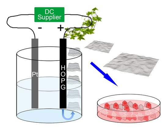
Metal Free Graphene Oxide (GO) Nanosheets and Pristine-Single Wall Carbon Nanotubes (p-SWCNTs) Biocompatibility Investigation: A Comparative Study in Different Human Cell Lines
Abstract
:1. Introduction
2. Results
2.1. Nanomaterials Characterization
2.2. Effects of GO and p-SWCNTs Treatments on Cell Lines
2.3. Morphological Changes in Cell Lines Treated with GO Nanosheets and p-SWCNTs
3. Discussion
4. Materials and Methods
4.1. Materials and Chemical Reagents
4.2. Determination of Metal Content of Nanotube Samples
4.3. Air Oxidation of p-SWCNTs
4.4. Synthesis and Characterization of GO Sheets
4.5. Cell Lines
4.6. LDH Assay
4.7. Statistics
Supplementary Materials
Author Contributions
Funding
Acknowledgments
Conflicts of Interest
References
- Marchesan, S.; Prato, M. Nanomaterials for Nanomedicine. ACS Med. Chem. Lett. 2013, 4, 147–149. [Google Scholar] [CrossRef] [PubMed]
- Krzysztof, A.; Tomaszewski, M.; Radomski, W.; Santos-Martinez, M.J. Review Nanodiagnostics, nanopharmacology and nanotoxicology of platelet–vessel wall interactions. Nanomedicine 2015, 10, 1451–1475. [Google Scholar]
- Zhang, X.; Hu, W.; Li, J.; Tao, L.; Wei, Y.A. comparative study of cellular uptake and cytotoxicity of multi-walled carbon nanotubes, graphene oxide, and nanodiamond. Toxicol. Res. 2012, 1, 62–68. [Google Scholar] [CrossRef]
- Conde, J.; Edelmana, E.R.; Artzia, N. Target-responsive DNA/RNA nanomaterials for microRNA sensing and inhibition: The jack-of-all-trades in cancer nanotheranostics? Adv. Drug Deliv. Rev. 2015, 81, 169–183. [Google Scholar] [CrossRef] [PubMed]
- Hollanda, L.M.; Lobo, A.O.; Lancellotti, M.; Berni, E.; Corat, E.J.; Zanin, H. Graphene and carbon nanotube nanocomposite for gene transfection. Mater. Sci. Eng. 2014, 39, 288–298. [Google Scholar] [CrossRef] [PubMed]
- Daniel, R.; Dreyer, S.; Park, C.; Bielawski, W.; Rodney, S.R. The chemistry of graphene oxide. Chem. Soc. Rev. 2010, 39, 228–240. [Google Scholar]
- Hernandez, Y.; Feng, X.; Müllen, K.; Chen, L. From Nanographene and Graphene Nanoribbons to Graphene Sheets: Chemical Synthesis. Angew. Chem. Int. Ed. 2012, 51, 7640–7654. [Google Scholar]
- Gollavelli, G.; Ling, Y.-C. Multi-functional graphene as an in vitro and in vivo imaging probe. Biomaterials 2012, 33, 2532–2545. [Google Scholar] [CrossRef] [PubMed]
- Wang, Y.; Li, Z.; Wang, J.; Li, J.; Lin, Y. Review: Graphene and graphene oxide: Biofunctionalization and applications in biotechnology. Trends Biotechnol. 2011, 29, 205–212. [Google Scholar] [CrossRef] [PubMed]
- Yang, K.; Feng, L.; Shi, X.; Liu, Z. Review Article Nano-graphene in biomedicine: Theranostic applications. Chem. Soc. Rev. 2013, 42, 530–547. [Google Scholar] [CrossRef] [PubMed]
- Yang, K.; Wan, J.; Zhang, S.; Zhang, Y.; Lee, S.-T.; Liu, Z. In Vivo Pharmacokinetics, Long-Term Biodistribution, and Toxicology of PEGylated Graphene in Mice. ACS Nano 2011, 5, 516–522. [Google Scholar] [CrossRef] [PubMed]
- Mehrali, M.; Moghaddam, E.; Shirazi, S.F.S.; Baradaran, S.; Mehrali, M.; Latibari, S.T.; Metselaar, H.S.C.; Kadri, N.A.; Zandi, K.; Azuan, N.; et al. Synthesis, Mechanical Properties, and in Vitro Biocompatibility with Osteoblasts of Calcium Silicate–Reduced Graphene Oxide Composites. ACS Appl. Mater. Interfaces 2014, 6, 3947–3962. [Google Scholar] [CrossRef] [PubMed]
- Wu, Q.; Zhao, Y.; Fang, J.; Wang, D. Immune response is required for the control of in vivo translocation and chronic toxicity of graphene oxide. Nanoscale 2014, 6, 5894–5906. [Google Scholar] [CrossRef] [PubMed]
- Yang, K.; Li, Y.; Tan, X.; Peng, R.; Liu, Z. Review Behavior and Toxicity of Graphene and Its Functionalized Derivatives in Biological Systems. Small 2013, 9, 1492–1503. [Google Scholar] [CrossRef] [PubMed]
- Fraczek, A.; Menaszek, E.; Paluszkiewicz, C.; Blazewicz, M. Comparative in vivo biocompatibility study of single- and multi-wall carbon nanotubes. Acta Biomater. 2008, 4, 1593–1602. [Google Scholar] [CrossRef] [PubMed]
- Lacerda, L.; Bianco, A.; Prato, M.; Kostarelos, K. Carbon nanotubes as nanomedicines: From toxicology to pharmacology. Adv. Drug Deliv. Rev. 2006, 58, 1460–1470. [Google Scholar] [CrossRef] [PubMed]
- Chen, G.; Seki, Y.; Kimura, H.; Sakurai, S.; Yumura, M.; Hata, K.; Futaba, D.N. Diameter control of single-walled carbon nanotube forests from 1.3–3.0 nm by arc plasma deposition. Sci. Rep. 2014, 4, 3804–3810. [Google Scholar] [CrossRef] [PubMed]
- Naahidi, S.; Jafari, M.; Edalat, F.; Raymond, K.; Khademhosseinic, A.; Chen, P. Review Biocompatibility of engineered nanoparticles for drug delivery. J. Control. Release 2013, 166, 182–194. [Google Scholar] [CrossRef] [PubMed]
- Pietroiusti, A.; Massimiani, M.; Fenoglio, I.; Colonna, M.; Valentini, F.; Palleschi, G.; Camaioni, A.; Magrini, A.; Siracusa, G.; Bergamaschi, A.; et al. Low Doses of Pristine and Oxidized Single-Wall Carbon Nanotubes Affect Mammalian Embryonic Development. ACS Nano 2011, 5, 4624–4633. [Google Scholar] [CrossRef] [PubMed] [Green Version]
- Li, N.; Zhang, Q.; Gao, S.; Song, Q.; Huang, R.; Wang, L.; Liu, L.; Dai, J.; Tang, M.; Cheng, G. Three-dimensional graphene foam as a biocompatible and conductive scaffold for neural stem cells. Sci. Rep. 2013, 3, 1604–1609. [Google Scholar] [CrossRef] [PubMed]
- Liu, Q.; Guo, B.; Rao, Z.; Zhang, B.; Gong, J.R. Strong Two-Photon-Induced Fluorescence from Photostable, Biocompatible Nitrogen-Doped Graphene Quantum Dots for Cellular and Deep-Tissue Imaging. Nano Lett. 2013, 13, 2436–2441. [Google Scholar] [CrossRef] [PubMed]
- Tu, Q.; Tian, C.; Ma, T.; Pang, L.; Wang, J. Click synthesis of quaternized poly (dimethylaminoethyl methacrylate) functionalized graphene oxide with improved antibacterial and antifouling ability. Colloids Surf. B Biointerfaces 2016, 141, 196–205. [Google Scholar] [CrossRef] [PubMed]
- Gurunathan, S.; Han, J.W.; Eppakayala, V.; Kim, J.-H. Biocompatibility of microbially reduced graphene oxide in primary mouse embryonic fibroblast cells. Colloids Surf. B Biointerfaces 2013, 105, 58–66. [Google Scholar] [CrossRef] [PubMed]
- Chang, Y.; Yang, S.-T.; Liu, J.-H.; Dong, E.; Wang, Y.; Cao, A.; Liu, Y.; Wang, H. In vitro toxicity evaluation of graphene oxide on A549 cells. Toxicol. Lett. 2011, 200, 201–210. [Google Scholar] [CrossRef] [PubMed]
- Liao, K.-H.; Lin, Y.-S.; Macosko, C.W.; Christy, L.H. Cytotoxicity of Graphene Oxide and Graphene in Human Erythrocytes and Skin Fibroblasts. ACS Appl. Mater. Interfaces 2011, 3, 2607–2615. [Google Scholar] [CrossRef] [PubMed]
- Visentin, S.; Barbero, N.; Musso, S.; Mussi, V.; Biale, C.; Ploeger, R.; Viscardi, G.A. Sensitive and Practical Fluorimetric Test for CNT Acidic Site Determination. Chem. Commun. 2010, 46, 1443–1445. [Google Scholar] [CrossRef] [PubMed]
- Li, J.; Zhang, W.; Chung, T.F.; Slipchenko, M.N.; Chen, Y.P.; Cheng, J.X.; Yang, C. Highly sensitive transient absorption imaging of graphene and graphene oxide in living cells and circulating blood. Sci. Rep. 2015, 5, 12394–12402. [Google Scholar] [CrossRef] [PubMed]
- Vimbela, G.V.; Ngo, S.M.; Fraze, C.; Yang, L.; Stout, D.A. Antibacterial properties and toxicity from metallic nanomaterials. Int. J. Nanomed. 2017, 12, 3941–3965. [Google Scholar] [CrossRef] [PubMed]
- Allegri, M.; Perivoliotis, D.K.; Bianchi, M.G.; Chiu, M.; Pagliaro, A.; Koklioti, M.A.; Trompeta, A.F.A.; Bergamaschi, E.; Bussolati, O.; Charitidis, C.A. Toxicity determinants of multi-walled carbon nanotubes: The relationship between functionalization and agglomeration. Toxicol. Rep. 2016, 3, 230–243. [Google Scholar] [CrossRef] [PubMed]
- Orlanducci, S.; Valentini, F.; Piccirillo, S.; Terranova, M.L.; Botti, S.; Ciardi, R.; Rossi, M.; Palleschi, G. Chemical/structural characterization of carbon nanoparticles produced by laser pyrolysis and used for nanotube growth. Mater. Chem. Phys. 2004, 87, 190–195. [Google Scholar] [CrossRef]
- Asbach, C.; Alexander, C.; Clavaguera, S.; Dahmann, D.; Dozol, H.; Faure, B.; Fierz, M.; Fontana, L.; Iavicoli, I.; Kaminski, H.; et al. Review of measurement techniques and methods for assessing personal exposure to airborne nanomaterials in workplaces. Sci. Total Environ. 2017, 603–604, 793–806. [Google Scholar] [CrossRef] [PubMed]
- Mari, E.; Mardente, S.; Morgante, E.; Tafani, M.; Lococo, E.; Fico, F.; Valentini, F.; Zicari, A. Graphene Oxide Nanoribbons Induce Autophagic Vacuoles in Neuroblastoma Cell Lines. Int. J. Mol. Sci. 2016, 17, 1995. [Google Scholar] [CrossRef] [PubMed]
- Bhamidipati, M.; Fabris, L. Multiparametric Assessment of Gold Nanoparticle Cytotoxicity in Cancerous and Healthy Cells: The Role of Size, Shape, and Surface Chemistry. Bioconjug. Chem. 2017, 28, 449–460. [Google Scholar] [CrossRef] [PubMed]
- Kane, A.B.; Hurt, R.H. The asbestos analogy revisited. Nat. Nanotoxycol. 2008, 3, 378–379. [Google Scholar] [CrossRef] [PubMed]
- Pei, S.; Wei, Q.; Huang, K.; Cheng, H.-M.; Ren, W. Green synthesis of graphene oxide by seconds timescale water electrolytic oxidation. Nat. Commun. 2018, 9, 145–154. [Google Scholar] [CrossRef] [PubMed]
- Valentini, F.; Amine, A.; Orlanducci, S.; Terranova, M.L.; Palleschi, G. Carbon nanotube purification: Preparation and characterization of carbon nanotube paste electrodes. Anal. Chem. 2003, 75, 5413–5421. [Google Scholar] [CrossRef] [PubMed]
- Lacerda, L.; Pastorin, G.; Gathercole, D.; Buddle, J.; Prato, M.; Bianco, A.; Kostarelos, K. Intracellular Trafficking of Carbon Nanotubes by Confocal Laser Scanning Microscopy. Adv. Mater. 2007, 19, 1480–1484. [Google Scholar] [CrossRef]


| Chemical Properties | Metallic Elements |
| Elemental analysis a (% w/w) | Si (n.d) S (n.d) Ca (n.d.) Cr (n.d.) Fe (n.d.) Co (n.d.) |
| Physical Properties | Range Values |
| Surface Area (µm2) b | 0.1–3.0 |
| Thickness (nm) b | 1.2 ± 0.3 |
| Number of Layer b | 2 (bilayer) |
| Weight loss % (TGA) c | 0.42 ± 0.40 |
| Acidic sites (nmol/mg) d | 4.02 ± 0.23 |
| Extent of defects (ID/IG) e | 0.10 |
| Peak BE (eV) | C1s At. % | Functional Groups |
|---|---|---|
| 284.4 | 59.0 | C-C |
| 285.4 | 16.0 | C-OH |
| 286.6 | 14.0 | C-O |
| 287.7 | 7.0 | C=O |
| 289.0 | 4.0 | C(O)O |
| 290.7 | 2.2 | π–π * |
| Chemical Properties | Metallic Elements |
| Elemental analysis a (% w/w) | Si 0.20 ± 0.06 S 0.47 ± 0.04 Ca 1.14 ± 0.31 Cr 1.03 ± 0.15 Fe 1.00 ± 0.25 Co 3.57 ± 0.56 |
| Physical Properties | Range Values |
| Diameter (nm) b | 2.35 ± 0.40 |
| Length (µm) b | 0.82 ± 0.42 |
| Weight loss % (TGA) c | 0.50 ±0.50 |
| Acidic sites (nmol/mg) d | 24.25 ± 1.10 |
| Extent of defects (ID/IG) e | 0.94 |
| A. EaHy926 | |||||
| Time (h) | CTRL | p-SWCNTs 0.2 μg/mL | p-SWCNTs 2 μg/mL | GO 0.2 μg/mL | GO 2 μg/mL |
| 6 | 99.40 | 94.40 | 84.32 | 95.50 | 95.20 |
| 12 | 98.20 | 80.20 | 78.20 | 92.60 | 91.60 |
| 24 | 98.20 | 70.20 | 62.50 | 92.60 | 89.40 |
| 48 | 98.20 | 56.60 | 47.30 | 92.00 | 88.60 |
| B. HEp-2 | |||||
| Time (h) | CTRL | p-SWCNTs 0.2 μg/mL | p-SWCNTs 2 μg/mL | GO 0.2 μg/mL | GO 2 μg/mL |
| 6 | 98.60 | 90.30 | 77.30 | 94.50 | 93.80 |
| 12 | 97.80 | 80.00 | 62.20 | 89.80 | 86.00 |
| 24 | 96.00 | 69.00 | 58.00 | 87.00 | 82.00 |
| 48 | 91.20 | 64.20 | 45.30 | 81.40 | 80.00 |
| C. HUVEC | |||||
| Time (h) | CTRL | p-SWCNTs 0.2 μg/mL | p-SWCNTs 2 μg/mL | GO 0.2 μg/mL | GO 2 μg/mL |
| 6 | 98.30 | 92.40 | 82.40 | 98.00 | 97.50 |
| 12 | 97.60 | 85.60 | 70.70 | 97.80 | 96.20 |
| 24 | 98.20 | 72.20 | 62.80 | 96.70 | 95.70 |
| 48 | 97.30 | 66.00 | 58.20 | 96.50 | 94.20 |
| D. HeLa | |||||
| Time (h) | CTRL | p-SWCNTs 0.2 μg/mL | p-SWCNTs 2 μg/mL | GO 0.2 μg/mL | GO 2 μg/mL |
| 6 | 97.20 | 85.70 | 84.60 | 96.30 | 95.20 |
| 12 | 98.50 | 75.30 | 73.90 | 96.60 | 94.20 |
| 24 | 97.60 | 72.20 | 65.00 | 95.40 | 91.60 |
| 48 | 96.70 | 67.00 | 65.00 | 93.20 | 89.20 |
| A. EaHy926 | |||||
| Time (h) | CTRL | p-SWCNTs 0.2 μg/mL | p-SWCNTs 2 μg/mL | GO 0.2 μg/mL | GO 2 μg/mL |
| 12 | 0.16 | 0.28 (12%) | 0.32 (15.4%) | 0.18 (1.9%) | 0.23 (6.7%) |
| 24 | 0.18 | 0.42 (23.5%) | 0.52 (33.3%) | 0.22 (3.9%) | 0.28 (9.8%) |
| 48 | 0.25 | 0.54 (30.5%) | 0.66 (43%) | 0.36 (11.6%) | 0.40 (15.8%) |
| B. HEp-2 | |||||
| Time (h) | CTRL | p-SWCNTs 0.2 μg/mL | p-SWCNTs 2 μg/mL | GO 0.2 μg/mL | GO 2 μg/mL |
| 12 | 0.18 | 0.38 (16.6%) | 0.42 (23.5%) | 0.19 (1.0%) | 0.25 (6.8%) |
| 24 | 0.21 | 0.41 (20.2%) | 0.50 (29.3%) | 0.24 (2.9%) | 0.27 (6%) |
| 48 | 0.26 | 0.46 (21.3%) | 1.22 (102%) | 0.29(3.2%) | 0.35 (9%) |
| C. HeLa | |||||
| Time (h) | CTRL | p-SWCNTs 0.2 μg/mL | p-SWCNTs 2 μg/mL | GO 0.2 μg/mL | GO 2 μg/mL |
| 12 | 0.14 | 0.18 (1.9%) | 0.30 (15%) | 0.14 (0%) | 0.15 (0.94%) |
| 24 | 0.15 | 0.28 (13.2%) | 0.31 (16%) | 0.16 (0.9%) | 0.22 (6.6%) |
| 48 | 0.16 | 0.49 (31.7%) | 0.59 (41.3%) | 0.18 (1.9%) | 0.28 (11.5%) |
| D. HUVEC | |||||
| Time (h) | CTRL | p-SWCNTs 0.2 μg/mL | p-SWCNTs 2 μg/mL | GO 0.2 μg/mL | GO 2 μg/mL |
| 12 | 0.12 | 0.31 (17.6%) | 0.45 (30.6%) | 0.13 (0.9%) | 0.22 (9.3%) |
| 24 | 0.14 | 0.34 (18.9%) | 0.50 (33.9%) | 0,.15 (0.9%) | 0.26 (11.3%) |
| 48 | 0.15 | 0.43 (26.6%) | 0.80 (61.9%) | 0.17 (1.9%) | 0.31 (15.4%) |
© 2018 by the authors. Licensee MDPI, Basel, Switzerland. This article is an open access article distributed under the terms and conditions of the Creative Commons Attribution (CC BY) license (http://creativecommons.org/licenses/by/4.0/).
Share and Cite
Valentini, F.; Mari, E.; Zicari, A.; Calcaterra, A.; Talamo, M.; Scioli, M.G.; Orlandi, A.; Mardente, S. Metal Free Graphene Oxide (GO) Nanosheets and Pristine-Single Wall Carbon Nanotubes (p-SWCNTs) Biocompatibility Investigation: A Comparative Study in Different Human Cell Lines. Int. J. Mol. Sci. 2018, 19, 1316. https://doi.org/10.3390/ijms19051316
Valentini F, Mari E, Zicari A, Calcaterra A, Talamo M, Scioli MG, Orlandi A, Mardente S. Metal Free Graphene Oxide (GO) Nanosheets and Pristine-Single Wall Carbon Nanotubes (p-SWCNTs) Biocompatibility Investigation: A Comparative Study in Different Human Cell Lines. International Journal of Molecular Sciences. 2018; 19(5):1316. https://doi.org/10.3390/ijms19051316
Chicago/Turabian StyleValentini, Federica, Emanuela Mari, Alessandra Zicari, Andrea Calcaterra, Maurizio Talamo, Maria Giovanna Scioli, Augusto Orlandi, and Stefania Mardente. 2018. "Metal Free Graphene Oxide (GO) Nanosheets and Pristine-Single Wall Carbon Nanotubes (p-SWCNTs) Biocompatibility Investigation: A Comparative Study in Different Human Cell Lines" International Journal of Molecular Sciences 19, no. 5: 1316. https://doi.org/10.3390/ijms19051316
APA StyleValentini, F., Mari, E., Zicari, A., Calcaterra, A., Talamo, M., Scioli, M. G., Orlandi, A., & Mardente, S. (2018). Metal Free Graphene Oxide (GO) Nanosheets and Pristine-Single Wall Carbon Nanotubes (p-SWCNTs) Biocompatibility Investigation: A Comparative Study in Different Human Cell Lines. International Journal of Molecular Sciences, 19(5), 1316. https://doi.org/10.3390/ijms19051316


