Article

CPR Gene Contributes to Integument Function and Ovary Development in a Rice Planthopper

1
Institute of Insect Sciences, Ministry of Agriculture Key Lab of Molecular Biology of Crop Pathogens and Insect Pests, College of Agriculture and Biotechnology, Zhejiang University, Hangzhou 310058, China
2
Key Laboratory of Biology of Crop Pathogens and Insects of Zhejiang Province, Zhejiang University, Hangzhou 310058, China
3
Zhejiang Provincial Key Laboratory of Biometrology and Inspection and Quarantine, College of Life Science, China Jiliang University, Hangzhou 310018, China
4
State Key Laboratory for Managing Biotic and Chemical Threats to the Quality and Safety of Agro-products, Key Laboratory of Biotechnology in Plant Protection of Ministry of Agriculture and Zhejiang Province, Institute of Plant Virology, Ningbo University, Ningbo 315211, China
*
Authors to whom correspondence should be addressed.
Int. J. Mol. Sci. 2022, 23(5), 2875; https://doi.org/10.3390/ijms23052875
Submission received: 20 January 2022 / Revised: 4 March 2022 / Accepted: 5 March 2022 / Published: 6 March 2022
(This article belongs to the Section Molecular Plant Sciences)

Abstract

:
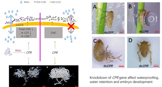
Nicotinamide adenine dinucleotide phosphate (NADPH)-cytochrome P450 reductase (CPR) is an essential enzyme that transfers electrons from NADPH to cytochrome P450 monooxygenases. CPR is involved in cuticular hydrocarbon (CHC) synthesis in insects and is vital for insect development and survival. Here, we clarify the physiological function of a CPR gene in Nilaparvata lugens, an important rice pest, by using RNA interference. CPR gene knockdown leads to the functional loss of waterproofing and water retention in the integument of female adults, which causes significantly reduced body weight and a lethal phenotype. Scanning electron microscopy shows that the lipid layer on the outermost surface of the abdominal cuticle becomes thin in dsCPR-injected adults. Furthermore, CHC profile analysis reveals that CPR knockdown significantly decreases the contents of CHCs with a carbon chain length ≥ C27 in adult females. Moreover, we find that CPR knockdown generates a deficient phenotype in ovaries with deformed oocytes and a complete failure of egg-laying. These findings suggest that CPR plays multiple functional roles in CHC biosynthesis and embryo development in insects.

Graphical Abstract

1. Introduction

Nicotinamide adenine dinucleotide phosphate (NADPH)-cytochrome P450 reductase (CPR) is a cofactor of cytochrome P450 monooxygenases (P450s) that donates electrons from NADPH to the heme center of P450s [1,2]. CPR belongs to the flavoprotein family and contains an N-terminal transmembrane region and three conserved binding domains for flavin mononucleotide (FMN), flavin adenine dinucleotide (FAD), and NADPH [3,4,5]. CPRs are present in all living organisms. Several CPRs have been identified and characterized in insects, such as Drosophila melanogaster [6,7], Helicoverpa armigera [8], Nilaparvata lugens [9], Rhopalosiphum padi (L.) [10], Laodelphax striatellus (fallén) [11], Locusta migratoria [12], Cnaphalocrocis medinalis [12], Spodoptera Litura [13], Bemisia tabaci [14], Acyrthosiphon pisum [15], and Diaphorina citri [16].
P450 enzymes catalyze the oxidative metabolism of various endogenous and xenobiotic substrates, including insecticides, steroids, and cuticular hydrocarbons (CHCs). As cofactors of P450s, CPRs are also essential for the oxidative metabolism of different substrates. It is known that CPRs are closely related to susceptibility to insecticides and CHC biosynthesis in insects [7,17].
Insect CHCs are composed of straight and methyl-branched saturated alkanes and unsaturated alkenes. The lipid layer covering insect cuticles is composed of CHCs, which provide resistance to chemical compounds and prevent the movement of water through insect cuticles [17,18,19]. CHCs are generated from fatty acids, which are catalyzed by fatty acid synthases (FAS), elongases (ELO), desaturases, fatty acyl-CoA reductases (FAR), and CYP4G oxidative decarbonylase [20,21]. CYP4Gs belong to a subfamily of P450 enzymes, which are required for transferring the electron from CPR in the last step of CHC synthesis [7]. CHCs participate in the formation of the lipid layer, which provides the function of waterproofing and water retention in insects [22,23]. CPRs have also been found to be expressed in Drosophila melanogaster antennae and embryos at high levels [6], indicating that CPR may play an important role in embryo development.
The brown planthopper, Nilaparvata lugens Stål (Hemiptera: Delphacidae), is one of the most destructive rice pests and is able to adapt to variable environments during long-distance migration [24,25]. By living in different humidities, such as extremely high humidities in paddy fields and volatile environments during migration, waterproofing and water retention are critical to N. lugens survival. Previous studies have reported that the knockdown of the CPR gene increases the susceptibility of N. lugens to insecticides [9]. However, it is unknown whether the CPR is involved in CHC synthesis during the development process in N. lugens.
In this study, we focus on the CPR gene to explore its functional roles in N. lugens. The expression profile of the CPR gene shows high transcript levels in the ovaries and eggs. Functional analysis using RNAi, waterproofing, and water retention testing, plus scanning electron microscopy combined with CHC profile analysis, revealed that CPR is involved in CHC biosynthesis and embryo development in N. lugens. Our findings are the first to report that CPR has important functions in oocyte development in female ovaries. Our data provide useful target molecules for the biological control of rice pests, which could be used to reduce pesticide usage in rice planting areas.

2. Results

2.1. Development Stage and Tissue Expression Specificities of the CPR Gene

To explore the functional roles of the CPR gene, we first analyzed the developmental-stage expression using qRT-PCR. Throughout all developmental stages, the CPR transcripts were detected at very high levels in eggs laid from 0 to 144 h (Figure 1A). Moreover, high transcript levels were found in the 1st-instar nymphs at 0–48 h. Intriguingly, extremely low levels were detected in the 2nd–4th instar nymphal stages, and these gradually increased in the 5th-instar nymphal stage at 0–72 h. High transcript levels were then achieved after eclosion in the adults. The high transcript levels in the eggs and adults implicated that CPR might play a role during egg development and reproduction. Subsequently, we investigated the tissue-specific expression of CPR in adults. The highest transcript levels were detected in the ovaries and oocytes, while relatively low levels were detected in the fat bodies, integuments, guts, and testes. These data suggest that CPR may play an important role during the oocyte development in female ovaries (Figure 1B). Along with the development expression patterns found, we hypothesize that CPR is involved in oocyte maturation in the ovaries and egg development after oviposition.

2.2. RNAi Result

To understand the functions of the CPR gene in waterproofing and water retention, we performed RNAi by injecting dsCPR in the 4th-instar nymphs to knock down the gene expression. Waterproofing assay showed that no water droplets remained on the cuticles of the dsGFP-treated nymphs after spaying water on the insects (Figure 2A), suggesting their good waterproofing function; meanwhile, the cuticles of the dsCPR-injected nymphs were covered with water, forming a big water droplet (Figure 2B) and indicating that the waterproofing function was lost. A similar phenotype was observed when we soaked the dsRNA-treated insects in water. The dsCPR-injected nymphs that had been submerged were not able to move and surface from the water (Video S1); meanwhile, the dsGFP-treated nymphs kept afloat on the water and could jump to the wall of the glass tube (Video S2). We observed that the dsCPR-injected nymphs had molting deficiency (Figure 2C). These insects could not complete molting as the exuviae were stuck to the abdomen and legs. Moreover, some individuals could complete molting but showed an abnormal phenotype with a shrunk abdomen, which led to a lethal phenotype (Figure 2D). In contrast, the dsGFP-injected nymphs successfully completed molting. Those insects showed normal abdomens (Figure 2A). The qRT-PCR result confirmed the high interference efficiency of the CPR gene (Figure 2E).
To understand the functions of the CRP gene in water retention, we measured the body weights of the dsRNA-injected insects at a relative humidity of 50 ± 5% and performed desiccation experiments by keeping the adult females under different humidity conditions at relative humidities of <5%, =50 ± 5%, and >95%. The body weights of the adult females that developed from dsCPR-injected 5th instar nymphs were significantly decreased when compared to those of the dsGFP-injected controls (Figure 3A). Under desiccation conditions at a relative humidity < 5%, the median survival time of dsCPR-treated insects, which was 2 h, was reduced by 2.5 times compared to that of the dsGFP-treated insects (6 h). Under the medium-humidity condition at a relative humidity of 50 ± 5%, the median survival time of the dsCPR-treated insects (6 h) was reduced by 2.17 times compared to that of the dsGFP-treated insects (13 h). Under the high-humidity condition at a relative humidity > 95%, 100% of the dsGFP-treated insects stayed alive for at least 16 h, while the dsCPR-treated insects started to die at 3 h and had a 20% mortality rate at 16 h (Figure 3B).
Subsequently, we observed the ovaries of dsRNA-injected female adults. The ovaries of dsCPR-treated females at 3 days displayed the deficient phenotype, especially in the oocytes (Figure 4A). The oocytes did not have the typical banana shapes in their ovaries, while the dsGFP-treated females had normal ovaries containing banana-shaped oocytes (Figure 4B). Then, we calculated the egg number of dsRNA-treated females which mated with non-injected males. The result showed each dsGFP-treated female laid 100–150 eggs within ten days but that the dsCPR-treated females laid very few eggs in the rice seedlings (Figure 4C).

2.3. Effect of CPR Knockdown on CHC Levels

To confirm the waterproofing and water retention functions of the CPR gene in the insect cuticle, we used scanning electron microscopy to observe the outermost layer of the abdominal cuticles of the dsGFP- and dsCPR-injected N. lugens. The images showed that a lipid layer covered the cuticle of dsGFP-treated N. lugens nymphal abdomen, suggesting its functions associated with the waterproofing and water retention (Figure 5A). However, the lipid layer was thin on the cuticle surface of the dsCPR-treated nymphal abdomen (Figure 5B). In the controls, the lipid layer was disrupted by n-hexane treatment in both dsGFP- and dsCPR-injected nymphs (Figure 5 C, D).
To further confirm whether the CPR gene knockdown caused a change in the CHC and a thinning in the lipid layer, we investigated the CHC levels in the dsGFP- and dsCPR-injected females 48 h after eclosion using gas chromatography–mass spectrometry (GC-MS). The results showed that the CHCs in adult females consisted of n-alkanes with various chain lengths, from C15 to C33, with C21 (n-heneicosane) used as an internal standard. The main component of CHCs in dsGFP-injected females was C29H60, which accounted for 32.55% of the total CHCs (Table 1). The dsCPR injection significantly decreased the total level of CHCs by 24.36%. The levels of C18, C19, and C20 were significantly increased by 11.11%, 13.04%, and 12.55%, and the levels of C27, C28, C29, C30, C31, and C33 were significantly decreased by 8.17%, 41.77%, 30.93%, 68.31%, 31.78%, 51.07%, and 21.20%, respectively (Figure 6). The proportion change of each CHC content was distinct. The proportion of n-alkanes with a chain length ≤ C26 increased by more than 20% (21.40–89.24%). However, the proportion of n-alkanes with a chain length of C27–C31 decreased. Among them, the proportion of C29 (as the main component) decreased by 58.1% (Table 1). The CHC profile changed dramatically, with the proportion of C15–C26 n-alkanes increasing from 53.94% to 75.48% and the proportion of C27–C33 n-alkanes decreasing from 46.06% to 24.52%.

3. Discussion

As an electron donor of P450s, the CPR gene plays a critical role in the metabolism of endogenous and xenobiotic substrates [5]. A previous study predicted the structure of the deduced CPR protein in N. lugens, which contains four conserved structural features: the N-terminal transmembrane region and the FMN-, FAD-, and NADP-binding domains [9]. N. lugens CPR shares a high sequence identity with the Drosophila melanogaster homolog. It is known that D. melanogaster CPR is involved in CHC biosynthesis and embryo development [6,7]. We suppose that N. lugens CPR may have potential functions in CHC biosynthesis and embryo development. In this study, we characterized the physiological function of the CPR gene in N. lugens by RNAi. The knockdown of the CPR transcript levels changed the CHC profile in female adults, leading to the function loss of waterproofing and water retention and inhibiting the embryo development in the female ovaries.
The CPR transcripts throughout all the developmental stages showed very high levels in the eggs, the 1st-instar nymphs, and the adults, suggesting that CPR might be involved in embryo development. The CPR transcripts in different tissues were at much higher levels in the ovaries and oocytes than in the other tissues, further indicating that CPR played an important role in oocyte maturity and development in adult ovaries.
To understand the functions of the CPR gene, we performed RNAi to knock down the transcript levels of CPR. The results showed that CPR knockdown led to waterproofing and water retention loss and generated lethal phenotypes, including molting failure and abdominal shrinkage. The body weights of CPR-knocked-down N. lugens were lighter than those of the dsGFP-treated controls, suggesting that the water retention function of the insect cuticle was lost. To better understand the physiological functions, we observed the survival of the dsRNA-injected N. lugens under different relative humidities. The survival time of the dsCPR-injected insects was much shorter than that of the dsGFP-injected ones. Under desiccation condition, the survival rates of dsCPR-injected insects dropped rapidly, and the median survival time was only 40% that of the control group. With the relative humidity increased to 50 ± 5%, the median survival time of dsRNA-treated N. lugens rose, but the median survival time of the dsCPR-injected ones was still much shorter than that of the control. Under a high relative humidity, no death was found in the dsGFP-injected N. lugens within 16 h, indicating that the water retention function was efficient. These results confirmed that CPR is involved in waterproofing and water retention functions. Previous studies have demonstrated that the CYP4G family is involved in the cuticle hydrocarbon biosynthesis [7,26]. As an indispensable component of the P450/CPR system, CPR may infect the CHC profile in a similar way and lead to the loss of waterproofing and water retention functions.
In the CHC profile assay, we first observed the lipid layer in the outermost cuticle of dsRNA-treated N. lugens insects. The lipid layer in the cuticle is the shield protecting the insects under humid or desiccated environments [23]. In this study, we found that the knockdown of CPR impaired the lipid layer and resulted in water loss under desiccation conditions and water being retained in the cuticle under humid conditions. Subsequently, we compared the CHC profiles of dsRNA-treated N. lugens female adults. CPR knockdown significantly increased the C18, C19, and C20 contents; increased the C16–C26 contents to a certain extent; and significantly decreased the C27, C28, C29, C30, C31, C33, and total CHC contents. The proportion of CHCs with a carbon chain length ≤ C26 increased by nearly 50%, and the proportion of C27–C33 n-alkanes decreased by nearly 50%. Our data indicated that the decreased total CHCs (especially C29, as the main component) and the change in the CHC profile caused the lipid layer to become thin and led to the failure of waterproofing and water retention. These results imply that the CHCs with a carbon chain length ≥ C27 may be the main functional aspects of waterproofing and water retention. The change in the CHC contents is likely to be a reason for the function loss of waterproofing and water retention.
In addition to the waterproofing and water retention functions, in this study we found for the first time that CPR knockdown generated deficient ovaries with deformed oocytes. These observations indicate that the CPR gene may participate in the regulation of embryo development and oviposition. We will clarify the function of CPR in female ovaries in future experiments to reveal the functional link between CPR and oocyte maturity and development in female ovaries.

4. Materials and Methods

4.1. Insects

N. lugens populations were obtained and maintained in our lab, as previously described [27]. The N. lugens populations were originally collected from a rice field in the Huajiachi Campus of Zhejiang University, Hangzhou, China. The insects were maintained for at least 50 generations at 26 ± 0.5 °C with a 50 ± 5% relative humidity on rice seedlings (Oryza sativa strain Xiushui 134) under a 16:8 h light/dark photoperiod.

4.2. Cloning of a N. lugens CPR cDNA

The CPR cDNA sequence was obtained by searching the N. lugens transcriptome database in the Sequence Read Archive database (http://www.ncbi.nlm.nih.gov/sra, accessed date: 7 January 2019) (SRA accession number SRX023419) [28]. Total RNAs were extracted from the whole bodies of the various developmental stages using RNAiso Plus Total RNA extraction reagent (Takara, Dalian, China). First-strand cDNA was synthesized using HiScript® II Q RT SuperMix for qPCR with gDNA wiper Mix (Vazyme Biotech, Nanjing, China). A pair of primers (sense: 5′-ATGGAGGTGGAGGCTGACTTAG-3′; antisense: 5′-TCAACTCCATACGTCGGCCG-3′) were designed to amplify the complete open reading frame (ORF) of the CPR gene. PCR was performed using 2 × Phanta® Max Master Mix (Vazyme Biotech, Nanjing, China). The PCR product was cloned into the pMD19-T vector (Takara, Dalian, China) and sequenced by Sanger sequencing (Tsingke Biotech, Beijing, China).

4.3. Quantitative Real-Time qPCR (qRT-PCR) Analysis

Developmental stage- and tissue-expression patterns of the CPR gene were examined by qRT-PCR, as previously described [27]. Briefly, total RNAs were extracted from different tissues of N. lugens adults (fat body, gut, integument, testis, ovary, and oocyte) and the different developmental stages (eggs, nymphs, and adult males and females). The RNAs were reverse transcribed using a HiScript® II Q RT SuperMix for qPCR with gDNA wiper Mix (Vazyme Biotech, Nanjing, China). qRT-PCR was performed in a 20 μL reaction volume containing 2 μL of 10-fold diluted cDNA, 0.6 μL of each primer and 10 μL of ChamQ SYBR Color qPCR Master Mix (Vazyme Biotech, Nanjing, China) using a Bio-Rad CFX96 real-time PCR system (Bio-Rad, Hercules, CA, USA). The gene-specific primers (sense: 5′-CTCGTGCCAGTCTTCATC-3′; antisense: 5′-CATACTCCTCAAGTTCCTCTT-3′) were used and the N. lugens 18S ribosomal RNA gene (GenBank No. JN662398) was used as an internal control to normalize the target gene expression using the following primers: 5′-CGCTACTACCGATTGAA-3′ (sense) and 5′-GGAAACCTTGTTACGACTT-3′ (antisense). The qRT-PCR reaction was performed using the following parameters: 95 °C for 30 s followed by 40 cycles at 95 °C for 5 s and 60 °C for 30 s. Three independent biological replicates were used, and each biological replicate included three technical replicates. The relative transcript levels were evaluated using the 2−ΔΔCt method [29].

4.4. RNAi

Double-stranded RNA (dsRNA) of the CPR gene and an Aequorea victoria green fluorescent protein (GFP) gene used for control were individually synthesized by in vitro transcription with PCR-generated DNA templates using a MEGAscript T7 Transcription Kit (Vazyme, Nanjing, China). The primers used for dsRNA synthesis were sense-1: 5′-taatacgactcactatagggagaATGCCAAGAACGATGATGA-3′ with antisense-1: 5′-taatacgactcactatagggagaGAATAGTAACGACACTGAAGG-3′ and sense-2: 5′-taatacgactcactatagggagaCCGACAAGAAGCCATCTC-3′ with antisense-2: 5′-taatacgactcactatagggagaGCCGATAACATTCCATAGTTC-3′. The quality and concentration of dsRNAs were determined using agarose gel electrophoresis and a Nanodrop 2000 spectrophotometer (Thermo Scientific, Wilmington, DE, USA). The microinjection of dsRNAs was carried out as previously described [25,30,31]. The 4th- and 5th-instar nymphs were used for RNAi. Both the 4th- and 5th-instar nymphs were injected with ~10 or 20 nL of the dsRNA (5 ng/μL). The treated insects were kept on fresh rice seedlings at 26 ± 0.5 °C with a 50 ± 5% humidity under a 16:8 h light/dark photoperiod. Ten insects were randomly collected to evaluate the RNAi efficiency 3 days after dsRNA injection using qRT-PCR. Other insects were used for the calculation of survival rates and for the observation of development.

4.5. Measurement of Body Weight in dsRNA-Injected Insects

The 5th-instar nymphs were injected with dsCPR or dsGFP. Ten adult females, 48 h after eclosion, were pooled and used as independent samples for the measurement of body weight. Nine biological replicates were used for each dsRNA treatment. The dsGFP-injected adults were used as controls. Statistical analyses of the body weights between the different groups were tested using the Student’s t-test. p < 0.01 was considered to be statistically significant.

4.6. Oviposition Experiment

Oviposition was observed according to Peng et al. [32]. Briefly, the 5th-instar nymphs were microinjected with dsCPR. dsGFP-injected nymphs were used as a control. The insects were reared on fresh rice seedlings until emergence. Mating was performed between a single newly emerged female that had been injected with dsCPR and a single non-injected male adult. As a control, mating was performed between a single newly emerged female that had been injected with dsGFP and a single non-injected male adult. The mating was performed in a long glass tube containing three leaf-stage fresh rice seedlings (6.5 ± 0.5 cm long) at 26 ± 0.5 °C with a 50% ± 5% relative humidity under a 16:8 h light/dark photoperiod for 5 days. Then, the female and male adults were moved to a new glass tube containing three leaf-stage fresh rice seedlings for laying eggs for 5 days. The eggs in the rice leaf sheaths were dissected and counted. Biological replicates were used for each mating (n = 8 ♀ × ♂). The statistical significance of the number of laid eggs in the different treated groups was tested by a Student’s t-test. p < 0.01 was considered statistically significant.

4.7. Waterproofing and Water Retention Assay

The 4th-instar nymphs were injected with dsCPR. dsGFP-injected nymphs were used as a control. The newly molted 5th instar nymphs were collected and kept on fresh rice seedlings in a plastic container at 26 °C ± 0.5, with a 50% ± 5 relative humidity, under a 16:8 h light/dark photoperiod, as described previously [20]. Water was sprayed onto the rice seedlings using a mini-sprayer. Insects were pressed into water contained in glass tubes. For a relative humidity treatment assay, the 5th-instar nymphs were injected with dsCPR and dsGFP. Nymphs were subjected to deferent relative humidity treatment using <5%, 50% ± 5, and >95% relative humidity at 26 °C ± 0.5, without food. The survival rates of the treated nymphs were calculated every 30 min.

4.8. Scanning Electron Microscope (SEM) Analysis

For the SEM analysis, the dsCPR- or dsGFP-injected 5th-instar nymphs were immersed in 200 μL of n-hexane or water for 1 min 48 h after molting. Then, insects were placed on a stub and dried in an ion sputter (Hitachi, Tokyo, Japan) under a vacuum [21,30]. After gold sputtering, the outermost layer of the insects was directly observed under SEM (TM-1000, Hitachi, Tokyo, Japan), as previously described. Three insects were observed for each treatment.

4.9. Extraction and Quantification of CHCs

The 5th-instar nymphs were injected with dsCPR. dsGFP-injected nymphs were used as a control. CHCs were extracted from the females at 48 h after eclosion following a procedure used in our previous study [20,21,33]. Briefly, 10 females were immersed in 200 μL of n-hexane with 500 ng of n-heneicosane (C21) as an internal standard. The solvent was stirred gently for 3 min and then drawn with a glass Pasteur pipette into a clean chromatography vial. We repeated this procedure twice, then finally used 200 μL of hexane to rinse the nymphs and vial. We combined all the extracted hexane and dried it to a volume of 200 μL under high-purity nitrogen gas. Then, the hexane extracts were loaded onto an ∼300 mg silica gel (70e230 mesh, Sigma-Aldrich, Louis, MO, USA) mini-column in a glass wool-stoppered Pasteur pipette. The HC fraction was eluted with 2 mL of hexane, left to dry absolutely under nitrogen gas, and resuspended in 100 μL of hexane. The samples were analyzed on a TRACE 1310 (Thermo Scientific, Waltham, MA, USA) gas chromatograph (GC) equipped with an ISQ single quadruple MS and interfaced with the Xcalibur 2.2 software. The constant flow of helium was 1 mL/min. A splitless injection of 1.0 μL was carried out in a 30 m × 0.32 mm × 0.25 μm HP-5MS UI capillary column (Agilent Technologies, Santa Clara, CA, USA). The temperature program was operated at 60 °C for 2 min, then at 5 °C/min to 320 °C, then held for 10 min. The temperatures of the injector and detector were set at 300 and 280 °C, respectively. Mass detection was run under EI mode with a 70 eV ionization potential and an effective m/z range of 45–650 at a scan rate of 5 scan/s.

5. Conclusions

In this study, we confirmed that CPR is a multifunctional enzyme that participates in various biological processes. We verified that CPR is essential for waterproofing and water retention in N. lugens. Our novel findings reveal that CPR is required for embryo development. These findings expand our knowledge of the role of CPR in CHC biosynthesis and embryo development. This work establishes a functional link between CHC biosynthesis and embryo development, which may be a potential molecular target for the biological control of rice pests in the future.

Supplementary Materials

The following supporting information can be downloaded at https://www.mdpi.com/article/10.3390/ijms23052875/s1: Video S1. Phenotype of RNAi. dsCPR was injected in the 4th-instar nymphs at 48 h after hatching. Video S2. Phenotype of RNAi. dsGFP was injected in the 4th-instar nymphs at 48 h after hatching.

Author Contributions

Conceptualization, Y.-Y.B., C.-X.Z. and Z.-C.W. Methodology, formal analysis and investigation, Z.-C.W., S.T., X.C. and D.-T.L. Writing—original draft preparation, Z.-C.W. Writing—review and editing, Y.-Y.B. and C.-X.Z. Funding acquisition, Y.-Y.B. and C.-X.Z. All authors have read and agreed to the published version of the manuscript.

Funding

This research was funded by the Zhejiang Provincial Natural Science Foundation of China under Grant No. LY22C140003 and the National Key Research and Development Program of China (No. 2021YFD1401100).

Institutional Review Board Statement

Not applicable.

Informed Consent Statement

Not applicable.

Data Availability Statement

Not applicable.

Acknowledgments

We thank Yong-Liang Fan of Northwest A&F University for his kind guidance and help in the work of CHC quantification.

Conflicts of Interest

The authors declare no conflict of interest.

References

  1. Louërat-Oriou, B.; Perret, A.; Pompon, D. Differential redox and electron-transfer properties of purified yeast, plant and human NADPH-cytochrome P-450 reductases highly modulate cytochrome P-450 activities. Eur. J. Biochem. 1998, 258, 1040–1049. [Google Scholar] [CrossRef] [PubMed]
  2. Paine, M.J.I.; Scrutton, N.S.; Munro, A.W.; Gutierrez, A.; Roberts, G.C.K.; Wolf, C.R. Electron Transfer Partners of Cytochrome P450. In Cytochrome P450; Ortiz de Montellano, P.R., Ed.; Springer: Boston, MA, USA, 2005. [Google Scholar]
  3. Vermilion, J.L.; Coon, M.J. Highly purified detergent-solubilized NADPH-cytochrome P-450 reductase from phenobarbital-induced rat liver microsomes. Biochem. Biophys. Res. Commun. 1974, 60, 1315–1322. [Google Scholar] [CrossRef] [Green Version]
  4. Porter, T.D.; Wilson, T.E.; Kasper, C.B. Expression of a functional 78,000 dalton mammalian flavoprotein, NADPH-cytochrome P-450 oxidoreductase, in Escherichia coli. Arch. Biochem. Biophys. 1987, 254, 353–367. [Google Scholar] [CrossRef]
  5. Ming, W.; Roberts, D.L. Three-dimensional structure of NADPH-cytochrome P450 reductase: Prototype for FMN- and FAD-containing enzymes. Proc. Natl. Acad. Sci. USA 1997, 94, 8411. [Google Scholar] [CrossRef] [Green Version]
  6. Hovemann, B.T.; Sehlmeyer, F.; Malz, J. Drosophila melanogaster NADPH–cytochrome P450 oxidoreductase: Pronounced expression in antennae may be related toodorantclearance. Gene 1997, 189, 213–219. [Google Scholar] [CrossRef]
  7. Qiu, Y.; Tittiger, C.; Wicker-Thomas, C.; Le Goff, G.; Young, S.; Wajnberg, E.; Fricaux, T.; Taquet, N.; Blomquist, G.J.; Feyereisen, R. An insect-specific P450 oxidative decarbonylase for cuticular hydrocarbon biosynthesis. Proc. Natl. Acad. Sci. USA 2012, 109, 14858–14863. [Google Scholar] [CrossRef] [Green Version]
  8. Liu, D.; Zhou, X.; Li, M.; Zhu, S.; Qiu, X. Characterization of NADPH-cytochrome P450 reductase gene from the cotton bollworm, Helicoverpa armigera. Gene 2014, 545, 262–270. [Google Scholar] [CrossRef]
  9. Liu, S.; Liang, Q.M.; Zhou, W.W.; Jiang, Y.D.; Zhu, Q.Z.; Yu, H.; Zhang, C.X.; Gurr, G.M.; Zhu, Z.R. RNA interference of NADPH-cytochrome P450 reductase of the rice brown planthopper, Nilaparvata lugens, increases susceptibility to insecticides. Pest Manag. Sci. 2015, 71, 32–39. [Google Scholar] [CrossRef]
  10. Wang, K.; Peng, X.; Zuo, Y.; Li, Y.; Chen, M. Molecular Cloning, Expression Pattern and Polymorphisms of NADPH-Cytochrome P450 Reductase in the Bird Cherry-Oat Aphid Rhopalosiphum padi (L.). PLoS ONE 2016, 11, e0154633. [Google Scholar] [CrossRef] [Green Version]
  11. Zhang, Y.; Wang, Y.; Wang, L.; Yao, J.; Guo, H.; Fang, J. Knockdown of NADPH-cytochrome P450 reductase results in reduced resistance to buprofezin in the small brown planthopper, Laodelphax striatellus (fallen). Pestic. Biochem. Physiol. 2016, 127, 21–27. [Google Scholar] [CrossRef]
  12. Zhang, Y.-X.; Li, S.-G.; Rao, X.-J.; Liu, S. Molecular characterization of a NADPH–cytochrome P450 reductase gene from the rice leaffolder, Cnaphalocrocis medinalis (Lepidoptera: Pyralidae). Appl. Entomol. Zool. 2017, 53, 19–27. [Google Scholar] [CrossRef]
  13. Ji, H.Y.; Staehelin, C.; Jiang, Y.P.; Liu, S.W.; Ma, Z.H.; Su, Y.J.; Zhang, J.E.; Wang, R.L. Tobacco Cutworm (Spodoptera Litura) Larvae Silenced in the NADPH-Cytochrome P450 Reductase Gene Show Increased Susceptibility to Phoxim. Int. J. Mol. Sci. 2019, 20, 14. [Google Scholar] [CrossRef] [PubMed] [Green Version]
  14. He, C.; Liang, J.J.; Liu, S.N.; Zeng, Y.; Wang, S.L.; Wu, Q.J.; Xie, W.; Zhang, Y.J. Molecular characterization of an NADPH cytochrome P450 reductase from Bemisia tabaci Q: Potential involvement in susceptibility to imidacloprid. Pestic. Biochem. Physiol. 2020, 162, 29–35. [Google Scholar] [CrossRef] [PubMed]
  15. Qiao, J.-W.; Fan, Y.-L.; Wu, B.-J.; Bai, T.-T.; Wang, Y.-H.; Zhang, Z.-F.; Wang, D.; Liu, T.-X. Downregulation of NADPH-cytochrome P450 reductase via RNA interference increases the susceptibility of Acyrthosiphon pisum to desiccation and insecticides. Insect Sci. 2021. [Google Scholar] [CrossRef]
  16. Yuan, C.Y.; Jing, T.X.; Li, W.; Liu, X.Q.; Liu, T.Y.; Liu, Y.; Chen, M.L.; Jiang, R.X.; Yuan, G.R.; Dou, W.; et al. NADPH-cytochrome P450 reductase mediates the susceptibility of Asian citrus psyllid Diaphorina citri to imidacloprid and thiamethoxam. Pest Manag. Sci. 2021, 77, 677–685. [Google Scholar] [CrossRef]
  17. Ginzel, M.D.; Blomquist, G.J. Insect Hydrocarbons: Biochemistry and Chemical Ecology. In Extracellular Composite Matrices in Arthropods; Cohen, E., Moussian, B., Eds.; Springer International Publishing: Cham, Switzerland, 2016; pp. 221–252. [Google Scholar]
  18. Gibbs, A.G. Thermodynamics of cuticular transpiration. J. Insect Physiol. 2011, 57, 1066–1069. [Google Scholar] [CrossRef]
  19. Quinlan, M.C.; Gibbs, A.G. Discontinuous gas exchange in insects. Respir. Physiol. Neurobiol. 2006, 154, 18–29. [Google Scholar] [CrossRef]
  20. Li, D.-T.; Chen, X.; Wang, X.-Q.; Moussian, B.; Zhang, C.-X. The fatty acid elongase gene family in the brown planthopper, Nilaparvata lugens. Insect Biochem. Mol. Biol. 2019, 108, 32–43. [Google Scholar] [CrossRef]
  21. Li, D.-T.; Chen, X.; Wang, X.-Q.; Zhang, C.-X. FAR gene enables the brown planthopper to walk and jump on water in paddy field. Sci. China Life Sci. 2019, 62, 1521–1531. [Google Scholar] [CrossRef]
  22. Chung, H.; Carroll, S.B. Wax, sex and the origin of species: Dual roles of insect cuticular hydrocarbons in adaptation and mating. BioEssays 2015, 37, 822–830. [Google Scholar] [CrossRef]
  23. Gibbs, A.G. Water-proofing properties of cuticular lipids. Am. Zool. 1998, 38, 471–482. [Google Scholar] [CrossRef]
  24. Xu, H.J.; Xue, J.; Lu, B.; Zhang, X.C.; Zhuo, J.C.; He, S.F.; Ma, X.F.; Jiang, Y.Q.; Fan, H.W.; Xu, J.Y.; et al. Two insulin receptors determine alternative wing morphs in planthoppers. Nature 2015, 519, 464–467. [Google Scholar] [CrossRef] [PubMed]
  25. Xue, J.; Zhou, X.; Zhang, C.X.; Yu, L.L.; Fan, H.W.; Wang, Z.; Xu, H.J.; Xi, Y.; Zhu, Z.R.; Zhou, W.W.; et al. Genomes of the rice pest brown planthopper and its endosymbionts reveal complex complementary contributions for host adaptation. Genome Biol. 2014, 15, 521. [Google Scholar] [CrossRef] [PubMed] [Green Version]
  26. Wang, S.Y.; Price, J.H.; Zhang, D. Hydrocarbons catalysed by TmCYP4G122 and TmCYP4G123 in Tenebrio molitor modulate the olfactory response of the parasitoid Scleroderma guani. Insect Mol. Biol. 2019, 28, 637–648. [Google Scholar] [CrossRef]
  27. Wang, W.; Yang, R.R.; Peng, L.Y.; Zhang, L.; Yao, Y.L.; Bao, Y.Y. Proteolytic activity of the proteasome is required for female insect reproduction. Open Biol. 2021, 11, 200251. [Google Scholar] [CrossRef] [PubMed]
  28. Xue, J.A.; Bao, Y.Y.; Li, B.L.; Cheng, Y.B.; Peng, Z.Y.; Liu, H.; Xu, H.J.; Zhu, Z.R.; Lou, Y.G.; Cheng, J.A.; et al. Transcriptome Analysis of the Brown Planthopper Nilaparvata lugens. PLoS ONE 2010, 5, 11. [Google Scholar] [CrossRef] [Green Version]
  29. Livak, K.J.; Schmittgen, T.D. Analysis of relative gene expression data using real-time quantitative PCR and the 2− ΔΔCT method. Methods 2001, 25, 402–408. [Google Scholar] [CrossRef]
  30. Huang, H.J.; Liu, C.W.; Xu, H.J.; Bao, Y.Y.; Zhang, C.X. Mucin-like protein, a saliva component involved in brown planthopper virulence and host adaptation. J. Insect Physiol. 2017, 98, 223–230. [Google Scholar] [CrossRef]
  31. Zhou, X.; Peng, L.Y.; Wang, Z.C.; Wang, W.; Zhu, Z.; Huang, X.H.; Chen, L.B.; Song, Q.S.; Bao, Y.Y. Identification of novel antimicrobial peptides from rice planthopper, Nilaparvata lugens. Insect Biochem. Mol. Biol. 2019, 113, 103215. [Google Scholar] [CrossRef]
  32. Peng, L.Y.; Dai, Z.W.; Yang, R.R.; Zhu, Z.; Wang, W.; Zhou, X.; Bao, Y.Y. NADPH Oxidase 5 Is Essential for Molting and Oviposition in a Rice Planthopper Nilaparvata lugens. Insects 2020, 11, 642. [Google Scholar] [CrossRef]
  33. Li, D.T.; Pei, X.J.; Ye, Y.X.; Wang, X.Q.; Wang, Z.C.; Chen, N.; Liu, T.X.; Fan, Y.L.; Zhang, C.X. Cuticular Hydrocarbon Plasticity in Three Rice Planthopper Species. Int. J. Mol. Sci. 2021, 22, 7733. [Google Scholar] [CrossRef] [PubMed]
Figure 1. Spatial–temporal expression patterns of the CPR gene. (A) Relative transcript levels of CPR throughout the developmental stages of N. lugens. The 1st, 2nd, 3rd, 4th, and 5th refer to the 1st–5th instar nymphs. Female and male refer to adults. Egg refers to the laid eggs in the rice seedlings. n = 100 laid eggs, with 20 nymphs and 10 adults. (B) Relative transcript levels of CPR in different tissues. The fat bodies, guts, integuments, testis, ovaries, and oocytes were isolated from N. lugens adults. n = 50–100. The relative transcript levels of CPR were normalized using N. lugens18S rRNA threshold cycle (Ct) values. Mean ± SE were calculated from three biological replicates.
Ijms 23 02875 g001
Figure 2. Phenotypes of RNAi. Each dsRNA was injected in the 4th-instar nymphs at 48 h after hatching. Phenotypes were observed in dsGFP- (A) and dsCPR-injected nymphs (BD). (E) RNAi efficiency of the CPR gene, the asterisk ** indicate significant differences at p < 0.01 (Student’s t-test), difference from dsGFP. Mean ± SE were calculated from three biological replicates.
Ijms 23 02875 g002
Figure 3. Observation of water retention in 5th-instar nymphs injected with dsRNA. (A) Measurement of body weights in dsRNA-treated adult females at 48 h after emergence under a relative humidity of 50 ± 5%. Body weights per ten insects were measured as a biological replicate. dsGFP-injected nymphs were used as a negative control. The data were calculated from nine biological replicates (mean ± SD). The asterisk *** indicate significant differences at p < 0.001 (Student’s t-test), difference from dsGFP. (B) Analysis of survival rates under different humidity treatments (relative humidity < 5%, =50 ± 5%, and > 95%) following dsRNA injection. Effect of different humidity treatments on the survival time of adult females of dsCPR or dsGFP in the 5th-instar nymphs at 48 h post injection. Fifty insect individuals were counted each 30 min in both treatments. Each humidity treatment showed p < 0.01 (Log-rank (Mantel–Cox) test), difference from dsGFP.
Ijms 23 02875 g003
Figure 4. Phenotypes of RNAi. Each dsRNA was injected in the 5th-instar nymphs at 48 h after hatching. Ovaries of female adults were observed in dsCPR- (A) and dsGFP-injected insects (B). (C) Oviposition analysis. A dsRNA-injected female was mated with a non-injected male in each replicate and oviposition for 10 days. The eggs in the rice leaf sheaths were dissected and counted. Biological replicates were carried out for each mating (n = 8 ♀ × ♂). The statistical significance of the number of the laid eggs between the different treated groups was tested by a Student’s t-test. The asterisk *** indicate significant differences at p < 0.001.
Ijms 23 02875 g004
Figure 5. Scanning electron microscope analysis of the outermost layer in the abdominal surface. The 4th-instar nymphs at 48 h after molting were injected with dsRNAs and the 5th-instar nymphs at 48 h after emergence were collected for SEM. (A) dsGFP-injected insects were washed with water; (B) dsCPR-injected insects were washed with water; (C) dsGFP-injected insects were washed with hexane; (D) dsCPR-injected insects were washed with hexane. The 4th abdominal segment of the nymphs are shown.
Ijms 23 02875 g005
Figure 6. Determination of CHC contents in N. lugens female adults following dsRNA injection. The contents of total hydrocarbons and straight-chain alkanes with different lengths (C15–C33) in female adults were analyzed by GC–MS. The results were calculated from five biological replicates (nanograms per milligram of fresh body mass ± SE). The asterisks ** and *** indicate significant differences at p < 0.01 and p < 0.001 between dsCPR and dsGFP-treatments (Student’s t-test).
Ijms 23 02875 g006
Table 1. CHC profile of N. lugens female adults.
CHCdsGFPdsCPRContent Change RatedsGFP Percentage of the TotaldsCPR Percentage of the TotalProportion Change Rate
C150.005789 ± 0.0051480.003344 ± 0.001431−42.25%0.74%0.57%−23.65%
C160.01252 ± 0.0023760.017922 ± 0.00625543.14%1.60%3.04%89.24%
C170.018235 ± 0.0048750.016934 ± 0.002075−7.14%2.34%2.87%22.77%
C180.093074 ± 0.0067880.103419 ± 0.00630311.11%11.93%17.52%46.90%
C190.011544 ± 0.0013550.013049 ± 0.00093813.04%1.48%2.21%49.44%
C200.089822 ± 0.0033630.101096 ± 0.0061912.55%11.51%17.13%48.80%
C220.06181 ± 0.0028390.064111 ± 0.0041323.72%7.92%10.86%37.13%
C230.041042 ± 0.002120.042432 ± 0.0042243.39%5.26%7.19%36.68%
C240.036321 ± 0.0024650.036459 ± 0.0049680.38%4.65%6.18%32.71%
C250.026527 ± 0.0044920.024414 ± 0.002927−7.96%3.40%4.14%21.68%
C260.02426 ± 0.0040570.022278 ± 0.002021−8.17%3.11%3.77%21.40%
C270.057761 ± 0.0102410.033636 ± 0.003238−41.77%7.40%5.70%−23.01%
C280.019489 ± 0.0021710.013462 ± 0.001666−30.93%2.50%2.28%−8.68%
C290.254038 ± 0.0305320.080512 ± 0.011656−68.31%32.55%13.64%−58.10%
C300.008287 ± 0.0013020.005653 ± 0.000758−31.78%1.06%0.96%−9.81%
C310.014764 ± 0.0028290.007224 ± 0.000978−51.07%1.89%1.22%−35.31%
C320.002795 ± 0.0002680.002523 ± 0.0004−9.73%0.36%0.43%19.34%
C330.002263 ± 0.0002370.001783 ± 0.000497−21.20%0.29%0.30%4.17%
Total0.780341 ± 0.0522790.590251 ± 0.032017−24.36%100.00%100.00%0.00%
Adult females at 48 h after emergence were collected for the analysis of the CHC profiles. The results were calculated from five biological replicates (nanograms per milligram of fresh body mass ± SE).
Publisher’s Note: MDPI stays neutral with regard to jurisdictional claims in published maps and institutional affiliations.