Article

Supplementation with Vitamin D3 Protects against Mitochondrial Dysfunction and Loss of BDNF-Mediated Akt Activity in the Hippocampus during Long-Term Dexamethasone Treatment in Rats

by
Daria Korewo-Labelle
,
Mateusz Jakub Karnia
,
Dorota Myślińska
and
Jan Jacek Kaczor
*
Department of Animal and Human Physiology, Faculty of Biology, University of Gdansk, 80-308 Gdansk, Poland
*
Author to whom correspondence should be addressed.
Int. J. Mol. Sci. 2023, 24(18), 13941; https://doi.org/10.3390/ijms241813941
Submission received: 2 August 2023 / Revised: 2 September 2023 / Accepted: 9 September 2023 / Published: 11 September 2023
(This article belongs to the Collection Feature Papers in Molecular Pharmacology)

Abstract

:
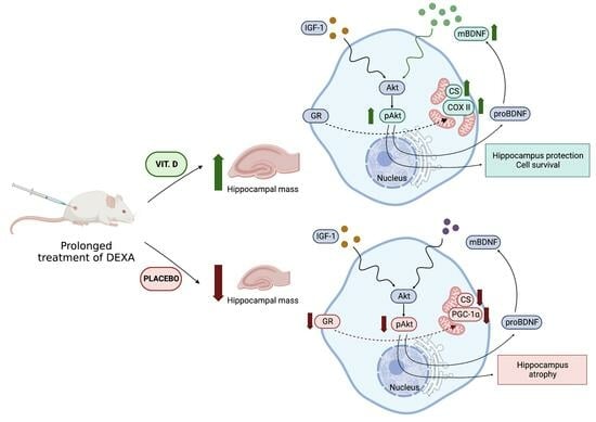
Dexamethasone (DEXA) is a commonly used steroid drug with immunosuppressive and analgesic properties. Unfortunately, long-term exposure to DEXA severely impairs brain function. This study aimed to investigate the effects of vitamin D3 supplementation during chronic DEXA treatment on neurogenesis, mitochondrial energy metabolism, protein levels involved in the BDNF-mediated Akt activity, and specific receptors in the hippocampus. We found reduced serum concentrations of 25-hydroxyvitamin D3 (25(OH)D3), downregulated proBDNF and pAkt, dysregulated glucocorticosteroid and mineralocorticoid receptors, impaired mitochondrial biogenesis, and dysfunctional mitochondria energy metabolism in the DEXA-treated group. In contrast, supplementation with vitamin D3 restored the 25(OH)D3 concentration to a value close to that of the control group. There was an elevation in neurotrophic factor protein level, along with augmented activity of pAkt and increased citrate synthase activity in the hippocampus after vitamin D3 administration in long-term DEXA-treated rats. Our findings demonstrate that vitamin D3 supplementation plays a protective role in the hippocampus and partially mitigates the deleterious effects of long-term DEXA administration. The association between serum 25(OH)D3 concentration and BDNF level in the hippocampus indicates the importance of applying vitamin D3 supplementation to prevent and treat pathological conditions.

Graphical Abstract

1. Introduction

Dexamethasone (DEXA) is a synthetic glucocorticosteroid (GC) with potent and long-lasting immunosuppressive and analgesic effects. Due to these properties, DEXA is widely used in medicine for recovery after surgery, during chemotherapy, and in treating severe acute respiratory syndrome coronavirus 2 (SARS-CoV-2) and autoimmune diseases [1,2,3,4].
Under natural conditions, GCs are produced by activating the hypothalamic–pituitary–adrenal (HPA) axis and play essential roles in neuroendocrine regulation. In addition, GCs control many physiological processes, such as carbohydrate metabolism, immune maintenance, and cardiovascular activity [5]. Moreover, GCs are responsible for normal brain function and mitochondrial energy metabolism regulation [6].
Overactivity of the HPA axis results in excessive GC secretion, adrenal hypertrophy, and atrophy of certain organs such as the thymus, spleen, and skeletal muscles [7,8,9]. Moreover, long-term treatment or high-dose exposure to GCs also affects the neurons, causing a reduction in mitochondrial oxidation, mitochondrial membrane potential, and calcium retention capacity in cortical neurons [10]. Furthermore, there is sufficient evidence of an association between high GC levels, psychiatric disorders, and neurodegenerative diseases such as Alzheimer’s disease (AD) and Parkinson’s disease (PD) [11,12], which are disorders where mitochondria play a crucial role [13]. Like prolonged stress, long-term DEXA treatment carries many adverse side effects and causes significant damage. Studies show that long-term DEXA treatment also has a negative impact on the central nervous system (CNS), including the hippocampus [14,15,16]. The hippocampus, a limbic system structure, appears to be particularly sensitive to the effects of GCs. High levels of GCs cause morphological, metabolic, and functional changes [17,18,19]. It is indicated that DEXA treatment causes changes in the differentiation [20] and morphology of neurons in the hippocampus [21]. Therefore, it may also lead to depression, mood disorders, decreased concentration, insomnia, abnormal behavior, memory deficits, and altered nervous activity [15,16]. Chronic exposure to GCs may negatively affect the brain, impair hippocampal function, and reduce the volume of this brain region [22].
Moreover, some studies have also shown that excessive flow of exogenous or endogenous GCs can induce neuroplasticity in the hippocampus, mediated by a brain-derived neurotrophic factor (BDNF). Chronic stress has been found to reduce BDNF in the hippocampus. Epigenetic, transcriptomic, and proteomic regulation, mediated by BDNF and/or its receptor Tropomyosin receptor kinase B (TrkB) expression, has been suggested to underlie the observed changes in rodents and humans [23,24]. Therefore, a reduction in the BDNF/TrkB signaling pathway in the hippocampus may be associated with synaptic loss, decreased neuronal plasticity, and regression network connectivity [25,26]. On the other hand, insulin-like growth factor 1 (IGF-1) is a crucial mediator of brain development in both young and adult individuals [27]. It is suggested that circulating IGF-1 triggers negative feedback amplification in the HPA axis. According to Baldini et al., an increase in IGF-1 levels may be correlated with an increase in GR expression [28]. Although the effects of DEX on IGF-1 are not well understood, it has been suggested that GCs may regulate IGF-1 signaling pathways in the hippocampus [29].
The hippocampus is equipped with two types of steroid receptors: glucocorticoid (GR) and mineralocorticoid (MR) receptors [30,31]. Both receptors play a crucial role in maintaining the proper structure and function of the hippocampus. Any changes in their activity or ratio may increase the intensity of hippocampal damage [32]. Furthermore, chronic exposure to GCs may alter the expression of GR and MR or the MR/GR ratio, which may also impact mitochondrial function in the brain. Depending on the dose and/or duration of exposure, GCs may either increase or decrease the levels of MR and GR in the hippocampus, resulting in contrasting effects [32,33,34]. GR translocation to mitochondria directly affects their function. During long-term administration, GCs may lead to respiratory chain dysfunction, reduced ATP production, and abnormal mitochondrial biogenesis, ultimately increasing the cell sensitivity to death [34].
Vitamin D, known for its pleiotropic effects, regulates calcium and phosphate metabolism and cares for the skin and skeletal health, and it is also an immunomodulatory hormone [35]. The active form of vitamin D, 1,25-dihydroxy vitamin D3 (1,25(OH)2D3), binds to the vitamin D receptor (VDR) and genetically regulates various cellular processes by promoting heterodimerization of this retinoid-X receptor (RXR). The activity of the VDR-RXR complex affects the gene expression of many tissues, including skeletal muscles and the brain [36]. Interestingly, VDR is also found in mitochondria, where it plays a crucial role in maintaining their integrity and proper function [37]. This characteristic is fundamental for optimal brain function and development [38]. Studies have demonstrated that excessive GC levels may induce a depression-like state and reduce the protein levels of VDR and cytochrome P450 proteins involved in vitamin D activation and catabolism in brain regions such as the hippocampus and prefrontal cortex [39,40]. Remarkably, deficiencies in vitamin D are associated with decreased hippocampal volume. However, supplementation with vitamin D3 has been shown to prevent tissue atrophy and memory deficits under conditions of GC exposure [8,41,42], suggesting its potential involvement in a protective mechanism against the negative effects of stress on the brain [39].
Therefore, this study aimed to determine whether vitamin D3 supplementation would positively affect the BDNF-mediated Akt activity pathway and mitochondria energy metabolism in the rat hippocampus during long-term DEXA treatment.

2. Results

2.1. Vitamin D Concentration

A 28-day treatment with DEXA reduced serum 25(OH)D3 concentration in the DEX group (6.02 ± 0.86) compared with the CON group (10.97 ± 1.71; p < 0.01). After vitamin D3 supplementation, a significantly higher 25(OH)D3 concentration (12.99 ± 1.42 ng/mL) was found in the DEX + VD group compared with the DEX group (p < 0.001; Figure 1).

2.2. Body and Hippocampus Mass Changes

After long-term treatment of DEXA, body weight was significantly lower in both groups treated with DEXA (p < 0.0001). The body weight of rats in the DEX group (245.83 ± 8.07 g) was significantly lower by 39% compared with the CON group (405.20 ± 18.98 g). In the DEX + VD group, body weight significantly increased by 11% (277.57 ± 7.49 g) compared with the DEX group (p < 0.05; Figure 2a). The weight of the hippocampus in the DEX group (0.06 ± 0.03 g; p < 0.05) decreased significantly compared with the CON group (0.14 ± 0.03). However, after vitamin D3 supplementation in the DEX + VD group, the weight of the hippocampus (0.12 ± 0.02 g) significantly increased compared with the DEX group (Figure 2b; p < 0.05). We also observed changes in the hippocampus/body weight ratio, where the ratio was significantly higher in the DEX + VD group (0.044 ± 0.006) compared with the DEX group after vitamin D3 supplementation. However, we did not observe any differences in the hippocampus/body weight ratio in the DEX group (0.027 ± 0.006) compared to the CON group (0.034 ± 0.007) (Figure 2c).

2.3. Hippocampal GR, MR, and VDR Levels

After 28 days of DEXA treatment, no significant differences in VDR and MR protein levels were observed (Figure 3a,b). We found a statistically significant reduction in the GR protein level by 50% in the DEX group compared with the CON (p < 0.05), but no changes were found in the DEX + VD group or between groups. However, a trend suggesting an increase in GR level was observed in the DEX + VD group versus the DEX group (p = 0.063; Figure 3c).

2.4. Assessment of BDNF-Akt Signaling Molecules

To determine the changes in prosurvival proteins in the rat hippocampus during long-term administration of DEXA, we measured the Akt protein level and phosphorylated Akt (pAkt). There were no significant changes in Akt protein levels between the groups (Figure 4a). However, the phosphorylation state of Akt (p-Akt) significantly increased in the DEX + VD group compared with the DEX group. Moreover, the p-Akt level was almost two-fold higher in the DEX + VD group compared with the CON group, but this was not statistically significant (Figure 4b). The pAkt/Akt ratio showed a significant increase in the DEX + VD group compared with the DEX group. Still, there was no significant difference between the treatment groups and the CON group (Figure 4c).
To evaluate neurogenesis, we measured the protein levels of the precursor of brain-derived neurotrophic factor (proBDNF) and mature BDNF (mBDNF). After 28 days of DEX treatment, the level of proBDNF in the DEX group significantly decreased compared with the CON group. In contrast, we did not observe any statistically significant changes in the DEX + VD group (Figure 4d). Furthermore, we observed significantly elevated mBDNF protein levels during prolonged exposure to DEX + VD in the hippocampal tissue compared with the DEX group (p < 0.05). This increase in mBDNF level was accompanied by an elevated phosphorylation state of Akt after vitamin D3 supplementation in the rat hippocampus during long-term DEXA treatment. However, there was no significant difference in values between the DEX and CON groups (Figure 4e). We did not observe any changes in the IGF-1 protein level between the groups (Figure 4f).

2.5. Assessment of Mitochondrial Biogenesis and Oxidative Metabolism

After 28 days of DEXA treatment, we found a higher level of cytochrome c oxidase subunit II coded by mitochondria (COX II) in the hippocampus in the DEX + VD group (1.019 ± 0.325), which increased by 37% compared with the DEX group (p < 0.05; Figure 5a). This may partially confirm earlier studies showing the influence of vitamin D in the modulation of mitochondrial metabolism [43,44,45]. There were no significant changes between groups compared to the CON group (0.732 ± 0.069 A.U.). The nuclear subunit IV of cytochrome c oxidase (COX IV) protein levels was not significantly different between groups (Figure 5b). Furthermore, long-term treatment with DEXA resulted in a decrease in the peroxisome proliferator-activated receptor gamma coactivator 1-alpha (PGC-1α) protein level in the DEX rats when compared with the CON group. At the same time, supplementation with 600 IU of vitamin D3 caused no significant changes in protein level versus the DEX + VD group (Figure 5c).

2.6. Mitochondrial Oxidative Metabolism Activity

After 28 days of daily DEXA treatment, the activity of citrate synthase (CS) was reduced in the hippocampus by 48% in the DEX group (1.37 ± 0.44) when compared with the CON group (2.50 ± 0.33 µmol/min/mg of protein; p < 0.05). Moreover, CS activity was also significantly elevated by 42% in the DEX + VD group (2.35 ± 0.73 µmol/min/mg of protein) compared with the DEX group (p < 0.05; Figure 6a). However, we noticed no statistically significant changes in cytochrome c oxidase (COX) activity in the rat hippocampus despite a downward trend in the DEX group (57.53 ± 29.14) compared to CON (93.32 ± 26.07) and the upward trend in COX activity in the DEX + VD group (81.46 ± 32.48 nmol/min/mg of protein; Figure 6b).

3. Discussion

In the present study, we showed that long-term treatment with DEXA reduced the body mass of rats, induced hippocampus atrophy, and decreased proBDNF and GR protein levels in the rat hippocampus. We also found attenuated serum concentrations of 25(OH)D3 in DEXA-treated rats. Additionally, we observed a reduction in BDNF signaling in the hippocampus, a corticolimbic area in the brain. This reduction in signaling was associated with inhibited pAkt activity, decreased PGC-1α protein level, and disrupted mitochondrial energy metabolism. According to our results, systematic vitamin D3 supplementation partially reversed the adverse effects of DEXA. Thus, supplementation with vitamin D3 led to increased body and hippocampus mass, as well as an increased hippocampus/body mass ratio in rats treated with prolonged DEXA. Furthermore, the present study showed a restoration of 25(OH)D3 concentration to a value close to the control group. There was an elevation in neurotrophic factor protein level, along with augmented activity of pAkt, increased subunit II (coded by mitochondria protein level, and higher CS activity in the hippocampus of rats chronically treated with DEXA. We did not observe any changes in the protein levels of IGF-1 between the groups. This may suggest that the BDNF-mediated activity Akt is more susceptible to DEXA’s negative action than the IGF signaling pathway. Our findings demonstrate that supplementation with vitamin D3 plays a protective role in the corticolimbic area and partially mitigates the deleterious effects caused by long-term DEXA administration.
Our findings indicate differences in the protective effects of vitamin D between the hippocampus and body weight, the full understanding of which requires further study. One of the side effects of GC therapy is weight loss, which may manifest as osteoporosis, muscle wasting, and changes in fat composition [46,47]. The variations in the effectiveness of vitamin D3 supplementation in terms of its protective role in the hippocampus versus the rest of the body seem to arise from the multifaceted impacts of DEXA on metabolism. These impacts primarily focus on reducing fat mass, inducing muscle wasting, causing intramuscular fat deposition, and leading to mitochondrial dysfunction [48,49]. The mechanism(s) of DEXA’s action resemble stress responses and are associated with hormone dysregulation, insulin resistance, hepatic hexokinase inhibition, and inhibition of glucose oxidation. The consequence of subsequent exposure to GCs leads to increased lipolysis and the release of free fatty acids (FFAs) from adipose tissue and elevated proteolysis of skeletal muscle (decrease in PI3K markers, PKB/Akt, GSK3) dependent on the activation/inhibition of AMP-activated protein kinase (AMPK) by GCs. Reported evidence after 6 weeks of vitamin D3 supplementation in patients with hypercortisolemia had an effect on lipid profile and insulin sensitivity but did not significantly affect baseline body weight (BMI, waist circumference) [50].
Recently, it was reported that long-term treatment with DEXA has a negative impact on the CNS [51]. Similar alterations were also observed during a chronic stress response [52]. These changes may be associated with an appropriate balance of GR and MR levels and activity, which are crucial for the proper function of the CNS. Notably, data on GR and MR levels after DEXA treatment are inconsistent. On the one hand, studies showed that long-term administration of GCs reduced GR expression, increased neuroinflammation, and caused neurodegeneration in the hippocampus [53,54]. Another study showed changes in mRNA expression in both receptors, revealing that GR was decreased while MR was increased in the hippocampus, striatum, and prefrontal cortex in DEXA-treated mice [55]. In line with this finding, our data showed similar trends in the hippocampus. On the other hand, the data reported inconsistent results regarding changes in GR and MR protein levels and gene expression in response to GCs action [10,34,56,57,58]. The results published by Zhe and coworkers showed that single-prolonged stress causes a reduced expression of MR and GR in CA1 of the hippocampus [32]. The differences between our results and those of other studies may be attributed to the dose and duration of GC exposure or other factors that can have different effects [10,59]. Short-term application of both high and low doses of GCs similarly affects GR in cortical neurons by increasing its mitochondrial localization, while during long-term supply, only a high dose decreases GR levels in mitochondria and negatively affects mitochondrial function in neurons [10]. In the current study, we found a lower GR protein level after long-term DEX treatment with the protective effect of vitamin D3 supplementation. Although supplementation with vitamin D3 partially reversed this adverse impact, the effect was not statistically significant.
In this study, we showed that the 28-day administration of DEXA at a dose of 2 mg/kg/day caused a decrease in the serum concentration of 25(OH)D3. A previous study found that DEXA affects VDR in a GR-dependent manner by increasing Vdr transcripts de novo [60]. Surprisingly, we did not find any substantial differences in VDR protein levels with DEX treatment alone or combined with D3 supplemented when compared to the control group. Our finding is partially in line with the report by Jiang et al. [39], which demonstrated dysregulation in vitamin D metabolism caused by a 10-day DEXA supply. The authors observed a decrease in CYP27B1, CYP24A1, and VDR expression not only in the hippocampus but also in the prefrontal cortex, heart, and kidneys. Moreover, VDR activity is essential at the cellular level for cell differentiation, cell growth, and apoptosis [61]. However, VDR silencing induces unbalanced metabolism and ultimately contributes to cytotoxicity, while VDR ablation induces many negative consequences, including mitochondrial damage and premature cell death [37]. Additionally, VDR may have an impact on the mitochondrial respiratory chain [62] by transcribing the mitochondrial (COX II) and nuclear (COX IV) subunits of cytochrome c oxidase, which are involved in ATP synthesis [37]. Our present study showed that DEXA significantly lowered the COX subunit II protein level, while vitamin D3 supplementation effectively reversed this alteration. Mitochondrial subunits play a crucial role in the catalytic cycle of the enzyme, with nuclear subunits being associated with regulating enzyme activity and ensuring the structural stability of the complex [63]. In our study, we observed a reduction in COX subunit II but not COX subunit IV. In addition, we did not find any changes in COX activity. Desquiret et al. [64] reported that chronic DEXA administration leads to a decrease in the activity of the electron transport chain (ETC) complex I and II while also increasing the activity of complex IV. To determine the function of mitochondria in the Krebs cycle, we examined the activity of citrate synthase (CS). CS is an enzyme localized in the mitochondrial matrix and is used as a quantitative enzymatic marker for the presence of intact mitochondria [65,66]. Our data indicate that long-term DEXA administration decreased CS activity in the hippocampus, while vitamin D3 supplementation contributed to maintaining proper enzyme activity.
According to our results, long-term treatment with DEXA reduces the activity of mitochondrial energy metabolism, while supplementation with vitamin D3 restores its proper function in the hippocampus. Vitamin D shows a potential protective role and causes reverse oxidative stress-induced cognitive impairment, which was also presented by Hajiluian and coworkers [67]. This translates into our study results, wherein, in the vitamin D3-supplemented group, there was a noticeable improvement in the function of the ETC and the citric acid cycle in the hippocampus. Vitamin D3 supplementation or administration of its metabolites reduces neurological damage and neurotoxicity. The detailed biochemical mechanism is still unclear, but it is known that vitamin D can act on multiple pathways, such as neuronal antioxidant pathways, immunomodulation, calcium regulation, and glutamatergic systems [68]. All these data demonstrate an antagonistic effect of vitamin D on GC activity [69]. Given that the impairment of mitochondrial function plays an important role in response to stress, brain aging, and the early stages of neurodegenerative diseases, we believe that enhancing mitochondrial brain energy metabolism with vitamin D may be an essential protective mechanism [66,70]. This study found a positive influence of vitamin D3 supplementation on the BDNF protein level, i.e., a higher serum concentration of 25(OH)D3 upregulated the BDNF level in the hippocampus of prolonged DEXA-treated rats. The association between serum concentration of 25(OH)D3 and BDNF level in the hippocampus reported in this study may indicate the importance of improving vitamin D with supplementation, which acts as a strategy for preventing and treating cognitive decline and increasing BDNF in excessive-prolonged flow of exogenous or endogenous GCs (chronic stress). On the one hand, Yosefian et al. reported that vitamin D3 supplementation did not affect BDNF concentration in the hippocampus in animal models of depression [71]. On the other hand, another study showed that the modulation of hippocampal BDNF with vitamin D might be an effective prevention strategy against depression in animal models of depression [72].
We are very far from any speculation, but our results strongly suggest that vitamin D3 supplementation may reverse the negative changes induced by long-term DEXA treatment. This reversal leads to the upregulation of BDNF and nearly restores GR protein levels. Based on previously reported results [73] and our findings, we assume the existence of crosstalk between GR and BDNF in the hippocampus. Therefore, the present study showed that the modulatory effects of vitamin D during long-term exposure to DEXA on GR restored mitochondrial function, and BDNF-mediated Akt activity reversed neuropathophysiological changes in the hippocampus. Our findings may have significant implications for neuroplastic processes, cognitive function, and several neuropsychiatric disorders. Moreover, our results provide further evidence of a key link between vitamin D and BDNF-mediated Akt activity, as well as the restoration of physiological function in the mitochondria through GR in the hippocampus. These findings may contribute to the neuroprotective and therapeutic potential (abilities) of vitamin D in patients with neuropathophysiological disorders.
This study has some limitations. First, we did not estimate TrkB, IGR, or other proteins involved in the signaling pathway, which could have directly confirmed our findings in the hippocampus of DEXA-treated rats. Second, we did not measure protein levels that can form complexes with GR, such as HSP70/90, Bcl-2, or proteins targeting mitochondria associated with chaperones that aid in their mitochondrial translocation. This would have helped to demonstrate the meaningful role of GR in the modification of mitochondria-coded protein expression. These limitations were primarily due to the lack of sufficient tissue for the analysis.
Overall, our findings showed that supplementation with vitamin D3 partially reversed negative changes in the hippocampus induced with long-term DEXA treatment in rats. We observed upregulation of BDNF and nearly restored GR protein level after vitamin D3 administration. Consequently, the present study demonstrated a higher activity of pAkt, elevated COX subunit II protein level, and augmented activity of CS in the hippocampus, suggesting the modulatory abilities of vitamin D during long-term exposure to DEXA. These findings have significant implications for neuroplastic processes, cognitive function, and neuropathophysiological conditions. Moreover, these findings may contribute to the understanding of the neuroprotective and therapeutic abilities of vitamin D in patients with neurophysiological disorders.

4. Materials and Methods

4.1. Animals

The Local Ethics Committee in Bydgoszcz, Poland (No. 10/2019), approved our animal studies. All procedures were carried out following European guidelines.
Male Wistar rats (56–70 days postnatal) weighing 300–400 g were used in this study. The animals were obtained from the Academic Experimental Animal House at the Medical University of Gdansk, Poland. The rats were housed 3–4 per cage in climate-controlled conditions (temperature: 22 ± 2 °C; humidity: 55 ± 2%) with a 12:12 h light/dark cycle and provided food and water ad libitum.

4.2. Experimental Procedure

The rats were habituated to the various experimental procedures daily for two weeks before the experiment began. The eighteen animals were randomly assigned to three groups: control (CON; n = 5), DEXA placebo (DEX; n = 6), and DEXA supplemented with vitamin D3 (DEX + VD; n = 7). The experiment lasted for 28 days. Throughout this time, the CON group was treated intraperitoneally with saline, and the two other groups were treated with dexamethasone at 2 mg/kg/day (dexamethasone D4902, DEXA, Sigma-Aldrich, St Paul, MN, USA) in the same manner. The DEX + VD group was orally supplemented with vitamin D3 (Juvit D3, PPF HASCO-LEK. SA., Wrocław, Poland) at 600 IU/kg/day, and the DEX group was orally supplemented with vegetable oil as a placebo. Blood was collected on Day 1 and Day 28 of the experiment from the tail vein under isoflurane anesthesia. On Day 29 of the experiment, all animals were sacrificed using decapitation. The brains were collected, and the hippocampus was dissected, weighed, frozen in liquid nitrogen, and stored at −80 °C for further analysis.

4.3. Tissue Preparation

4.3.1. Blood Collection

To determine the serum 25(OH)D3 concentration, blood samples were centrifuged for 10 min at 2000× g at 4 °C. The serum was collected and frozen at −80 °C for further analysis.

4.3.2. Western Blot

A part of the hippocampus was homogenized in RIPA buffer (89901; Thermo Scientific, Waltham, MA, USA) in the presence of protease and phosphatase inhibitors (1:100; 1861280; Thermo Scientific, Waltham, MA, USA). The 8% tissue homogenates were centrifuged at 750× g for 10 min at 4 °C. Then, the collected supernatant was centrifuged again at 12,000× g (10 min at 4 °C), aliquoted to cryogenic microfuge tubes, and stored at −80 °C for further analysis.

4.3.3. Enzymes Activity

The rest of the hippocampi were homogenized in buffer containing 50 mM Tris-HCl (T3253; Sigma-Aldrich, USA), 150 mM NaCl, 1 mM EDTA (ED3SS; Sigma-Aldrich, USA), 0.5 mM DTT (443853B; VWR International; Radnor, PA, USA) pH 7.2, and 0.2% HALT protease inhibitors cocktail (P834; Sigma Aldrich, STL, USA). The 4% tissue homogenates were centrifuged at 750× g for 10 min at 4 °C. The supernatant was frozen and stored at −80 °C for further analysis.

4.4. Vitamin D3 Metabolite Concentration

The concentration of 25(OH)D3 in serum was measured using liquid chromatography coupled to tandem mass spectrometry (LC-MS/MS), according to the procedure of Rola and coworkers [74]. Briefly, serum was analyzed using the Eksigent Exion LC HPLC system with a CTC PAL autosampler (Zwinger, Hofstetten bei Brienz, Switzerland) coupled to a QTRAP® 4500 MS/MS system (Sciex, Framingham, MA, USA).

4.5. Protein Expression

Western Blot Analysis
Samples (approximately 25–30 µg) were prepared from the collected supernatants and mixed with RIPA buffer. The protein samples were denatured with heating at 95 °C for 5 min, cooled to room temperature, and then separated into 10% and 12% mini protean TGX precast Protein Gels (4561035, 4561045; BioRad, Hercules, CA, USA). Membranes of thickness 0.2 μm were used for semi-dry transfer. The membranes were blocked with Every Blot Blocking Buffer (12010020; BioRad, CA, USA) or 5% nonfat milk in TBST (for the determination of PGC-1α). The primary antibodies used were: VDR (ab3508; diluted 1:1000, Abcam, Cambridge, UK), MR (ab64457; diluted 1:1000, Abcam, UK), GR (ab183127; diluted 1:500, Abcam, UK), BDNF (ab108319; diluted 1:1000, Abcam, UK), Akt (C67E7; diluted 1:1000, Cell Signaling, Danvers, MA, USA), pAkt (D25E6; diluted 1:1000, Cell Signaling, MA, USA), IGF-1 (ab9572; diluted 1:1000, Abcam, UK), COX II (NBP2-94364; diluted 1:1000, Novusbio, Centennial, CO, USA), COX IV (4D11-B3-E8; diluted 1:1000, Cell Signaling, MA, USA), PGC-1α (ab191838; diluted 1:500, Abcam, UK), and ß-tubulin. (AF7011; diluted 1:1000, Affinity, Beachwood, OH, USA). The membranes were treated with secondary rabbit anti-mouse (ab6728; Abcam) and goat anti-rabbit (111-035-003; Jackson ImmunoResearch, Ely, UK) antibodies (both diluted 1:1000–1:3000). The antibodies were prepared according to the instructions provided by the manufacturer. The results were visualized using Clarity Western ECL Substrate (1705061; BioRad, CA, USA) and imaged using the ChemiDoc MP imaging system (BioRad, CA, USA). Protein concentration was measured with the Pierce™ BCA protein assay method.

4.6. Enzyme Activities

4.6.1. Citrate synthase Activity

Citrate synthase (CS) activity was determined according to Dzik and coworkers [43]. In brief, 3 μL of the supernatant was combined with 181 μL of buffer (50 mM TRIS-HCl with 5 mM EDTA, pH 8.1), 20 μL of freshly made DTNB (1 mM), 2 μL of acetyl-coenzyme A (15 mM), and 2 μL of freshly made oxaloacetate acid (10 mM) to initiate the reaction. The results were obtained in 2 min by measuring the absorbance change at 412 nm using a multimode microplate reader (Varioskan Flash-Spectral Scanning Multimode Microplate Reader 183, Thermo Fisher Scientific, MA, USA) at 37 °C. The CS activity was measured in duplicate and expressed as µmol/min/mg of protein.

4.6.2. Cytochrome c Oxidase Activity

Cytochrome c oxidase (COX) activity was measured according to [75] using a microplate combining 5 μL of supernatant, 192 μL of K phosphate buffer (50 mM, 1 mM EDTA, pH 7.2), and 3 μL of 2 mM reduced cytochrome c (c2037; Sigma-Aldrich; USA; reduced with ascorbic acid 20:1) to initiate the reaction. The COX activity was measured in duplicate at 37 °C, with an absorbance of 550 nm using a microplate reader (Varioskan Flash-Spectral Scanning Multimode Microplate Reader 183, Thermo Scientific, MA, USA). The results were expressed as nmol/min/mg of protein. Protein concentration was measured with the Bradford method.

4.7. Statistical Analysis

All results were analyzed using the GraphPad Prism 8.3 software program (GraphPad Software, San Diego, CA, USA). A one-way ANOVA with least significant difference (LSD) post hoc test was used to analyze body and hippocampus weight, 25(OH)D3 concentration, and the results obtained from the Western Blot with chemiluminescence using Image Lab 6.1.0 software. A one-way ANOVA with a Tukey post hoc test was used for CS and COX enzyme activity. p-values < 0.05 were considered significant. Data are presented as mean ± SEM.

Author Contributions

Conceptualization, D.K.-L., M.J.K. and J.J.K.; data curation, D.K.-L. and J.J.K.; formal analysis, D.K.-L., M.J.K. and J.J.K.; funding acquisition, M.J.K. and J.J.K.; investigation, D.K.-L., M.J.K., D.M. and J.J.K.; methodology, D.K.-L., D.M. and J.J.K.; project administration, M.J.K. and J.J.K.; supervision J.J.K.; writing—original draft, D.K.-L. and J.J.K.; writing—review and editing, D.K.-L., M.J.K., D.M. and J.J.K. All authors have read and agreed to the published version of the manuscript.

Funding

This study was supported by grants from the National Science Centre in Poland (grant number: 2018/31/N/NZ7/03680) and the Faculty of Biology, University of Gdansk, Poland (DS No. 531-D080-D248-23).

Institutional Review Board Statement

This study was conducted in accordance with the Declaration of Helsinki and approved by the Local Ethics Committee in Bydgoszcz, Poland (No. 10/2019).

Informed Consent Statement

Not applicable.

Data Availability Statement

The datasets used and/or analyzed in the current study are available from the corresponding author upon reasonable request.

Conflicts of Interest

The authors declared no conflict of interest regarding the publication of this article. The funders had no role in the design of this study; in the collection, analyses, or interpretation of data; in the writing of this manuscript, or in the decision to publish the results.

References

  1. Giles, A.J.; Hutchinson, M.N.D.; Sonnemann, H.M.; Jung, J.; Fecci, P.E.; Ratnam, N.M.; Zhang, W.; Song, H.; Bailey, R.; Davis, D.; et al. Dexamethasone-Induced Immunosuppression: Mechanisms and Implications for Immunotherapy. J. Immunother. Cancer 2018, 6, 51. [Google Scholar] [CrossRef] [PubMed]
  2. Tokuda, E.; Watanabe, S.; Okawa, E.; Ono, S. Regulation of Intracellular Copper by Induction of Endogenous Metallothioneins Improves the Disease Course in a Mouse Model of Amyotrophic Lateral Sclerosis. Neurotherapeutics 2015, 12, 461–476. [Google Scholar] [CrossRef]
  3. Johnson, D.B.; Lopez, M.J.; Kelley, B. Dexamethasone. In Statpearls; StatPearls Publishing: Treasure Island, FL, USA, 2022. [Google Scholar]
  4. Wiersinga, W.J.; Rhodes, A.; Cheng, A.C.; Peacock, S.J.; Prescott, H.C. Pathophysiology, Transmission, Diagnosis, and Treatment of Coronavirus Disease 2019 (COVID-19): A Review. JAMA 2020, 324, 782–793. [Google Scholar] [CrossRef] [PubMed]
  5. Manoli, I.; Alesci, S.; Blackman, M.R.; Su, Y.A.; Rennert, O.M.; Chrousos, G.P. Mitochondria as Key Components of the Stress Response. Trends Endocrinol. Metab. 2007, 18, 190–198. [Google Scholar] [CrossRef] [PubMed]
  6. Chung, S.; Son, G.H.; Kim, K. Circadian Rhythm of Adrenal Glucocorticoid: Its Regulation and Clinical Implications. Biochim. Biophys. Acta 2011, 1812, 581–591. [Google Scholar] [CrossRef] [PubMed]
  7. Herman, J.P.; McKlveen, J.M.; Ghosal, S.; Kopp, B.; Wulsin, A.; Makinson, R.; Scheimann, J.; Myers, B. Regulation of the Hypothalamic-Pituitary-Adrenocortical Stress Response. Compr. Physiol. 2016, 6, 603–621. [Google Scholar]
  8. Karnia, M.J.; Korewo, D.; Myslinska, D.; Ciepielewski, Z.M.; Puchalska, M.; Konieczna-Wolska, K.; Kowalski, K.; Kaczor, J.J. The Positive Impact of Vitamin D on Glucocorticoid-Dependent Skeletal Muscle Atrophy. Nutrients 2021, 13, 936. [Google Scholar] [CrossRef]
  9. Karnia, M.J.; Myslinska, D.; Dzik, K.P.; Flis, D.J.; Ciepielewski, Z.M.; Podlacha, M.; Kaczor, J.J. The Electrical Stimulation of the Bed Nucleus of the Stria Terminalis Causes Oxidative Stress in Skeletal Muscle of Rats. Oxid. Med. Cell. Longev. 2018, 2018, 4671213. [Google Scholar] [CrossRef]
  10. Du, J.; Wang, Y.; Hunter, R.; Wei, Y.; Blumenthal, R.; Falke, C.; Khairova, R.; Zhou, R.; Yuan, P.; Machado-Vieira, R.; et al. Dynamic Regulation of Mitochondrial Function by Glucocorticoids. Proc. Natl. Acad. Sci. USA 2009, 106, 3543–3548. [Google Scholar] [CrossRef]
  11. Ouanes, S.; Popp, J. High Cortisol and the Risk of Dementia and Alzheimer’s Disease: A Review of the Literature. Front. Aging Neurosci. 2019, 11, 43. [Google Scholar] [CrossRef]
  12. van Wamelen, D.J.; Leta, V.; Johnson, J.; Ocampo, C.L.; Podlewska, A.M.; Rukavina, K.; Rizos, A.; Martinez-Martin, P.; Chaudhuri, K.R. Drooling in Parkinson’s Disease: Prevalence and Progression from the Non-Motor International Longitudinal Study. Dysphagia 2020, 35, 955–961. [Google Scholar] [CrossRef] [PubMed]
  13. Monzio Compagnoni, G.; Di Fonzo, A.; Corti, S.; Comi, G.P.; Bresolin, N.; Masliah, E. The Role of Mitochondria in Neurodegenerative Diseases: The Lesson from Alzheimer’s Disease and Parkinson’s Disease. Mol. Neurobiol. 2020, 57, 2959–2980. [Google Scholar] [CrossRef] [PubMed]
  14. Xu, J.; Wang, R.; Liu, Y.; Wang, W.; Liu, D.; Jiang, H.; Pan, F. Short- and Long-Term Alterations of Fkbp5-Gr and Specific Micrornas in the Prefrontal Cortex and Hippocampus of Male Rats Induced by Adolescent Stress Contribute to Depression Susceptibility. Psychoneuroendocrinology 2019, 101, 204–215. [Google Scholar] [CrossRef] [PubMed]
  15. Ciriaco, M.; Ventrice, P.; Russo, G.; Scicchitano, M.; Mazzitello, G.; Scicchitano, F.; Russo, E. Corticosteroid-Related Central Nervous System Side Effects. J. Pharmacol. Pharmacother. 2013, 4 (Suppl. 1), S94–S98. [Google Scholar] [CrossRef] [PubMed]
  16. Edelmann, M.N.; Ogg, R.J.; Scoggins, M.A.; Brinkman, T.M.; Sabin, N.D.; Pui, C.H.; Srivastava, D.K.; Robison, L.L.; Hudson, M.M.; Krull, K.R. Dexamethasone Exposure and Memory Function in Adult Survivors of Childhood Acute Lymphoblastic Leukemia: A Report from the Sjlife Cohort. Pediatr. Blood Cancer 2013, 60, 1778–1784. [Google Scholar] [CrossRef] [PubMed]
  17. Sapolsky, R.M. Stress and the Brain: Individual Variability and the Inverted-U. Nat. Neurosci. 2015, 18, 1344–1346. [Google Scholar] [CrossRef]
  18. Sapolsky, R.M. Glucocorticoids and Hippocampal Atrophy in Neuropsychiatric Disorders. Arch. Gen. Psychiatry 2000, 57, 925–935. [Google Scholar] [CrossRef]
  19. Starkman, M.N.; Gebarski, S.S.; Berent, S.; Schteingart, D.E. Hippocampal Formation Volume, Memory Dysfunction, and Cortisol Levels in Patients with Cushing’s Syndrome. Biol. Psychiatry 1992, 32, 756–765. [Google Scholar] [CrossRef]
  20. Levone, B.R.; Codagnone, M.G.; Moloney, G.M.; Nolan, Y.M.; Cryan, J.F.; OF, L. Adult-Born Neurons from the Dorsal, Intermediate, and Ventral Regions of the Longitudinal Axis of the Hippocampus Exhibit Differential Sensitivity to Glucocorticoids. Mol. Psychiatry 2021, 26, 3240–3252. [Google Scholar] [CrossRef]
  21. Silva-Gómez, A.B.; Aguilar-Salgado, Y.; Reyes-Hernández, D.O.; Flores, G. Dexamethasone Induces Different Morphological Changes in the Dorsal and Ventral Hippocampus of Rats. J. Chem. Neuroanat. 2013, 47, 71–78. [Google Scholar] [CrossRef]
  22. MacPherson, A.; Dinkel, K.; Sapolsky, R. Glucocorticoids Worsen Excitotoxin-Induced Expression of Pro-Inflammatory Cytokines in Hippocampal Cultures. Exp. Neurol. 2005, 194, 376–383. [Google Scholar] [CrossRef] [PubMed]
  23. Tsankova, N.; Renthal, W.; Kumar, A.; Nestler, E.J. Author Correction: Epigenetic Regulation in Psychiatric Disorders. Nat. Rev. Neurosci. 2019, 20, 187–188. [Google Scholar] [CrossRef]
  24. Numata, S.; Ishii, K.; Tajima, A.; Iga, J.; Kinoshita, M.; Watanabe, S.; Umehara, H.; Fuchikami, M.; Okada, S.; Boku, S.; et al. Blood Diagnostic Biomarkers for Major Depressive Disorder Using Multiplex DNA Methylation Profiles: Discovery and Validation. Epigenetics 2015, 10, 135–141. [Google Scholar] [CrossRef] [PubMed]
  25. Berton, O.; McClung, C.A.; Dileone, R.J.; Krishnan, V.; Renthal, W.; Russo, S.J.; Graham, D.; Tsankova, N.M.; Bolanos, C.A.; Rios, M.; et al. Essential Role of Bdnf in the Mesolimbic Dopamine Pathway in Social Defeat Stress. Science 2006, 311, 864–868. [Google Scholar] [CrossRef]
  26. Taliaz, D.; Loya, A.; Gersner, R.; Haramati, S.; Chen, A.; Zangen, A. Resilience to Chronic Stress Is Mediated by Hippocampal Brain-Derived Neurotrophic Factor. J. Neurosci. 2011, 31, 4475–4483. [Google Scholar] [CrossRef] [PubMed]
  27. Wrigley, S.; Arafa, D.; Tropea, D. Insulin-Like Growth Factor 1: At the Crossroads of Brain Development and Aging. Front. Cell. Neurosci. 2017, 11, 14. [Google Scholar] [CrossRef]
  28. Baldini, S.; Restani, L.; Baroncelli, L.; Coltelli, M.; Franco, R.; Cenni, M.C.; Maffei, L.; Berardi, N. Enriched Early Life Experiences Reduce Adult Anxiety-Like Behavior in Rats: A Role for Insulin-Like Growth Factor 1. J. Neurosci. 2013, 33, 11715–11723. [Google Scholar] [CrossRef]
  29. Santi, A.; Bot, M.; Aleman, A.; Penninx, B.; Aleman, I.T. Circulating Insulin-Like Growth Factor I Modulates Mood and Is a Biomarker of Vulnerability to Stress: From Mouse to Man. Transl. Psychiatry 2018, 8, 142. [Google Scholar] [CrossRef]
  30. Barrientos, R.M.; Thompson, V.M.; Kitt, M.M.; Amat, J.; Hale, M.W.; Frank, M.G.; Crysdale, N.Y.; Stamper, C.E.; Hennessey, P.A.; Watkins, L.R.; et al. Greater Glucocorticoid Receptor Activation in Hippocampus of Aged Rats Sensitizes Microglia. Neurobiol. Aging 2015, 36, 1483–1495. [Google Scholar] [CrossRef]
  31. Herman, J.P.; Patel, P.D.; Akil, H.; Watson, S.J. Localization and Regulation of Glucocorticoid and Mineralocorticoid Receptor Messenger Rnas in the Hippocampal Formation of the Rat. Mol. Endocrinol. 1989, 3, 1886–1894. [Google Scholar] [CrossRef]
  32. Zhe, D.; Fang, H.; Yuxiu, S. Expressions of Hippocampal Mineralocorticoid Receptor (Mr) and Glucocorticoid Receptor (Gr) in the Single-Prolonged Stress-Rats. Acta Histochem. Cytochem. 2008, 41, 89–95. [Google Scholar] [CrossRef] [PubMed]
  33. Sapolsky, R.M.; Krey, L.C.; McEwen, B.S. Stress Down-Regulates Corticosterone Receptors in a Site-Specific Manner in the Brain. Endocrinology 1984, 114, 287–292. [Google Scholar] [CrossRef] [PubMed]
  34. Kokkinopoulou, I.; Moutsatsou, P. Mitochondrial Glucocorticoid Receptors and Their Actions. Int. J. Mol. Sci. 2021, 22, 6054. [Google Scholar] [CrossRef] [PubMed]
  35. Charoenngam, N.; Holick, M.F. Immunologic Effects of Vitamin D on Human Health and Disease. Nutrients 2020, 12, 2097. [Google Scholar] [CrossRef]
  36. An, B.S.; Tavera-Mendoza, L.E.; Dimitrov, V.; Wang, X.; Calderon, M.R.; Wang, H.J.; White, J.H. Stimulation of Sirt1-Regulated Foxo Protein Function by the Ligand-Bound Vitamin D Receptor. Mol. Cell. Biol. 2010, 30, 4890–4900. [Google Scholar] [CrossRef]
  37. Ricca, C.; Aillon, A.; Bergandi, L.; Alotto, D.; Castagnoli, C.; Silvagno, F. Vitamin D Receptor Is Necessary for Mitochondrial Function and Cell Health. Int. J. Mol. Sci. 2018, 19, 1672. [Google Scholar] [CrossRef]
  38. Son, G.; Han, J. Roles of Mitochondria in Neuronal Development. BMB Rep. 2018, 51, 549–556. [Google Scholar] [CrossRef]
  39. Jiang, P.; Zhang, W.Y.; Li, H.D.; Cai, H.L.; Liu, Y.P.; Chen, L.Y. Stress and Vitamin D: Altered Vitamin D Metabolism in Both the Hippocampus and Myocardium of Chronic Unpredictable Mild Stress Exposed Rats. Psychoneuroendocrinology 2013, 38, 2091–2098. [Google Scholar] [CrossRef]
  40. Camargo, A.; Dalmagro, A.P.; Platt, N.; Rosado, A.F.; Neis, V.B.; Zeni, A.L.B.; Kaster, M.P.; Rodrigues, A.L.S. Cholecalciferol Abolishes Depressive-Like Behavior and Hippocampal Glucocorticoid Receptor Impairment Induced by Chronic Corticosterone Administration in Mice. Pharmacol. Biochem. Behav. 2020, 196, 172971. [Google Scholar] [CrossRef]
  41. Al-Amin, M.; Bradford, D.; Sullivan, R.K.P.; Kurniawan, N.D.; Moon, Y.; Han, S.H.; Zalesky, A.; Burne, T.H.J. Vitamin D Deficiency Is Associated with Reduced Hippocampal Volume and Disrupted Structural Connectivity in Patients with Mild Cognitive Impairment. Hum. Brain Mapp. 2019, 40, 394–406. [Google Scholar] [CrossRef]
  42. Bakhtiari-Dovvombaygi, H.; Izadi, S.; Zare, M.; Hassanlouei, E.A.; Dinpanah, H.; Ahmadi-Soleimani, S.M.; Beheshti, F. Vitamin D3 Administration Prevents Memory Deficit and Alteration of Biochemical Parameters Induced by Unpredictable Chronic Mild Stress in Rats. Sci. Rep. 2021, 11, 16271. [Google Scholar] [CrossRef] [PubMed]
  43. Dzik, K.P.; Skrobot, W.; Kaczor, K.B.; Flis, D.J.; Karnia, M.J.; Libionka, W.; Antosiewicz, J.; Kloc, W.; Kaczor, J.J. Vitamin D Deficiency Is Associated with Muscle Atrophy and Reduced Mitochondrial Function in Patients with Chronic Low Back Pain. Oxid. Med. Cell. Longev. 2019, 2019, 6835341. [Google Scholar] [CrossRef] [PubMed]
  44. Dzik, K.P.; Kaczor, J.J. Mechanisms of Vitamin D on Skeletal Muscle Function: Oxidative Stress, Energy Metabolism and Anabolic State. Eur. J. Appl. Physiol. 2019, 119, 825–839. [Google Scholar] [CrossRef] [PubMed]
  45. Consiglio, M.; Viano, M.; Casarin, S.; Castagnoli, C.; Pescarmona, G.; Silvagno, F. Mitochondrial and Lipogenic Effects of Vitamin D on Differentiating and Proliferating Human Keratinocytes. Exp. Dermatol. 2015, 24, 748–753. [Google Scholar] [CrossRef] [PubMed]
  46. Otsuka, Y.; Egawa, K.; Kanzaki, N.; Izumo, T.; Rogi, T.; Shibata, H. Quercetin Glycosides Prevent Dexamethasone-Induced Muscle Atrophy in Mice. Biochem. Biophys. Rep. 2019, 18, 100618. [Google Scholar] [CrossRef]
  47. Aru, M.; Alev, K.; Pehme, A.; Purge, P.; Onnik, L.; Ellam, A.; Kaasik, P.; Seene, T. Changes in Body Composition of Old Rats at Different Time Points after Dexamethasone Administration. Curr. Aging Sci. 2019, 11, 255–260. [Google Scholar] [CrossRef]
  48. Alev, K.; Aru, M.; Vain, A.; Pehme, A.; Kaasik, P.; Seene, T. Short-Time Recovery Skeletal Muscle from Dexamethasone-Induced Atrophy and Weakness in Old Female Rats. Clin. Biomech. 2022, 100, 105808. [Google Scholar] [CrossRef]
  49. Koorneef, L.L.; van der Meulen, M.; Kooijman, S.; Sanchez-Lopez, E.; Scheerstra, J.F.; Voorhoeve, M.C.; Ramesh, A.N.N.; Rensen, P.C.N.; Giera, M.; Kroon, J.; et al. Dexamethasone-Associated Metabolic Effects in Male Mice Are Partially Caused by Depletion of Endogenous Corticosterone. Front. Endocrinol. 2022, 13, 960279. [Google Scholar] [CrossRef]
  50. Guarnotta, V.; Di Gaudio, F.; Giordano, C. Vitamin D Deficiency in Cushing’s Disease: Before and after Its Supplementation. Nutrients 2022, 14, 973. [Google Scholar] [CrossRef]
  51. Xu, J.; Zhang, X.; Feng, S.; Zhao, N.; Hu, X.; Cheng, Y.; Wu, Y.; Zhou, L.; Tong, J.; Zheng, C. Clinical Efficacy of High-Dose Dexamethasone with Sequential Prednisone Maintenance Therapy for Newly Diagnosed Adult Immune Thrombocytopenia in a Real-World Setting. J. Int. Med. Res. 2021, 49, 3000605211007322. [Google Scholar] [CrossRef]
  52. Eshkevari, L.; Mulroney, S.E.; Egan, R.; Lao, L. Effects of Acupuncture, Ru-486 on the Hypothalamic-Pituitary-Adrenal Axis in Chronically Stressed Adult Male Rats. Endocrinology 2015, 156, 3649–3660. [Google Scholar] [CrossRef] [PubMed]
  53. Unemura, K.; Kume, T.; Kondo, M.; Maeda, Y.; Izumi, Y.; Akaike, A. Glucocorticoids Decrease Astrocyte Numbers by Reducing Glucocorticoid Receptor Expression in Vitro and in Vivo. J. Pharmacol. Sci. 2012, 119, 30–39. [Google Scholar] [CrossRef] [PubMed]
  54. Zhang, B.; Zhang, Y.; Xu, T.; Yin, Y.; Huang, R.; Wang, Y.; Zhang, J.; Huang, D.; Li, W. Chronic Dexamethasone Treatment Results in Hippocampal Neurons Injury Due to Activate Nlrp1 Inflammasome in Vitro. Int. Immunopharmacol. 2017, 49, 222–230. [Google Scholar] [CrossRef] [PubMed]
  55. Skupio, U.; Tertil, M.; Sikora, M.; Golda, S.; Wawrzczak-Bargiela, A.; Przewlocki, R. Behavioral and Molecular Alterations in Mice Resulting from Chronic Treatment with Dexamethasone: Relevance to Depression. Neuroscience 2015, 286, 141–150. [Google Scholar] [CrossRef]
  56. Froger, N.; Palazzo, E.; Boni, C.; Hanoun, N.; Saurini, F.; Joubert, C.; Dutriez-Casteloot, I.; Enache, M.; Maccari, S.; Barden, N.; et al. Neurochemical and Behavioral Alterations in Glucocorticoid Receptor-Impaired Transgenic Mice after Chronic Mild Stress. J. Neurosci. 2004, 24, 2787–2796. [Google Scholar] [CrossRef]
  57. Kunugi, H.; Hori, H.; Adachi, N.; Numakawa, T. Interface between Hypothalamic-Pituitary-Adrenal Axis and Brain-Derived Neurotrophic Factor in Depression. Psychiatry Clin. Neurosci. 2010, 64, 447–459. [Google Scholar] [CrossRef]
  58. Yau, J.L.; Seckl, J.R. Local Amplification of Glucocorticoids in the Aging Brain and Impaired Spatial Memory. Front/Aging Neurosci. 2012, 4, 24. [Google Scholar] [CrossRef]
  59. Koning, A.; Habets, P.C.; Bogaards, M.; Kroon, J.; van Santen, H.M.; de Bont, J.M.; Meijer, O.C. Mineralocorticoid Receptor Status in the Human Brain after Dexamethasone Treatment: A Single Case Study. Endocr. Connect. 2022, 11, e210425. [Google Scholar] [CrossRef]
  60. Hidalgo, A.A.; Deeb, K.K.; Pike, J.W.; Johnson, C.S.; Trump, D.L. Dexamethasone Enhances 1alpha,25-Dihydroxyvitamin D3 Effects by Increasing Vitamin D Receptor Transcription. J. Biol. Chem. 2011, 286, 36228–36237. [Google Scholar] [CrossRef]
  61. Zenata, O.; Vrzal, R. Fine Tuning of Vitamin D Receptor (Vdr) Activity by Post-Transcriptional and Post-Translational Modifications. Oncotarget 2011, 8, 35390–35402. [Google Scholar] [CrossRef]
  62. Consiglio, M.; Destefanis, M.; Morena, D.; Foglizzo, V.; Forneris, M.; Pescarmona, G.; Silvagno, F. The Vitamin D Receptor Inhibits the Respiratory Chain, Contributing to the Metabolic Switch That Is Essential for Cancer Cell Proliferation. PLoS ONE 2014, 9, e115816. [Google Scholar] [CrossRef] [PubMed]
  63. Srinivasan, S.; Avadhani, N.G. Cytochrome C Oxidase Dysfunction in Oxidative Stress. Free Radic. Biol. Med. 2012, 53, 1252–1263. [Google Scholar] [CrossRef] [PubMed]
  64. Desquiret, V.; Gueguen, N.; Malthièry, Y.; Ritz, P.; Simard, G. Mitochondrial Effects of Dexamethasone Imply Both Membrane and Cytosolic-Initiated Pathways in Hepg2 Cells. Int. J. Biochem. Cell Biol. 2008, 40, 1629–1641. [Google Scholar] [CrossRef] [PubMed]
  65. Scaini, G.; Rochi, N.; Benedet, J.; Ferreira, G.K.; Teodorak, B.P.; Comim, C.M.; Lde, S.C.; Vuolo, F.; Constantino, L.C.; Quevedo, J.; et al. Inhibition of Brain Citrate Synthase Activity in an Animal Model of Sepsis. Rev. Bras. De Ter. Intensiv. 2011, 23, 158–163. [Google Scholar] [CrossRef]
  66. Meng, L.B.; Hu, G.F.; Shan, M.J.; Zhang, Y.M.; Yu, Z.M.; Liu, Y.Q.; Xu, H.X.; Wang, L.; Gong, T.; Liu, D.P. Citrate Synthase and Ogdh as Potential Biomarkers of Atherosclerosis under Chronic Stress. Oxid. Med. Cell. Longev. 2021, 2021, 9957908. [Google Scholar] [CrossRef]
  67. Hajiluian, G.; Farhangi, M.A.; Nameni, G.; Shahabi, P.; Megari-Abbasi, M. Oxidative Stress-Induced Cognitive Impairment in Obesity Can Be Reversed by Vitamin D Administration in Rats. Nutr. Neurosci. 2018, 21, 744–752. [Google Scholar] [CrossRef]
  68. AlJohri, R.; AlOkail, M.; Haq, S.H. Neuroprotective Role of Vitamin D in Primary Neuronal Cortical Culture. eNeurologicalSci 2019, 14, 43–48. [Google Scholar] [CrossRef]
  69. Obradovic, D.; Gronemeyer, H.; Lutz, B.; Rein, T. Cross-Talk of Vitamin D and Glucocorticoids in Hippocampal Cells. J. Neurochem. 2006, 96, 500–509. [Google Scholar] [CrossRef]
  70. Hagl, S.; Asseburg, H.; Heinrich, M.; Sus, N.; Blumrich, E.M.; Dringen, R.; Frank, J.; Eckert, G.P. Effects of Long-Term Rice Bran Extract Supplementation on Survival, Cognition and Brain Mitochondrial Function in Aged Nmri Mice. Neuromolecular. Med. 2016, 18, 347–363. [Google Scholar] [CrossRef]
  71. Yousefian, Z.; Khaleghian, A.; Parsaei, H.; Vafaei, A.A.; Rashidy-Pour, A.; Sedaghat, K. Effect of Vitamin D on Hippocampus Brain-Derived Neurotrophic Factor Level in Chronic Mild Stress Model of Depression in Rats. Middle East J. Rehabil. Health 2018, 5, e63901. [Google Scholar] [CrossRef]
  72. Xu, Y.; Liang, L. Vitamin D3/Vitamin D Receptor Signaling Mitigates Symptoms of Post-Stroke Depression in Mice by Upregulating Hippocampal Bdnf Expression. Neurosci. Res. 2021, 170, 306–313. [Google Scholar] [CrossRef] [PubMed]
  73. Chen, H.; Amazit, L.; Lombes, M.; Le Menuet, D. Crosstalk between Glucocorticoid Receptor and Early-Growth Response Protein 1 Accounts for Repression of Brain-Derived Neurotrophic Factor Transcript 4 Expression. Neuroscience 2019, 399, 12–27. [Google Scholar] [CrossRef] [PubMed]
  74. Rola, R.; Kowalski, K.; Bieńkowski, T.; Studzińska, S. Improved Sample Preparation Method for Fast Lc-Ms/Ms Analysis of Vitamin D Metabolites in Serum. J. Pharm. Biomed. Anal. 2020, 190, 113529. [Google Scholar] [CrossRef] [PubMed]
  75. Gianni, P.; Jan, K.J.; Douglas, M.J.; Stuart, P.M.; Tarnopolsky, M.A. Oxidative Stress and the Mitochondrial Theory of Aging in Human Skeletal Muscle. Exp. Gerontol. 2004, 39, 1391–1400. [Google Scholar] [CrossRef] [PubMed]
Figure 1. The concentration of 25(OH)D3 in the serum in the control group (CON, n = 5), the DEXA-treated group (DEX, n = 6), and the group supplemented with vitamin D3 (DEX + VD, n = 7) after 28 days of the experiment. Results are expressed as mean ± SEM. ** p < 0.01; *** p < 0.001.
Ijms 24 13941 g001
Figure 2. The body weight (a), hippocampus mass (b), and the hippocampus to body mass ratio (c) of rats in the CON (n = 5), DEX (n = 6), and DEX + VD (n = 7) groups. Results are expressed as mean ± SEM. * p < 0.05; **** p < 0.0001.
Ijms 24 13941 g002
Figure 3. The protein levels of VDR (a), MR (b), and GR (c) in the hippocampus of rats in the control group (CON, n = 4), the DEXA-treated group (DEX, n = 4), and the group supplemented with vitamin D3 (DEX + VD, n = 5). Results are expressed as the mean ± SEM; * p < 0.05.
Ijms 24 13941 g003
Figure 4. The protein levels of Akt (a), pAkt (b), proBDNF (d), mBDNF (e), and IGF-1 (f) and the pAkt/Akt ratio (c) in rat hippocampus in the control group (CON) and groups treated with DEXA and supplemented with vitamin D3 (DEX + VD). Results are expressed as the mean ± SEM. * p < 0.05.
Ijms 24 13941 g004
Figure 5. The protein levels of COX subunits (mitochondrial-II and nuclear-IV) (a,b) and peroxisome proliferator-activated receptor gamma coactivator 1-alpha (PGC-1α) (c) in rats from the control group (CON, n = 4), the DEXA-treated group (DEX, n = 4), and the group supplemented with vitamin D3 (DEX + VD, n = 4). Results are expressed as the mean ± SEM. * p < 0.05.
Ijms 24 13941 g005
Figure 6. The CS (a) and COX (b) activity in the hippocampus of rats from the control group (CON, n = 5), the DEXA-treated group (DEX, n = 6), and the group supplemented with vitamin D3 (DEX + VD, n = 7). Results are expressed as the mean ± SEM. * p < 0.05.
Ijms 24 13941 g006
Disclaimer/Publisher’s Note: The statements, opinions and data contained in all publications are solely those of the individual author(s) and contributor(s) and not of MDPI and/or the editor(s). MDPI and/or the editor(s) disclaim responsibility for any injury to people or property resulting from any ideas, methods, instructions or products referred to in the content.