Multiclass Classification of Hepatic Anomalies with Dielectric Properties: From Phantom Materials to Rat Hepatic Tissues


Abstract
:
1. Introduction
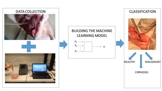
- Application of multiclass ML algorithms to in vivo dielectric property data: Performed by applying multiclass ML algorithms to in vivo rat dielectric properties collected from rat hepatic tissues including healthy, cirrhosis, and malignant tissues. This approach reveals the ability of the technique to discriminate different pathological stages of a diseased tissue in a realistic scenario.
- Potential proliferation of the data with phantoms: In vivo data collection is laborious, costly, requires facilities, and subject to strict ethical regulations. On the other hand, ML algorithms thrives with large amount of data. Therefore, there is a need to proliferate the data. One option is to acquire the data from phantom materials. This was performed by first, collecting dielectric property data from phantom materials mimicking the dielectric properties of liver tissues. Next, classifying the collected dielectric property data with multiclass ML algorithms. Finally, generalizing the model to in vivo dielectric property measurements.
2. Materials and Methods
2.1. In Vivo Dielectric Property Measurements
2.1.1. Experiment Samples
2.1.2. Measurement Setup
2.1.3. In Vivo Measurements
2.2. Phantom Characterization and Measurements
2.2.1. Phantom Materials
2.2.2. Measurement Setup
2.3. Uncertainty Analysis
- Repeatability: Dielectric property measurements of 0.1 M NaCl solution were collected at different sessions. To calculate uncertainty due to repeatability, standard deviation from the mean (SDM) was calculated from a total of 54 measurements.
- System drift: It was calculated by taking the difference between the dielectric property measurement after calibration and dielectric property measurement 30 min after calibration. Thirty min was chosen since during in vivo rat liver measurements, the system was re-calibrated every thirty minutes. To calculate the system drift, 30 measurements were used.
- Cable movements: It was characterized by calculating the difference between measured dielectric properties of 0.1 M NaCl solutions for different cable positions. Note that the most extreme cable movements that could occur during in vivo measurements were considered. A total of 68 measurements were used to calculate the uncertainty due to cable movements.
2.4. Multiclass Classification
2.4.1. k-Nearest Neighbors (kNN)
2.4.2. Logistic Regression (LR)
2.4.3. Random Forest (RF)
3. Results
3.1. Uncertainty Calculation with 0.1 M NaCl Solution
3.2. Dielectric Properties of Healthy and Diseased Rat Liver Tissues
3.3. Statistical Analysis
3.4. Dielectric Properties of Tissue Mimicking Materials
3.5. Multiclass Classification
4. Discussion
5. Conclusions
Funding
Conflicts of Interest
References
- Popovic, D.; McCartney, L.; Beasley, C.; Lazebnik, M.; Okoniewski, M.; Hagness, S.C.; Booske, J.H. Precision open-ended coaxial probes for in vivo and ex vivo dielectric spectroscopy of biological tissues at microwave frequencies. IEEE Trans. Microw. Theory Tech. 2005, 53, 1713–1722. [Google Scholar] [CrossRef] [Green Version]
- Anderson, W. Microwave Biopsy Probe. U.S. Patent Application No. 10/961,812, 8 October 2004. [Google Scholar]
- Keysight Technologies. Probe Characteristics and Specifications, Keysight N1501A, Dielectric Probe Kit 10 MHz to 50 GHz. Available online: https://literature.cdn.keysight.com/litweb/pdf/5992-0264EN.pdf?id=2605692 (accessed on 26 July 2019).
- Hashimshony, D.; Aharonowitz, G.; Cohen, G.; Geltner, I. Tissue-Characterization System and Method. U.S. Patent 8,147,423, 3 April 2012. [Google Scholar]
- Hashimshony, D.; Cohen, G. Tissue Sampling for Pathological Study. U.S. Patent 9,217,739, 22 December 2015. [Google Scholar]
- Foster, K.R.; Schwan, H.P. Dielectric Properties of Tissues Handbook of Biological Effects of Electromagnetic Fields; CRC Press: Boca Raton, FL, USA, 1995; Volume 2, pp. 25–102. [Google Scholar]
- O’rourke, A.P.; Lazebnik, M.; Bertram, J.M.; Converse, M.C.; Hagness, S.C.; Webster, J.G.; Mahvi, D.M. Dielectric properties of human normal, malignant and cirrhotic liver tissue: In vivo and ex vivo measurements from 0.5 to 20 GHz using a precision open-ended coaxial probe. Phys. Med. Biol. 2007, 52, 4707–4719. [Google Scholar] [CrossRef] [PubMed] [Green Version]
- Peyman, A.; Rezazadeh, A.A.; Gabriel, C. Changes in the dielectric properties of rat tissue as a function of age at microwave frequencies. Phys. Med. Biol. 2001, 46, 1617. [Google Scholar] [CrossRef] [PubMed]
- Farrugia, L.; Schembri Wismayer, P.; Zammit Mangion, L.; Sammut, C.V. Accurate in vivo dielectric properties of liver from 500 MHz to 40 GHz and their correlation to ex vivo measurements. Electromagn. Biol. Med. 2016, 35, 365–373. [Google Scholar] [CrossRef] [PubMed]
- Shahzad, A.; Khan, S.; Jones, M.; Dwyer, R.M.; O’Halloran, M. Investigation of the effect of dehydration on tissue dielectric properties in ex vivo measurements. Biomed. Phys. Eng. Express 2017, 3, 045001. [Google Scholar] [CrossRef] [Green Version]
- Balidemaj, E.; van den Berg, C.A.T.; Trinks, J.; van Lier, A.L.H.M.W.; Nederveen, A.J.; Stalpers, L.J.A.; Crezee, H.; Remis, R.F. CSI-EPT: A Contrast Source Inversion Approach for Improved MRI-Based Electric Properties Tomography. IEEE Trans. Med. Imaging 2015, 34, 1788–1796. [Google Scholar] [CrossRef] [PubMed]
- Bevacqua, M.T.; Bellizzi, G.G.; Crocco, L.; Isernia, T. A method for quantitative imaging of electrical properties of human tissues from only amplitude electromagnetic data. Inverse Probl. 2019, 35, 025006. [Google Scholar] [CrossRef]
- Yilmaz, T.; Kilic, M.A.; Erdogan, M.; Cayoren, M.; Tunaoglu, D.; Kurtoglu, I.; Yaslan, Y.; Çayören, H.; Arıkan, A.E.; Teksöz, S.; et al. Machine learning aided diagnosis of hepatic malignancies through in vivo dielectric measurements with microwaves. Phys. Med. Biol. 2016, 61, 5089–5102. [Google Scholar] [CrossRef]
- Yilmaz, T.; Foster, R.; Hao, Y. Broadband tissue mimicking phantoms and a patch resonator for evaluating noninvasive monitoring of blood glucose levels. IEEE Trans. Antennas Propag. 2014, 62, 3064–3075. [Google Scholar] [CrossRef]
- Taylor, B.N.; Kuyatt, C.E. Guidelines for Evaluating and Expressing the Uncertainty of NIST Measurement Results; United States Department of Commerce: Washington, DC, USA, 1994.
- ISO Guide to the Expression of Uncertainty in Measurement; ISO: Geneva, Switzerland, 1995.
- Gabriel, C.; Peyman, A. Dielectric measurement: Error analysis and assessment of uncertainty. Phys. Med. Biol. 2006, 51, 6033–6046. [Google Scholar] [CrossRef]
- Fornes-Leal, A.; Cardona, N.; Frasson, M.; Castelló-Palacios, S.; Nevárez, A.; Pons Beltrán, V.; Garcia-Pardo, C. Dielectric Characterization of In Vivo Abdominal and Thoracic Tissues in the 0.5–26.5 GHz Frequency Band for Wireless Body Area Networks. IEEE Access 2019, 7, 31854–31864. [Google Scholar] [CrossRef]
- Büchner, R.; Hefter, G.T.; May, P.M. Dielectric relaxation of aqueous NaCl solutions. J. Phys. Chem. A 1999, 103, 1–9. [Google Scholar] [CrossRef]
- Peyman, A.; Gabriel, C.; Grant, E.H. Complex permittivity of sodium chloride solutions at microwave frequencies. Bioelectromagn. J. Bioelectromagn. Soc. 2007, 28, 264–274. [Google Scholar] [CrossRef] [PubMed]
- Jiang, S.; Pang, G.; Wu, M.; Kuang, L. An improved K-nearest-neighbor algorithm for text categorization. Expert Syst. Appl. 2012, 39, 1503–1509. [Google Scholar] [CrossRef]
- Banu, S.; Aydınalp, C.; Cansız, G.; Joof, S.; Yilmaz, T.; Çayören, M.; Önal, B.; Akduman, I. Microwave dielectric property based classification of renal calculi: Application of a kNN algorithm. Comput. Biol. Med. 2019, 112, 103366. [Google Scholar]
- Breiman, L. Random forests. Mach. Learn. 2001, 45, 5–32. [Google Scholar] [CrossRef] [Green Version]
- Biau, G. Analysis of a random forests model. J. Mach. Learn. Res. 2012, 13, 1063–1095. [Google Scholar]
- Azadeh, P.; Kos, B.; Djokić, M.; Trotovšek, B.; Limbaeck-Stokin, C.; Serša, G.; Miklavčič, D. Variation in dielectric properties due to pathological changes in human liver. Bioelectromagnetics 2015, 36, 603–612. [Google Scholar]
- Gabriel, S.; Lau, R.W.; Gabriel, C. The dielectric properties of biological tissues: II. Measurements in the frequency range 10 Hz to 20 GHz. Phys. Med. Biol. 1996, 41, 2251. [Google Scholar] [CrossRef] [Green Version]
- Mariya, L.; Converse, M.C.; Booske, J.H.; Hagness, S.C. Ultrawideband temperature-dependent dielectric properties of animal liver tissue in the microwave frequency range. Phys. Med. Biol. 2006, 51, 1941. [Google Scholar]
- Lourdes, A.; Sammut, C.; Mangion, L.Z. Dielectric properties of muscle and liver from 500 MHz–40 GHz. Electromagn. Biol. Med. 2013, 32, 244–252. [Google Scholar]


| Dielectric Properties | Repeatability | Difference from the Literature [19,20] | System Drift | Cable Movement | Combined Uncertainty | Expanded Uncertainty |
|---|---|---|---|---|---|---|
| 0.37 | 0.58 | 0.53 | 0.33 | 0.93 | 1.86 | |
| 1.27 | 3.17 | 2.19 | 0.58 | 4.09 | 8.18 | |
| 0.15 | 0.36 | 0.23 | 0.10 | 0.47 | 0.94 |
| Frequency (GHz) | Dielectric Properties | Malignant | Cirrhosis | Healthy |
|---|---|---|---|---|
| 54.97 ± 6.71 | 52.95 ± 4.23 | 46.20 ± 5.79 | ||
| 21.89 ± 5.65 | 20.19 ± 4.51 | 17.74 ± 4.93 | ||
| 1.22 ± 0.49 | 1.12 ± 0.46 | 0.99 ± 0.47 | ||
| 51.18 ± 6.78 | 49.33 ± 4.30 | 42.15 ± 5.49 | ||
| 14.06 ± 4.39 | 13.80 ± 4.29 | 12.29 ± 4.50 | ||
| 2.35 ± 0.55 | 2.30 ± 0.53 | 2.05 ± 0.58 | ||
| 48.99 ± 6.61 | 47.19 ± 4.40 | 40.31 ± 5.42 | ||
| 15.66 ± 4.53 | 16.11 ± 5.00 | 13.99 ± 4.94 | ||
| 4.36 ± 0.78 | 4.48 ± 0.98 | 3.89 ± 0.95 |
| Frequency (GHz) | Dielectric Properties | Malignant | Cirrhosis | Healthy | p-Value |
|---|---|---|---|---|---|
| 44.33 | 53.41 | 50.81 | <0.01 | ||
| 12.88 | 15.12 | 14.39 | <0.01 | ||
| 41.26 | 50.65 | 48.27 | <0.01 | ||
| 13.27 | 14.98 | 14.77 | <0.01 | ||
| 38.98 | 48.38 | 45.72 | <0.01 | ||
| 45.72 | 17.40 | 18.01 | <0.01 |
| Model | Accuracy (%) | Precision (%) | Recall (%) | F1 score (%) |
|---|---|---|---|---|
| LR | 48 | 48 | 48 | 47 |
| kNN | 50 | 54 | 50 | 50 |
| Tissue Type | Reference | Species | Condition | ||
|---|---|---|---|---|---|
| Malignant | 3.64 | 53.91 | O’Rourke et al. [7] | human | ex vivo |
| 18.17 | 13.31 | Peyman et al. [25] | human | ex vivo | |
| Cirrhosis | 2.14 | 13.81 | O’Rourke et al. [7] | human | ex vivo |
| 14.8 | 19.12 | Peyman et al. [25] | human | ex vivo | |
| Healthy | 1.58 | 3.85 | Gabriel et al. [26] | human | N/A |
| 4.66 | 35.12 | O’Rourke et al. [7] | human | ex vivo | |
| 9.10 | 10.29 | Lazebnik et al. [27] | human | ex vivo | |
| 4.13 | 13.81 | Abdilla et al. [28] | porcine | ex vivo |
| Model | Accuracy (%) | Precision (%) | Recall (%) | F1 Score (%) |
|---|---|---|---|---|
| LR | 50 | 50 | 50 | 46 |
| kNN | 44 | 53 | 44 | 47 |
© 2020 by the author. Licensee MDPI, Basel, Switzerland. This article is an open access article distributed under the terms and conditions of the Creative Commons Attribution (CC BY) license (http://creativecommons.org/licenses/by/4.0/).
Share and Cite
Yilmaz, T. Multiclass Classification of Hepatic Anomalies with Dielectric Properties: From Phantom Materials to Rat Hepatic Tissues. Sensors 2020, 20, 530. https://doi.org/10.3390/s20020530
Yilmaz T. Multiclass Classification of Hepatic Anomalies with Dielectric Properties: From Phantom Materials to Rat Hepatic Tissues. Sensors. 2020; 20(2):530. https://doi.org/10.3390/s20020530
Chicago/Turabian StyleYilmaz, Tuba. 2020. "Multiclass Classification of Hepatic Anomalies with Dielectric Properties: From Phantom Materials to Rat Hepatic Tissues" Sensors 20, no. 2: 530. https://doi.org/10.3390/s20020530
APA StyleYilmaz, T. (2020). Multiclass Classification of Hepatic Anomalies with Dielectric Properties: From Phantom Materials to Rat Hepatic Tissues. Sensors, 20(2), 530. https://doi.org/10.3390/s20020530