
Toxic Metals and Non-Communicable Diseases in HIV Population: A Systematic Review


 ,
, 
Abstract
:1. Introduction
2. Materials and Methods
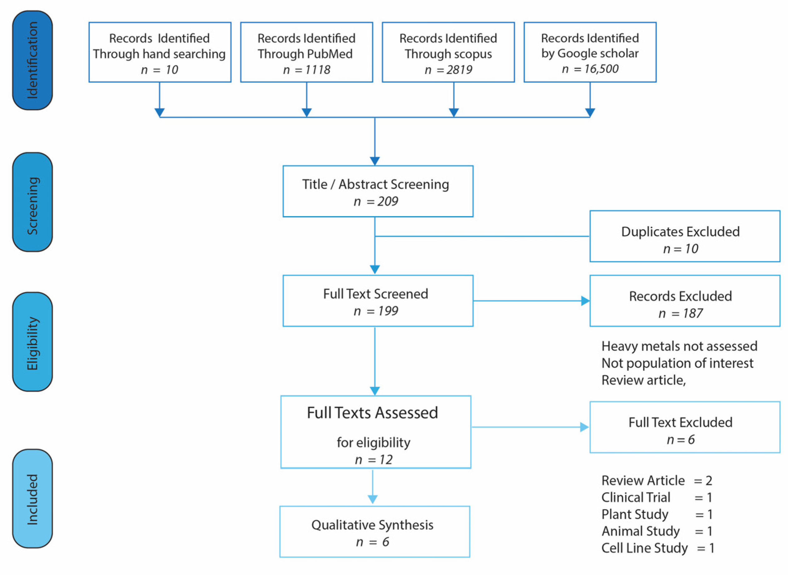
2.1. Search Strategy and Information Sources
2.2. Eligibility Criteria
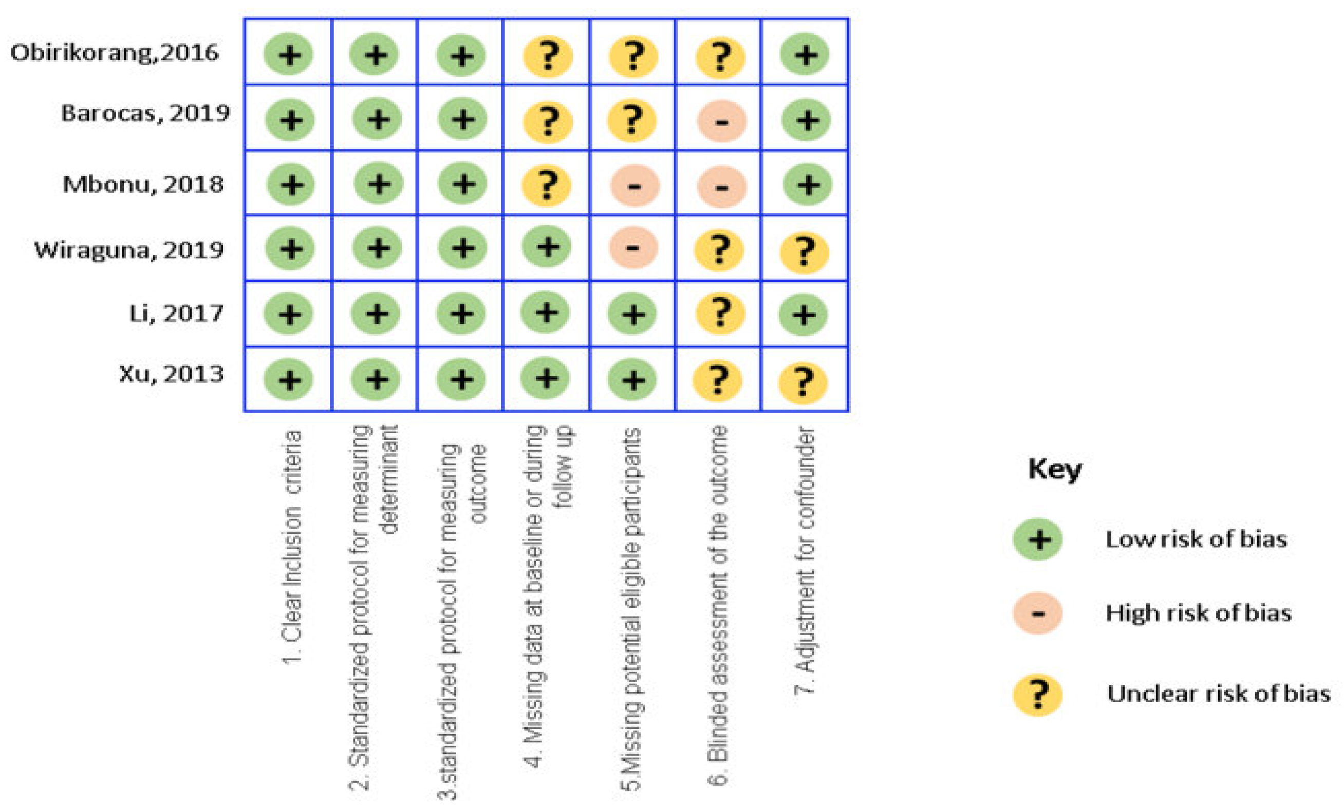
2.3. Study Quality Assessment
2.4. Data Extraction and Synthesis
2.5. Critical Appraisal
3. Results
3.1. Characteristics of Included Studies
3.2. Study Findings
3.2.1. Biological Essential Heavy Metals
3.2.2. Non-Biological Essential Heavy Metals
4. Discussion
4.1. Summary of Evidence and Considerations
4.2. Limitations
5. Conclusions
Author Contributions
Funding
Institutional Review Board Statement
Informed Consent Statement
Data Availability Statement
Conflicts of Interest
References
- Piot, P.; Bartos, M.; Ghys, P.D.; Walker, N.; Schwartländer, B. The global impact of HIV/AIDS. Nat. Cell Biol. 2001, 410, 968–973. [Google Scholar] [CrossRef]
- The Global HIV/AIDS Epidemic. 2019. Available online: https://www.hiv.gov/hiv-basics/overview/data-and-trends/global-statistics (accessed on 13 April 2020).
- Kharsany, A.B.; Karim, Q.A. HIV Infection and AIDS in Sub-Saharan Africa: Current Status, Challenges and Opportunities. Open AIDS J. 2016, 10, 34–48. [Google Scholar] [CrossRef] [Green Version]
- Bloomfield, G.S.; Khazanie, P.; Morris, A.; Rabadán-Diehl, C.; Benjamin, L.A.; Murdoch, D.; Radcliff, V.S.; Velazquez, J.E.; Hicks, C. HIV and Non-Communicable Cardiovascular and Pulmonary Diseases in Low–and Middle-Income Countries in the ART Era: What We Know and Best Directions for Future Research. J. Acquir. Immune. Defic. Syndr. 2014, 67, S40–S53. [Google Scholar] [CrossRef] [PubMed] [Green Version]
- Kalyesubula, R.; Wearn, N.; Semitala, F.C.; Bowa, K. HIV-associated renal and genitourinary comorbidities in Africa. JAIDS J. Acquir. Immune Defic. Syndr. 2014, 67, S68–S78. [Google Scholar] [CrossRef] [PubMed] [Green Version]
- Narayan, K.V.; Miotti, P.G.; Anand, N.P.; Kline, L.M.; Harmston, C.; Gulakowski, R., III; Vermund, S.H. HIV and Noncommunicable Disease Comorbidities in the Era of Antiretroviral Therapy: A Vital Agenda for Research in Low- and Middle-Income Country Settings. J. Acquir. Immune. Defic. Syndr. 2014, 67, S2–S7. [Google Scholar] [CrossRef] [PubMed] [Green Version]
- Maciel, R.A.; Klück, H.M.; Durand, M.; Sprinz, E. Comorbidity is more common and occurs earlier in persons living with HIV than in HIV-uninfected matched controls, aged 50 years and older: A cross-sectional study. Int. J. Infect. Dis. 2018, 70, 30–35. [Google Scholar] [CrossRef] [Green Version]
- Schouten, J.; Wit, F.W.; Stolte, I.G.; Kootstra, N.A.; Van Der Valk, M.; Geerlings, S.E.; Prins, M.; Reiss, P.; Kooij, K.W.; Van Zoest, R.A.; et al. Cross-sectional Comparison of the Prevalence of Age-Associated Comorbidities and Their Risk Factors Between HIV-Infected and Uninfected Individuals: The AGEhIV Cohort Study. Clin. Infect. Dis. 2014, 59, 1787–1797. [Google Scholar] [CrossRef]
- Lucas, G.M.; Ross, M.J.; Stock, P.G.; Shlipak, M.G.; Wyatt, C.M.; Gupta, S.K.; Atta, M.G.; Wools-Kaloustian, K.K.; Pham, P.A.; Bruggeman, L.A.; et al. Clinical Practice Guideline for the Management of Chronic Kidney Disease in Patients Infected with HIV: 2014 Update by the HIV Medicine Association of the Infectious Diseases Society of America. Clin. Infect. Dis. 2014, 59, e96–e138. [Google Scholar] [CrossRef] [Green Version]
- Vos, A.G.; Idris, N.S.; Barth, R.E.; Klipstein-Grobusch, K.; Grobbee, D.E. Pro-Inflammatory Markers in Relation to Cardiovascular Disease in HIV Infection. A Systematic Review. PLoS ONE 2016, 11, e0147484. [Google Scholar] [CrossRef] [Green Version]
- Syed, F.F.; Sani, M.U. Recent advances in HIV-associated cardiovascular diseases in Africa. Heart 2013, 99, 1146–1153. [Google Scholar] [CrossRef]
- Adebamowo, C.A.; Casper, C.; Bhatia, K.; Mbulaiteye, S.M.; Sasco, A.J.; Phipps, W.; Vermund, S.H.; Krown, S.E. Challenges in the Detection, Prevention, and Treatment of HIV-Associated Malignancies in Low- and Middle-Income Countries in Africa. JAIDS J. Acquir. Immune Defic. Syndr. 2014, 67, S17–S26. [Google Scholar] [CrossRef] [PubMed] [Green Version]
- Grogg, K.L.; Miller, R.F.; Dogan, A. HIV infection and lymphoma. J. Clin. Pathol. 2006, 60, 1365–1372. [Google Scholar] [CrossRef] [Green Version]
- Zaid, D.; Greenman, Y. Human Immunodeficiency Virus Infection and the Endocrine System. Endocrinol. Metab. 2019, 34, 95–105. [Google Scholar] [CrossRef] [PubMed]
- Everall, I.P.; Luthert, P.J.; Lantos, P.L. Neuronal number and volume alterations in the neocortex of HIV infected individuals. J. Neurol. Neurosurg. Psychiatry 1993, 56, 481–486. [Google Scholar] [CrossRef] [PubMed]
- Rao, V.R.; Ruiz, A.P.; Prasad, V.R. Viral and cellular factors underlying neuropathogenesis in HIV associated neurocognitive disorders (HAND). AIDS Res. Ther. 2014, 11, 13. [Google Scholar] [CrossRef] [Green Version]
- Jensen, B.K.; Monnerie, H.; Mannell, M.V.; Gannon, P.J.; Espinoza, C.A.; Erickson, M.A.; Bruce-Keller, A.J.; Gelman, B.B.; Briand, L.A.; Pierce, R.C.; et al. Altered Oligodendrocyte Maturation and Myelin Maintenance: The Role of Antiretrovirals in HIV-Associated Neurocognitive Disorders. J. Neuropathol. Exp. Neurol. 2015, 74, 1093–1118. [Google Scholar] [CrossRef] [PubMed] [Green Version]
- Lanman, T.; Letendre, S.; Ma, Q.; Bang, A.; Ellis, R. CNS Neurotoxicity of Antiretrovirals. J. Neuroimmune Pharmacol. 2021, 16, 130–143. [Google Scholar] [CrossRef]
- Hung, K.-M.; Chen, P.-C.; Hsieh, H.-C.; Calkins, M.J. Mitochondrial defects arise from nucleoside/nucleotide reverse transcriptase inhibitors in neurons: Potential contribution to HIV-associated neurocognitive disorders. Biochim. Biophys. Acta (BBA) Mol. Basis Dis. 2017, 1863, 406–413. [Google Scholar] [CrossRef]
- Collazos, J.; Martinez, E.; Mayo, J.; Ibarra, S. Sexual hormones in HIV-infected patients: The influence of antiretroviral therapy. AIDS 2002, 16, 934–937. [Google Scholar] [CrossRef] [PubMed]
- Turner, M.D.; Nedjai, B.; Hurst, T.; Pennington, D.J. Cytokines and chemokines: At the crossroads of cell signalling and inflammatory disease. Biochim. Biophys. Acta 2014, 1843, 2563–2582. [Google Scholar] [CrossRef] [Green Version]
- Orisakwe, O.E. Lead and Cadmium in public health in Nigeria: Physicians neglect and pitfall in patient management. N. Am. J. Med. Sci. 2014, 6, 61–70. [Google Scholar] [CrossRef] [Green Version]
- Savarino, A.; Pescarmona, G.P.; Boelaert, J.R. Iron metabolism and HIV infection: Reciprocal interactions with potentially harmful consequences? Cell Biochem. Funct. 1999, 17, 279–287. [Google Scholar] [CrossRef]
- Sanchez, T. Effects of Mercury, Lead, Arsenic and Zinc to Human Renal Oxidative Stress and Functions: A Review. Arch. Med. 2018, 4, 2. [Google Scholar] [CrossRef]
- Valko, M.; Rhodes, C.; Moncol, J.; Izakovic, M.; Mazur, M. Free radicals, metals and antioxidants in oxidative stress-induced cancer. Chem. Interact. 2006, 160, 1–40. [Google Scholar] [CrossRef]
- Fasinu, P.S.; Orisakwe, O.E. Heavy metal pollution in sub-Saharan Africa and possible implications in cancer epidemiology. Asian Pac. J. Cancer Prev. 2013, 14, 3393–3402. [Google Scholar] [CrossRef] [PubMed] [Green Version]
- Chowdhury, R.; Ramond, A.; O’Keeffe, L.M.; Shahzad, S.; Kunutsor, S.K.; Muka, T.; Gregson, J.; Willeit, P.; Warnakula, S.; Khan, H.; et al. Environmental toxic metal contaminants and risk of cardiovascular disease: Systematic review and meta-analysis. BMJ 2018, 362, k3310. [Google Scholar] [CrossRef] [Green Version]
- Frumkin, H.; Haines, A. Global Environmental Change and Noncommunicable Disease Risks. Annu. Rev. Public Heal. 2019, 40, 261–282. [Google Scholar] [CrossRef] [PubMed] [Green Version]
- Wiraguna, A.A.G.P.; Andriani, P.I.; Adiguna, M.S. Comparison of Plasma Zinc Levels among HIV+ ans HIV- Subjects Infected with Condyloma Acuminata. Asian Pac. J. Cancer Prev. 2019, 20, 943–949. [Google Scholar] [CrossRef] [PubMed]
- Xu, X.; Hu, H.; Dailey, A.B.; Kearney, G.; Talbott, E.O.; Cook, R.L. Potential Health Impacts of Heavy Metals on HIV-Infected Population in USA. PLoS ONE 2013, 8, e74288. [Google Scholar] [CrossRef] [Green Version]
- Barocas, J.A.; So-Armah, K.; Cheng, D.M.; Lioznov, D.; Baum, M.; Gallagher, K.; Fuster, D.; Gnatienko, N.; Krupitsky, E.; Freiberg, M.S.; et al. Zinc deficiency and advanced liver fibrosis among HIV and hepatitis C co-infected anti-retroviral naïve persons with alcohol use in Russia. PLoS ONE 2019, 14, e0218852. [Google Scholar] [CrossRef] [Green Version]
- Obirikorang, C.; Issahaku, R.G.; Osakunor, D.N.M.; Osei-Yeboah, J. Anaemia and Iron Homeostasis in a Cohort of HIV-Infected Patients: A Cross-Sectional Study in Ghana. AIDS Res. Treat. 2016, 2016, 1–8. [Google Scholar] [CrossRef] [PubMed] [Green Version]
- Li, R.; Zhao, L.; Hou, Z.; Zhang, D.; Wan, L.; Wei, L.; Yang, Y.; Lv, J.; Ma, M.; Zhu, Y. A Preliminary Study about the Potential Effects of Heavy Metals on the Human Male Reproductive Parameters in HIV-Infected Population in China. Biol. Trace Element Res. 2017, 180, 39–47. [Google Scholar] [CrossRef] [PubMed]
- Ma, E.; Mbonu, I. Blood levels of some toxic metals in Human Immunodeficiency Virus (HIV) Type 1- infection. Ann. Health Res. 2018, 4, 75–81. [Google Scholar]
- Pooter, D.D. Heavy Metals. 2002. Available online: http://www.coastalwiki.org/wiki/Heavy_metals (accessed on 15 May 2020).
- Firkin, F. Diagnostic Tests: Interpretation of biochemical tests for iron deficiency: Diagnostic difficulties related to limitations of individual tests. Aust. Prescr. 1997, 20, 74–76. [Google Scholar] [CrossRef]
- Orisakwe, O.E.; Amadi, C.N.; Frazzoli, C. Management of Iron Overload in Resource Poor Nations: A Systematic Review of Phlebotomy and Natural Chelators. J. Toxicol. 2020, 2020, 1–14. [Google Scholar] [CrossRef]
- Shen, X.; Lee, K.; König, R. Effects of heavy metal ions on resting and antigen-activated CD4(+) T cells. Toxicology 2001, 169, 67–80. [Google Scholar] [CrossRef]
- Annesi-Maesano, I.; Pollitt, R.; King, G.; Bousquet, J.; Hellier, G.; Sahuquillo, J.; Huel, G. In utero exposure to lead and cord blood total IgE. Is there a connection? Allergy 2003, 58, 589–594. [Google Scholar] [CrossRef] [PubMed]
- Wells, E.M.; Bonfield, T.L.; Dearborn, D.G.; Jackson, L.W. The relationship of blood lead with immunoglobulin E, eosinophils, and asthma among children: NHANES 2005–2006. Int. J. Hyg. Environ. Health 2014, 217, 196–204. [Google Scholar] [CrossRef]
- Metryka, E.; Chibowska, K.; Gutowska, I.; Falkowska, A.; Kupnicka, P.; Barczak, K.; Chlubek, D.; Baranowska-Bosiacka, I. Lead (Pb) Exposure Enhances Expression of Factors Associated with Inflammation. Int. J. Mol. Sci. 2018, 19, 1813. [Google Scholar] [CrossRef] [Green Version]
- Heo, Y.; Parsons, P.J.; Lawrence, D.A. Lead differentially modifies cytokine production in vitro and in vivo. Toxicol. Appl. Pharmacol. 1996, 138, 149–157. [Google Scholar] [CrossRef]
- Yücesoy, B.; Turhan, A.; Üre, M.; Imir, T.; Karakaya, A. Effects of occupational lead and cadmium exposure on some immunoregulatory cytokine levels in man. Toxicology 1997, 123, 143–147. [Google Scholar] [CrossRef]
- Vagaska, B.; New SE, P.; Alvarez-Gonzalez, C.; D’Acquisto, F.; Gomez, S.G.; Bulstrode, N.W.; Madrigal, A.; Ferretti, P. MHC-class-II are expressed in a subpopulation of human neural stem cells in vitro in an IFN–independent fashion and during development. Sci. Rep. 2016, 6, 2425. [Google Scholar] [CrossRef] [PubMed]
- Lawrence, D.A. Posited mechanisms of metal immunotoxicity. Hum. Exp. Toxicol. 1995, 14, 114–116. [Google Scholar] [CrossRef]
- Heo, Y.; Lee, W.T.; Lawrence, D.A. Differential Effects of Lead and cAMP on Development and Activities of Th1- and Th2-Lymphocytes. Toxicol. Sci. 1998, 43, 172–185. [Google Scholar] [CrossRef] [PubMed]
- Miller, T.E.; Golemboski, K.A.; Ha, R.S.; Bunn, T.; Sanders, F.S.; Dietert, R.R. Developmental exposure to lead causes persistent immunotoxicity in Fischer 344 rats. Toxicol. Sci. 1998, 42, 129–135. [Google Scholar] [CrossRef]
- Mishra, K. Lead exposure and its impact on immune system: A review. Toxicol. Vitr. 2009, 23, 969–972. [Google Scholar] [CrossRef] [PubMed]
- Guo, T.L.; Mudzinski, S.P.; Lawrence, D.A. The heavy metal lead modulates the expression of both TNF-α and TNF-α receptors in lipopolysaccharide-activated human peripheral blood mononuclear cells. J. Leukoc. Biol. 1996, 59, 932–939. [Google Scholar] [CrossRef]
- Farzan, S.F.; Korrick, S.; Li, Z.; Enelow, R.; Gandolfi, A.J.; Madan, J.; Nadeau, K.; Karagas, M.R. In utero arsenic exposure and infant infection in a United States cohort: A prospective study. Environ. Res. 2013, 126, 24–30. [Google Scholar] [CrossRef] [Green Version]
- Cao, J.J.; Xu, X.; Hylkema, M.N.; Zeng, E.Y.; Sly, P.D.; Suk, W.A.; Bergman, Å.; Huo, X. Early-life Exposure to Widespread Environmental Toxicants and Health Risk: A Focus on the Immune and Respiratory Systems. Ann. Glob. Health 2016, 82, 119–131. [Google Scholar] [CrossRef]
- Ayatollahi, M.O. Study of the impact of blood lead level on humoral immunity in humans. Toxicol. Ind. Health 2002, 18, 39–44. [Google Scholar] [CrossRef]
- Ritz, B.; Heinrich, J.; Wjst, M.; Wichmann, E.; Krause, C. Effect of Cadmium Body Burden on Immune Response of School Children. Arch. Environ. Health Int. J. 1998, 53, 272–280. [Google Scholar] [CrossRef]
- Miro, J.M.; Cofán, F.; Trullas, J.C.; Manzardo, C.; Cervera, C.; Tuset, M.; Oppenheimer, F.; Brunet, M.; Moreno, A.; Campistol, J.M.; et al. Renal Dysfunction in the Setting of HIV/AIDS. Curr. HIV/AIDS Rep. 2012, 9, 187–199. [Google Scholar] [CrossRef] [PubMed]
- Moon, J. The role of vitamin D in toxic metal absorption: A review. J. Am. Coll. Nutr. 1994, 13, 559–564. [Google Scholar] [CrossRef] [PubMed]
- Schwalfenberg, G.K.; Genuis, S.J. Vitamin D, Essential Minerals, and Toxic Elements: Exploring Interactions between Nutrients and Toxicants in Clinical Medicine. Sci. World J. 2015, 2015, 1–8. [Google Scholar] [CrossRef] [PubMed] [Green Version]
- Kemp, F.W.; Neti, P.V.; Howell, R.W.; Wenger, P.; Louria, D.B.; Bogden, J.D. Elevated Blood Lead Concentrations and Vitamin D Deficiency in Winter and Summer in Young Urban Children. Environ. Health Perspect. 2007, 115, 630–635. [Google Scholar] [CrossRef] [PubMed] [Green Version]
- Fischbein, A.; Tsang, P.; Luo, J.-C.J.; Roboz, J.P.; Jiang, J.D.; Bekesi, J. Phenotypic Aberrations of CD3+ and CD4+ Cells and Functional Impairments of Lymphocytes at Low-Level Occupational Exposure to Lead. Clin. Immunol. Immunopathol. 1993, 66, 163–168. [Google Scholar] [CrossRef]
- Ündeḡer, Ü.; Başaran, N.; Canpmar, H.; Kansu, E. Immune alterations in lead-exposed workers. Toxicology 1996, 109, 167–172. [Google Scholar] [CrossRef]
- Li, S.; Zhengyan, Z.; Rong, L.; Hanyun, C. Decrease of CD4+ T-Lymphocytes in Children Exposed to Environmental Lead. Biol. Trace Element Res. 2005, 105, 19–26. [Google Scholar] [CrossRef]
- Baum, M.K.; Adriana, C.; Shengan, L.; Hong, L.; Page, J. Zinc Status in Human Immunodeficiency Virus Type 1 Infection and Illicit Drug Use. Clin. Infect. Dis. 2003, 37, S117–S123. Available online: http://www.jstor.org/stable/4462563 (accessed on 20 May 2020). [CrossRef]
- Telisman, S.; Cvitković, P.; Jurasović, J.; Pizent, A.; Gavella, M.; Rocić, B. Semen quality and reproductive endocrine function in relation to biomarkers of lead, cadmium, zinc, and copper in men. Environ. Health Perspect. 2000, 108, 45–53. [Google Scholar] [CrossRef] [PubMed]
- Pant, N.; Upadhyay, G.; Pandey, S.; Mathur, N.; Saxena, D.; Srivastava, S. Lead and cadmium concentration in the seminal plasma of men in the general population: Correlation with sperm quality. Reprod. Toxicol. 2003, 17, 447–450. [Google Scholar] [CrossRef]
- Bujan, L.; Sergerie, M.; Moinard, N.; Martinet, S.; Porte, L.; Massip, P.; Pasquier, C.; Daudin, M. Decreased Semen Volume and Spermatozoa Motility in HIV-1-Infected Patients Under Antiretroviral Treatment. J. Androl. 2006, 28, 444–452. [Google Scholar] [CrossRef]
- Dulioust, E.; Le Du, A.; Costagliola, D.; Guibert, J.; Kunstmann, J.-M.; Heard, I.; Juillard, J.-C.; Salmon, D.; Leruez-Ville, M.; Mandelbrot, L.; et al. Semen alterations in HIV-1 infected men. Hum. Reprod. 2002, 17, 2112–2118. [Google Scholar] [CrossRef] [Green Version]
- Krieger, J.N.; Coombs, R.W.; Collier, A.C.; Koehler, J.K.; Ross, S.O.; Chaloupka, K.; Murphy, V.L.; Corey, L. Fertility Parameters in Men Infected with Human Immunodeficiency Virus. J. Infect. Dis. 1991, 164, 464–469. [Google Scholar] [CrossRef] [PubMed]
- Haraguchi, Y.; Sakurai, H.; Hussain, S.; Anner, B.M.; Hoshino, H. Inhibition of HIV-1 infection by zinc group metal compounds. Antivir. Res. 1999, 43, 123–133. [Google Scholar] [CrossRef]
- Baum, M.K.; Shor-Posner, G.; Lu, Y.; Rosner, B.; Sauberlich, H.E.; Fletcher, M.A.; Szapocznik, J.; Eisdorfer, C.; Buring, J.E.; Hennekens, C.H. Micronutrients and HIV-1 disease progression. AIDS 1995, 9, 1051–1056. [Google Scholar] [CrossRef]
- Martinez, S.S.; Campa, A.; Li, Y.; Fleetwood, C.; Stewart, T.; Ramamoorthy, V.; Baum, M.K. Low Plasma Zinc Is Associated with Higher Mitochondrial Oxidative Stress and Faster Liver Fibrosis Development in the Miami Adult Studies in HIV Cohort. J. Nutr. 2017, 147, 556–562. [Google Scholar] [CrossRef] [PubMed] [Green Version]
- Johannessen, A.; Naman, E.; Ngowi, B.J.; Sandvik, L.; I Matee, M.; E Aglen, H.; Gundersen, S.G.; Bruun, J.N. Predictors of mortality in HIV-infected patients starting antiretroviral therapy in a rural hospital in Tanzania. BMC Infect. Dis. 2008, 8, 52. [Google Scholar] [CrossRef]
- Erhabor, O.; Ejele, O.A.; Nwauche, C.A.; Buseri, F.I. Some haematological parameters in Human Immunodeficiency Virus (HIV) infected Africans: The Nigerian perspective. Niger. J. Med. 2006, 14, 33–38. [Google Scholar] [CrossRef]
- Gebremedhin, K.B.; Haye, T.B. Factors Associated with Anemia among People Living with HIV/AIDS Taking ART in Ethiopia. Adv. Hematol. 2019, 2019, 1–8. [Google Scholar] [CrossRef]
- Frazzoli, C.; Mazzanti, F.; Achu, M.B.; Pouokam, G.B.; Fokou, E. Elements of kitchen toxicology to exploit the value of traditional (African) recipes in the diet of HIV+/AIDS subjects: The case of Egusi okra meal. Toxicol. Rep. 2017, 4, 474–483. [Google Scholar] [CrossRef]
- Frazzoli, C.; Mantovani, A. Toxicological risk factors in the burden of malnutrition: The case of nutrition (and risk) transition in sub-Saharan Africa. Food Chem. Toxicol. 2020, 146, 111789. [Google Scholar] [CrossRef]
- Amadi, C.N.; Offor, S.J.; Frazzoli, C.; Orisakwe, O.E. Natural antidotes and management of metal toxicity in developing nations. Environ. Sci. Pollut. Res. Int. 2019, 26, 18032–18052. [Google Scholar] [CrossRef] [PubMed]
- Amadi, C.N.; Frazzoli, C.; Orisakwe, O.E. Nigerian foods of probiotic potentials and relevance to chronic metal expo-sure: A systematic review. Environ. Sci. Pollut. Res. 2020, 27, 19285–19297. [Google Scholar]
- Oluwaseun Odukoya, J.; Olayemi Odukoya, J.; Tantoh Ndinteh, D. Elemental measurements and health risk assessment of sub-Saharan African medicinal plants used for cardiovascular diseases’ and related risk factors’ treatment. J. Trace. Elem. Med. Biol. 2021, 65, 126725. [Google Scholar] [CrossRef] [PubMed]
- More, G.; Makola, R.; Prinsloo, G. In Vitro Evaluation of Anti-Rift Valley Fever Virus, Antioxidant and Anti-Inflammatory Activity of South African Medicinal Plant Extracts. Viruses 2021, 13, 221. [Google Scholar] [CrossRef] [PubMed]
- Pouokam, G.B.; Hamed, H.; Ngwafor, R.; Frazzoli, C. Toxicovigilance systems and practices in Africa. Toxics 2016, 4, 13. [Google Scholar]
- Frazzoli, C. The vulnerable and the susceptible: The weight of evidenza to stop exploiting activities generating toxic expo-sures in unprotected and deprived countries. J. Glob. Health 2021, 11, 03046. [Google Scholar] [CrossRef] [PubMed]
- Acquavia, M.A.; Foti, L.; Pascale, R.; Nicolò, A.; Brancaleone, V.; Cataldi, T.R.; Martelli, G.; Scrano, L.; Bianco, G. Detection and quantification of Covid-19 antiviral drugs in biological fluids and tissues. Talanta 2021, 224, 121862. [Google Scholar] [CrossRef]


| Demographic Indicators | Xu et al., 2013 | Obirikorang et al., 2016 | Li et al., 2017 | Ma et al., 2018 | Wiraguna et al., 2019 | Barocas et al., 2019 |
|---|---|---|---|---|---|---|
| Number of participants | 11,761 | 319 | 59 | 300 | 45 | 204 |
| Country | USA | Ghana | China | Nigeria | Indonesia | Russia |
| Age(years) | 18–49 | >18 | 23–44 | 30–35 | 18–60 | 18–70 |
| Sex(n%) | ||||||
| Female | 6184 (52.5) | 217 (68) | 162 (54) | 17 (37.8) | 50 (24.5) | |
| Male | 5577 (47.4) | 102 (32) | 50(100) | 138 (46) | 28 (62) | 154 (75.5) |
| Ethnicity | N/A | N/A | N/A | N/A | N/A | |
| White | 4983 (42.4) | 0 | ||||
| Black | 2567 (21.8) | 300 | ||||
| Other | 4211 (35.8) | 0 | ||||
| Level of Education | N/A | N/A | N/A | N/A | N/A | |
| <High School | 3190 | 92 (28.8) | ||||
| High School | 2939 | 65 (20.4) | ||||
| >High School | 5624 | 56 (17.6) | ||||
| Abuse (Yes) | N/A | N/A | N/A | N/A | N/A | 179 (88.2) |
| Heavy drinking | 190 (93.1) | |||||
| Moderate drinking | 14 (6.9) | |||||
| Current Cocaine Use Yes (%) | 4 (2.0) | |||||
| Marital Status n (%) | N/A | N/A | N/A | N/A | N/A | |
| Married | 198 (62.1) | |||||
| Single | 121 (37.9) | |||||
| Employment Status n (%) | N/A | N/A | N/A | N/A | N/A | |
| Formal | 73 (22.9) | |||||
| Informal | 175 (54.9) | |||||
| Unemployed | 71 (22.3) | |||||
| HIV Status | ||||||
| HIV | 60 (0.51) | 319 (100) | 50 (100) | 200 (66.7) | 18 (40) | 204 (100) |
| Non HIV | 11,701 (99.5) | 0 (0) | 0 (0) | 100 (33.3) | 27 (60) | 0 |
| Author | Study Type | Population (n) | Sample Type | Markers Assessed/Method | Statistical Analysis |
|---|---|---|---|---|---|
| Xu et al., 2013 | Cross-sectional | HIV positive (60), HIV negative (11,701) | Blood (Serum/Plasma) | Cadmium, lead, mercury-plasma mass spectrometry, HIV antibody-enzyme immunoassay (EIA) (Bio-Rad Laboratories, Hercules, CA, USA). Western blot (Calypte Biomedical Corporation, Rockville, MD, USA) Serum cotinine-ID HPLC-APCI MS/MS | Two-sided student t-tests Wald chi-square analysis Multivariate linear regression models (evaluate the associations between HIV status and each heavy metal). Statistical analysis performed using SAS Institute Inc., Cary, NC, USA) |
| Obirikorang et al., 2016 | Comparative cross-sectional | HAART-treated (219), HAART-naïve (100) | Blood (Serum) | CD4/CD3 lymphocyte count-flow cytometry by flow cytometry (BD FACSCOUNT, Becton Dickenson and Company, San Diego, CA, USA) haemoglobin and white cell indices. (Mindray BC 3000 Plus Mind ray Company, Shenzhen, China). serum iron, ferritin, transferrin, and transferrin saturation (TSAT)-Flexor XL analyzer from vital scientific serum CRP-semi quantitative immune-chromatographic method | Unpaired t-test (compare means of continuous variables), Fisher’s exact test/chi-square, one-way ANOVA. Data analyzed using Graph pad Prism version 6.0 for windows (Graph pad software, San Diego, CA, USA). |
| Li et al., 2017 | Cross-sectional | HIV positive men (50) | serum plasma, urine, semen |
| ANOVA Spearman’s rank correlation data analyzed using SPSS 16.0 for windows (SPSS Inc., Chicago, IL, USA) |
| Ma, et al., 2018 | Cross-sectional | HIV HAART-treated (100), HIV HAART-naïve (100), HIV-negative Controls (100) | Blood(Plasma) | HIV Screening-Unigold, Determine, CD-4 T count- Cyflow counter flow cytometer (Facs Flow Cytometer count system, Lincolnshire, IL, USA). Plasma levels of Pb, Cd, Hg, and Ni- Inductively Coupled Plasma Mass Spectrometer (ICP-MS), Agilent 7500, Norwalk, CT, USA. | Student’s t-test, ANOVA, Pearson’s correlation coefficient Statistical software SPSS version IBM 21 (SPSS Inc., Chicago, IL, USA) |
| Wiraguna et al., 2019 | Cross-sectional | HIV positive (18), HIV negative (27) | Blood (Plasma) |
| Bivariate analysis, chi-square test, unpaired t-test, Shapiro–Wilk, SPSS 23.0 |
| Barocas et al., 2019 | Cross-sectional | HIV positive (204) | Blood (Plasma) | Zn level testing (ImmunoBioService laboratory, St. Petersburg) Laboratory assays: ALT, AST, platelet count (St. Petersburg Pasteur Institute Central Clinical Diagnostic Laboratory, Northwestern Federal District, Russia) liver stiffness-Elastography (Fibro scan) | Generalized additive models (GAMs), Multiple linear regression models, chi-square, Fisher’s exact test (comparison of groups for categorical variables), two-tailed tests t-tests and Wilcoxon tests (for continuous variables) Analysis performed using SAS version 9.3 (SAS Institute, Inc., Cary, NC, USA) |
| Heavy Metal Marker | Author | Sample Size | Assessment Method | Findings | Outcome Measured/Method | Associations between Heavy Metal Markers and Outcome Measured |
|---|---|---|---|---|---|---|
| Lead (Pb) | Xu et al., 2013 | 11,761 | ICP-MS | HIV:1.43 (1.17–1.75), Non-HIV Negative:1.11 (1.09–1.14) p = 0.02 | Elevated prevalence of heavy metals in HIV patients | Pb levels were higher in HIV-infected patients aged 18–34; who reported being neither Hispanic, white, or black; who had only graduated from high school compared to non-HIV, p < 0.05. Female subjects with HIV infection had higher levels of blood lead compared to females without HIV. |
| Li et al., 2017 | 50 | graphite furnace atomic absorption spectrophotometer | Seminal Pb: 8.57 ± 0.86 μg/L Urine Pb: 5.34 ± 0.41 μg/L Serum Pb: 6.40 ± 0.45 | Effects on reproductive parameters: FSH, LH, Testosterone | HIV-1 viral loads were significantly associated with increased seminal Pb. Seminal Pb positively correlated with LH; Serum Pb negatively correlated with FSH. | |
| Ma et al., 2018 | 300 | ICP-MS | HIV-Positive: 1.22 ± 1.00 HIV-Negative: 0.57 ± 0.41 HAART-Naive: 1.07 ± 0.85 μg/dL HAART-Treated: 1.38 ± 1.16 HIV-Neg Controls: 0.57 ± 0.41 μg/dL p < 0.001 | Comparison of heavy metal concentration in HIV-Positive HAART-treated and HIV-Positive HAART-naive | Blood level of Pb decreased with increasing CD4 count. | |
| Cadmium (Cd) | Xu et al., 2013 | 11,761 | ICP-MS | HIV:0.47 μg/dL (0.38–0.59) Non-HIV:0.34 μg/dL (0.33–0.35) p < 0.01 | Elevated prevalence of heavy metals in HIV patients | HIV individuals had higher Cd levels compared with control. Female subjects with HIV infection had higher levels of blood Cd compared to females without HIV. Cd blood level was higher in male HIV-infected subjects aged 35 to 49. |
| Li et al., 2017 | 50 | graphite furnace atomic absorption spectrophotometer | Seminal Cd: 1.69 ± 0.33 μg/dL Urine Cd: 1.41 ± 0.17 μg/dL Serum Cd: 0.33 ± 0.44 μg/dL | Effects on reproductive parameters: FSH, LH, Testosterone | Seminal Cd negatively correlated with motile sperm and motile sperm rate and positively correlated with immotile rate and immotile sperm count. Urine Cd was negatively correlated with serum testosterone. Serum Cd was negatively correlated with progressively motile sperm. Cd was significantly correlated with semen quality and serum hormone in HIV-infected samples. | |
| Ma et al., 2018 | 300 | ICP-MS | HIV Subjects: 0.62 ± 0.27 μg/dL HIV-Neg: 0.10 ± 0.01 p < 0.001 | Comparison of heavy metal concentration in HIV-Positive HAART-treated and HIV Positive HAART-naive | Blood level of Cd decreased with increasing CD4 count. | |
| HAART-Naive: 0.55 ± 0.26 μg/dL HAART-Treated: 0.68 ± 0.04 μg/dL HIV-Neg Controls: 0.10 ± 0.01 p < 0.001 | ||||||
| Zinc (Zn) | Barocas et al., 2019 | 204 | Zn level testing (ImmunoBioService laboratory, St. Petersburg) | Adjusted Odd’s ratio(95%CI):1.25(0.62–2.53) | Impact of Zn deficiency on occurrence of liver fibrosis among ART-naive young HIV/HCV co-infected persons | No significant association was found between continuous zinc level and FIB-4 score. |
| Wiraguna et al., 2019 | 45 | ICP-MS | HIV-Infected with CA: 57.27 ± 8.32 HIV Non-Infected with CA: 64.59 ± 8.20 p = 0.006 | Comparison of mean plasma Zn levels in condyloma acuminata patients with HIV and without HIV infection. | The mean plasma Zn levels in condyloma acuminata patients with HIV were significantly lower than those without HIV infection. | |
| Mercury (Hg) | Xu et al., 2013 | ICP-MS | HIV:1.04 μg/dL(0.69–1.55) Non-HIV:0.91 μg/dL (0.86–0.96) p = 0.50 | Elevated heavy metal concentration in HIV patients | Subjects with HIV had significantly higher but not statistically significant different levels of total Hg (1.04 vs. 0.91 μg/dL, p = 0.5) than HIV-uninfected population. | |
| Ma et al., 2018 | 300 | ICP-MS | HIV-Positive: 0.08 ± 0.00 μg/dL HIV-Negative: 0.04 ± 0.00 μg/dL p < 0.001 HAART-Naive: 0.06 ± 0.02 μg/dL HAART-Treated: 0.09 ± 0.01 μg/dL HIV-Negative Controls: 0.04 ± 0.00 μg/dL p < 0.001 | Comparison of heavy metal concentration in HIV-positive HAART-treated and HIV-positive HAART-naive | Mean blood levels of Hg in HIV-positive subjects was significantly higher than in the control subjects (p < 0.001). Blood level of Hg decreased with increasing CD4 count. | |
| Iron (Fe) | Obirikorang et al., 2016 | 319 | Flexor XL analyzer from vital scientific | Fe(μmol/L): Total−13.63 ± 11.73, HAART-Treated: 14.51 ± 12.40 HAART-Naive: 9.70 ± 3.94 p = 0.0187 Ferritin(μg/L): Total: 255 ± 51.48 HAART-Treated: 265.202 ± 89.96HAART-Naive: 238.10 ± 57.45 p = 0.0691 Transferrin (mg/dL): Total:203.90 ± 36.81, HAART-treated:199.60 ± 30.28, HAART-Naive: 223.20: ± 54.05 p = 0.0002 TIBC(dL): Total: 259 ± 46.75HAART-Naive: 253.50 ± 38.45 p = 0.0002 %TSAT: Total:30.82 ± 27.08, HAART-Treated:33 ± 18.57 HAART-Naive:21.06 ± 10.85 p = 0.0114 | To determine the prevalence of anaemia and evaluated markers of iron homeostasis in a cohort of HIV patients | Serum iron(p < 0.0019), ferritin (p < 0.0021), and TSAT (p < 0.0002) were significantly higher in anaemic than non anaemic patients. Serum transferrin (p < 0.0001) and TIBC (p < 0.0001) were, however, higher in anaemic than non-anaemic patients. Participants with anaemia had a significantly lower CD4/CD3 lymphocyte count (p < 0.0001). Serum ferritin(p = 0.9022),transferrin (p = 0.0143), and TIBC (p = 0.0143). |
| Nickel (Ni) | Ma et al., 2018 | 300 | ICP-MS | HIV-Positive:0.89 ± 1.19 μg/dL HIV Negative:0.11 ± 0.01 p < 0.001 HAART-naive:0.95 ± 1.51 μg/dL HAART-treated: 0.84 ± 0.11 μg/dL HIV-Negative Controls: 0.11 ± 0.01 p < 0.001 | Comparison of heavy metal concentration in HIV-positive HAART-treated and HIV-positive HAART-naive | Mean blood levels of nickel were significantly higher in HIV-positive patients compared to controls. Ni level increased with increasing CD4 count but without statistical significance. |
Publisher’s Note: MDPI stays neutral with regard to jurisdictional claims in published maps and institutional affiliations. |
© 2021 by the authors. Licensee MDPI, Basel, Switzerland. This article is an open access article distributed under the terms and conditions of the Creative Commons Attribution (CC BY) license (https://creativecommons.org/licenses/by/4.0/).
Share and Cite
Folorunso, O.M.; Frazzoli, C.; Chijioke-Nwauche, I.; Bocca, B.; Orisakwe, O.E. Toxic Metals and Non-Communicable Diseases in HIV Population: A Systematic Review. Medicina 2021, 57, 492. https://doi.org/10.3390/medicina57050492
Folorunso OM, Frazzoli C, Chijioke-Nwauche I, Bocca B, Orisakwe OE. Toxic Metals and Non-Communicable Diseases in HIV Population: A Systematic Review. Medicina. 2021; 57(5):492. https://doi.org/10.3390/medicina57050492
Chicago/Turabian StyleFolorunso, Opeyemi M., Chiara Frazzoli, Ifeyinwa Chijioke-Nwauche, Beatrice Bocca, and Orish E. Orisakwe. 2021. "Toxic Metals and Non-Communicable Diseases in HIV Population: A Systematic Review" Medicina 57, no. 5: 492. https://doi.org/10.3390/medicina57050492
APA StyleFolorunso, O. M., Frazzoli, C., Chijioke-Nwauche, I., Bocca, B., & Orisakwe, O. E. (2021). Toxic Metals and Non-Communicable Diseases in HIV Population: A Systematic Review. Medicina, 57(5), 492. https://doi.org/10.3390/medicina57050492