
Morphometry of the Entire Internal Carotid Artery on CT Angiography


Abstract
:1. Introduction
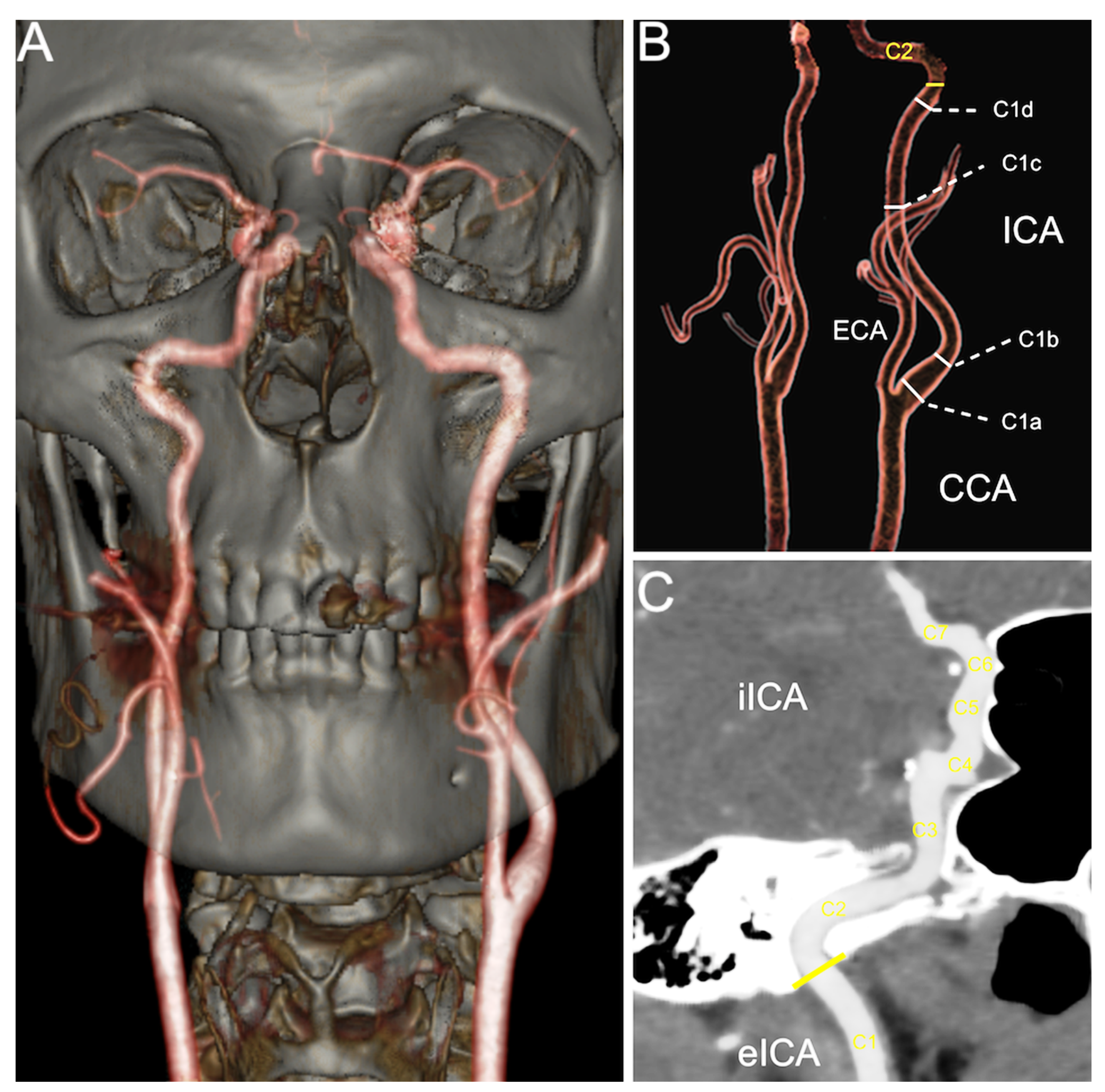
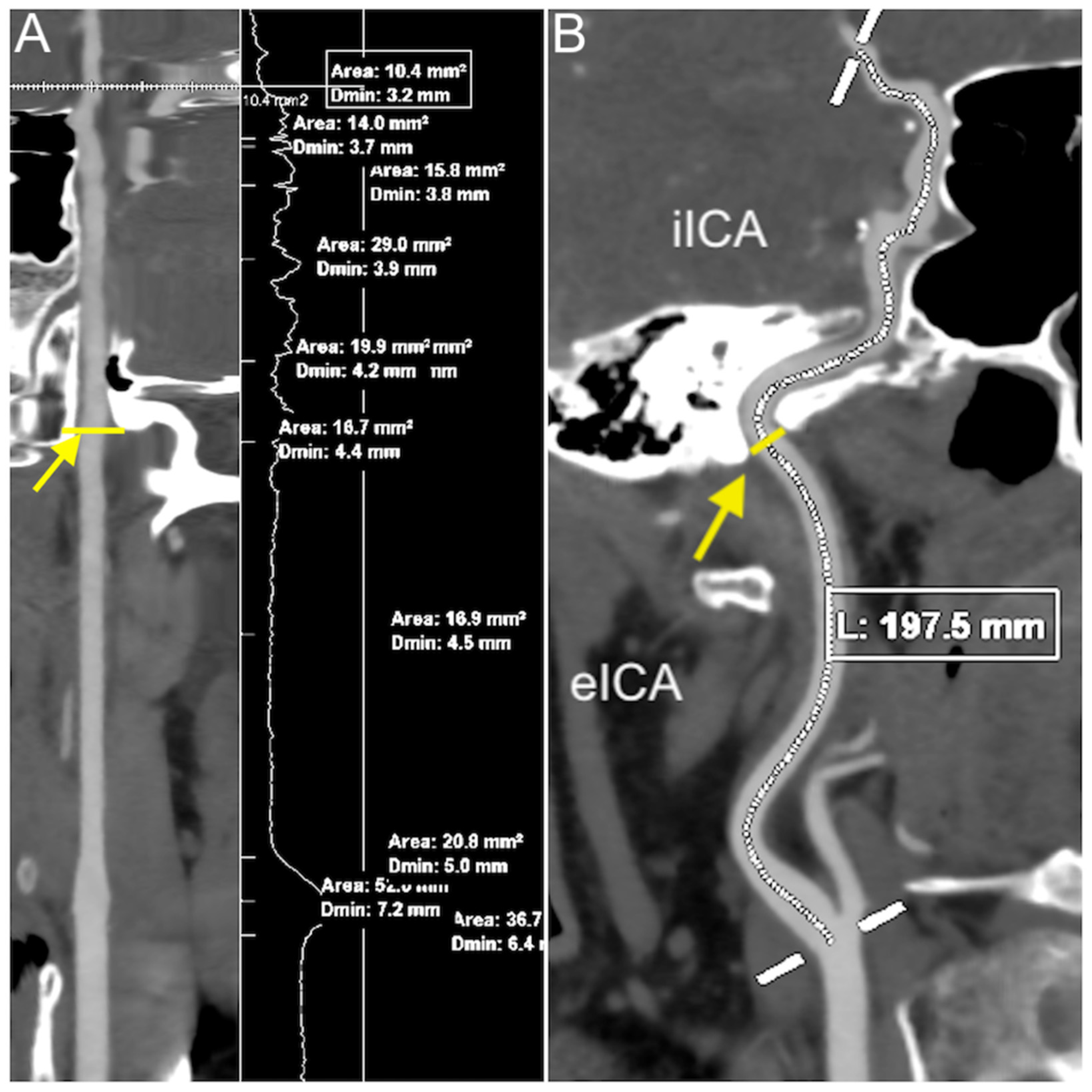
2. Materials and Methods
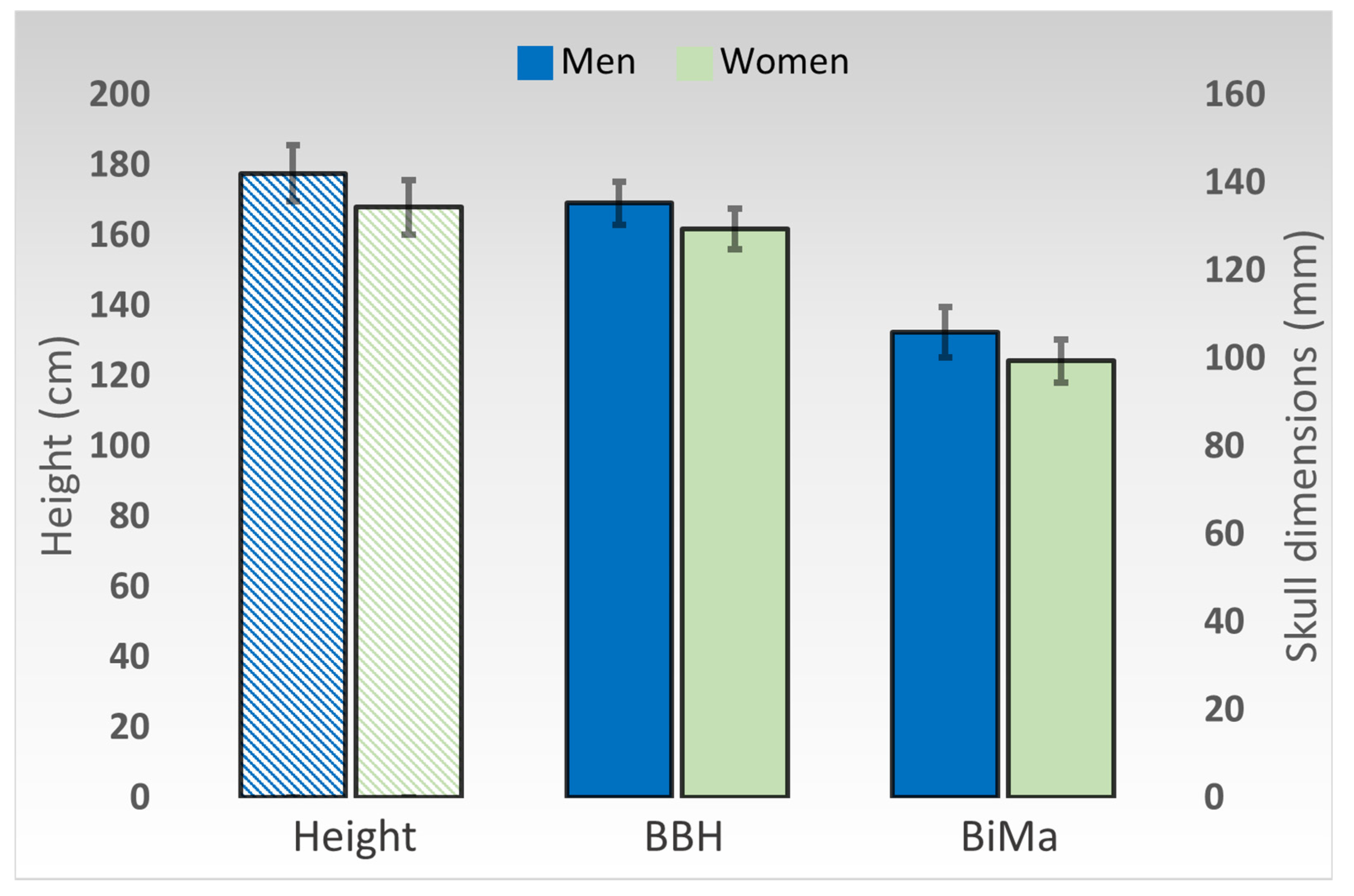
3. Results
4. Discussion
5. Conclusions
Author Contributions
Funding
Institutional Review Board Statement
Informed Consent Statement
Data Availability Statement
Conflicts of Interest
References
- Benjamin, E.J.; Blaha, M.J.; Chiuve, S.E.; Cushman, M.; Das, S.R.; Deo, R.; de Ferranti, S.D.; Floyd, J.; Fornage, M.; Gillespie, C.; et al. Heart Disease and Stroke Statistics-2017 Update: A Report From the American Heart Association. Circulation 2017, 135, e146–e603. [Google Scholar] [CrossRef] [PubMed]
- Virani, S.S.; Alonso, A.; Aparicio, H.J.; Benjamin, E.J.; Bittencourt, M.S.; Callaway, C.W.; Carson, A.P.; Chamberlain, A.M.; Cheng, S.; Delling, F.N.; et al. Heart Disease and Stroke Statistics—2021 Update. Circulation 2021, 143, e254–e743. [Google Scholar] [CrossRef] [PubMed]
- Gupta, A.; Gialdini, G.; Lerario, M.P.; Baradaran, H.; Giambrone, A.; Navi, B.B.; Marshall, R.S.; Iadecola, C.; Kamel, H. Magnetic resonance angiography detection of abnormal carotid artery plaque in patients with cryptogenic stroke. J. Am. Heart Assoc. 2015, 4, e002012. [Google Scholar] [CrossRef] [PubMed] [Green Version]
- Bartlett, E.S.; Walters, T.D.; Symons, S.P.; Fox, A.J. Quantification of carotid stenosis on CT angiography. Am. J. Neuroradiol. 2006, 27, 13–19. [Google Scholar]
- Pretterklieber, B.; Pretterklieber, M.L. A Phylogenetic and Ontogenetic Perspective of the Unique Accumulation of Arterial Variations in One Human Anatomic Specimen. Medicina 2020, 56, 449. [Google Scholar] [CrossRef]
- Cobiella, R.; Quinones, S.; Aragones, P.; León, X.; Abramovic, A.; Vazquez, T.; Ramón Sanudo, J.; Maranillo, E.; Olewnik, L.; Simon de Blas, C.; et al. Anatomic mapping of the collateral branches of the external carotid artery with regard to daily clinical practice. Ann. Anat. 2021, 238, 151789. [Google Scholar] [CrossRef]
- Bijari, P.B.; Wasserman, B.A.; Steinman, D.A. Carotid bifurcation geometry is an independent predictor of early wall thickening at the carotid bulb. Stroke 2014, 45, 473–478. [Google Scholar] [CrossRef]
- Wollschlaeger, P.B.; Wollschlaeger, G. Anterior cerebral-internal carotid artery and middle cerebral-internal carotid artery ratios. Acta Radiol. Diagn. 1966, 5, 615–620. [Google Scholar] [CrossRef]
- Spanos, K.; Petrocheilou, G.; Karathanos, C.; Labropoulos, N.; Mikhailidis, D.; Giannoukas, A. Carotid Bifurcation Geometry and Atherosclerosis. Angiology 2017, 68, 757–764. [Google Scholar] [CrossRef]
- Krejza, J.; Arkuszewski, M.; Kasner, S.E.; Weigele, J.; Ustymowicz, A.; Hurst, R.W.; Cucchiara, B.L.; Messe, S.R. Carotid artery diameter in men and women and the relation to body and neck size. Stroke 2006, 37, 1103–1105. [Google Scholar] [CrossRef] [Green Version]
- Limbu, Y.R.; Gurung, G.; Malla, R.; Rajbhandari, R.; Regmi, S.R. Assessment of carotid artery dimensions by ultrasound in non-smoker healthy adults of both sexes. Nepal Med. Coll. J. 2006, 8, 200–203. [Google Scholar]
- Koskinen, S.M.; Soinne, L.; Valanne, L.; Silvennoinen, H. The normal internal carotid artery: A computed tomography angiographic study. Neuroradiology 2014, 56, 723–729. [Google Scholar] [CrossRef]
- White, J.H.; Bartlett, E.S.; Bharatha, A.; Aviv, R.I.; Fox, A.J.; Thompson, A.L.; Bitar, R.; Symons, S.P. Reproducibility of semi-automated measurement of carotid stenosis on CTA. Can. J. Neurol. Sci. 2010, 37, 498–503. [Google Scholar] [CrossRef] [Green Version]
- Lloyd, K.D.; Barinas-Mitchell, E.; Kuller, L.H.; Mackey, R.H.; Wong, E.A.; Sutton-Tyrrell, K. Common carotid artery diameter and cardiovascular risk factors in overweight or obese postmenopausal women. Int. J. Vasc. Med. 2012, 2012, 169323. [Google Scholar] [CrossRef] [PubMed] [Green Version]
- Kato, M.; Dote, K.; Habara, S.; Takemoto, H.; Goto, K.; Nakaoka, K. Clinical implications of carotid artery remodeling in acute coronary syndrome: Ultrasonographic assessment of positive remodeling. J. Am. Coll. Cardiol. 2003, 42, 1026–1032. [Google Scholar] [CrossRef]
- Sasaki, R.; Yamano, S.; Yamamoto, Y.; Minami, S.; Yamamoto, J.; Nakashima, T.; Takaoka, M.; Hashimoto, T. Vascular remodeling of the carotid artery in patients with untreated essential hypertension increases with age. Hypertens. Res. 2002, 25, 373–379. [Google Scholar] [CrossRef] [PubMed] [Green Version]
- Deng, Y.; Wang, X.M.; Wu, L.B.; Sun, C.; Duan, Y.H.; Cheng, Z.P.; Wu, D.W. Significance of the preoperative guidance of dual-source CT in carotid body tumor. Chin. Med. J. 2010, 123, 2816–2819. [Google Scholar]
- Bouthillier, A.; van Loveren, H.R.; Keller, J.T. Segments of the internal carotid artery: A new classification. Neurosurgery 1996, 38, 425–433. [Google Scholar] [CrossRef]
- Martin, R.; Saller, K. Lehrbuch der Anthropologie II; Gustav Fischer: Stuttgart, Germany, 1957. [Google Scholar]
- Ring, B.A.; Waddington, M.M. Intraluminal diameters of the intracranial arteries. Vasc. Surg. 1967, 1, 137–151. [Google Scholar] [CrossRef]
- Gabrielsen, T.O.; Greitz, T. Normal size of the internal carotid, middle cerebral and anterior cerebral arteries. Acta Radiol. Diagn. 1970, 10, 1–10. [Google Scholar] [CrossRef]
- Markert, M.S.; Della-Morte, D.; Cabral, D.; Roberts, E.L., Jr.; Gardener, H.; Dong, C.; Wright, C.B.; Elkind, M.S.; Sacco, R.L.; Rundek, T. Ethnic differences in carotid artery diameter and stiffness: The Northern Manhattan Study. Atherosclerosis 2011, 219, 827–832. [Google Scholar] [CrossRef] [Green Version]
- Zhang, Y.; Zhang, X.; Chang, R.; Cang, P.; Liu, X.; Xia, Q. Diameter measurements of cerebral arteries on three-dimensional time-of-flight MR angiograms. Chin. J. Radiol. 2003, 37, 394–398. [Google Scholar]
- Williams, M.A.; Nicolaides, A.N. Predicting the normal dimensions of the internal and external carotid arteries from the diameter of the common carotid. Eur. J. Vasc. Surg. 1987, 1, 91–96. [Google Scholar] [CrossRef]
- Choudhry, F.A.; Grantham, J.T.; Rai, A.T.; Hogg, J.P. Vascular geometry of the extracranial carotid arteries: An analysis of length, diameter, and tortuosity. J. Neurointerv. Surg. 2016, 8, 536–540. [Google Scholar] [CrossRef] [PubMed]
- Mujagić, S. The inner diameter of arteries of the Circle of Willis regarding gender and age on Magnetic Resonance Angiography. Acta Med. Salin. 2013, 42, 6–12. [Google Scholar]
- Takegoshi, H.; Kikuchi, S. An anatomic study of the horizontal petrous internal carotid artery: Sex and age differences. Auris Nasus Larynx 2007, 34, 297–301. [Google Scholar] [CrossRef] [PubMed]
- Vijaywargiya, M.; Deopujari, R.; Athavale, S.A. Anatomical study of petrous and cavernous parts of internal carotid artery. Anat. Cell Biol. 2017, 50, 163–170. [Google Scholar] [CrossRef] [PubMed] [Green Version]
- Arat, Y.O.; Arat, A.; Aydin, K. Angiographic Morphometry of Internal Carotid Artery Circulation in Turkish Children. Turk. Neurosurg. 2015, 25, 608–616. [Google Scholar] [CrossRef] [Green Version]
- McNamara, J.R.; Fulton, G.J.; Manning, B.J. Three-dimensional computed tomographic reconstruction of the carotid artery: Identifying high bifurcation. Eur. J. Vasc. Endovasc. Surg. 2015, 49, 147–153. [Google Scholar] [CrossRef] [Green Version]
- Toda, T.; Tsuda, N.; Nishimori, I.; Leszczynski, D.E.; Kummerow, F.A. Morphometrical analysis of the aging process in human arteries and aorta. Acta Anat. 1980, 106, 35–44. [Google Scholar] [CrossRef]
- Kamenskiy, A.V.; Pipinos, I.I.; Carson, J.S.; MacTaggart, J.N.; Baxter, B.T. Age and disease-related geometric and structural remodeling of the carotid artery. J. Vasc. Surg. 2015, 62, 1521–1528. [Google Scholar] [CrossRef] [Green Version]
- Sterpetti, A.V. Eversion endarterectomy of the internal carotid artery combined with open endarterectomy of the common carotid artery. Am. J. Surg. 2010, 200, e44–e47. [Google Scholar] [CrossRef] [PubMed]
- Darling, R.C., 3rd; Mehta, M.; Roddy, S.P.; Paty, P.S.; Kreienberg, P.B.; Ozsvath, K.J.; Chang, B.B.; Shah, D.M. Eversion carotid endarterectomy: A technical alternative that may obviate patch closure in women. Cardiovasc. Surg. 2003, 11, 347–352. [Google Scholar] [CrossRef]
- Morr, S.; Lin, N.; Siddiqui, A.H. Carotid artery stenting: Current and emerging options. Med. Devices 2014, 7, 343–355. [Google Scholar] [CrossRef] [Green Version]


| Segment | Right Side | Left Side | p-Value | Male | Female | p-Value |
|---|---|---|---|---|---|---|
| Carotid bulb (C1a) | 7.56 (±0.97) | 7.61 (±1.01) | 0.6092 | 7.94 (±1.05) | 7.27 (±0.82) | 0.0001 |
| Post-bulbar section (C1b) | 5.49 (±0.64) | 5.52 (±0.62) | 0.6183 | 5.74 (±0.67) | 5.31 (±0.50) | 0.0001 |
| Midpoint of C1 (C1c) | 4.84 (±0.54) | 4.85 (±0.53) | 0.7750 | 5.01 (±0.56) | 4.70 (±0.46) | 0.0006 |
| Endpoint of C1 (C1d) | 4.65 (±0.46) | 4.69 (±0.47) | 0.4373 | 4.81 (±0.50) | 4.55 (±0.40) | 0.0013 |
| C2 | 4.53 (±0.43) | 4.53 (±0.50) | 0.8731 | 4.66 (±0.50) | 4.42 (±0.41) | 0.0026 |
| C3 | 4.32 (±0.41) | 4.34 (±0.49) | 0.7105 | 4.48 (±0.48) | 4.20 (±0.38) | 0.0002 |
| C4 | 4.25 (±0.41) | 4.28 (±0.48) | 0.5618 | 4.43 (±0.46) | 4.13 (±0.38) | 0.0001 |
| C5 | 4.13 (±0.46) | 4.17 (±0.50) | 0.3886 | 4.34 (±0.46) | 3.98 (±0.43) | <0.0001 |
| C6 | 2.89 (±0.38) | 2.88 (±0.41) | 0.4700 | 2.91 (±0.41) | 2.86 (±0.38) | 0.5307 |
| C7 | 2.72 (±0.35) | 2.70 (±0.38) | 0.2770 | 2.73 (±0.37) | 2.70 (±0.37) | 0.6190 |
| Extracranial length | 86.33 (±18.76) | 85.87 (±18.53) | 0.5540 | 90.21 (±20.66) | 82.43 (±15.76) | 0.0151 |
| Intracranial length | 69.40 (±8.95) | 69.33 (±8.42) | 0.8370 | 71.29 (±7.97) | 67.64 (±8.93) | 0.0122 |
| Total ICA length | 154.89 (±26.17) | 155.20 (±23.57) | 0.7895 | 160.62 (±28.56) | 150.07 (±19.83) | 0.0141 |
| Segment | Measurement 1 | Height | BBH 2 | BiMa 3 |
|---|---|---|---|---|
| Carotid bulb (C1a) | 7.59 (±0.99) | p = 0.0005 | p = 0.0080 | p = 0.0226 |
| Post-bulbar section (C1b) | 5.51 (±0.63) | p = 0.0356 | p = 0.4014 | p = 0.5521 |
| Midpoint of C1 (C1c) | 4.85 (±0.53) | p = 0.0365 | p = 0.0730 | p = 0.1512 |
| Endpoint of C1 (C1d) | 4.67 (±0.47) | p = 0.0224 | p = 0.1031 | p = 0.1539 |
| C2 | 4.53 (±0.47) | p = 0.0598 | p = 0.3029 | p = 0.2993 |
| C3 | 4.33 (±0.45) | p = 0.0150 | p = 0.1846 | p = 0.1744 |
| C4 | 4.27 (±0.45) | p = 0.0075 | p = 0.0889 | p = 0.0984 |
| C5 | 4.15 (±0.48) | p = 0.0027 | p = 0.0166 | p = 0.0506 |
| C6 | 2.88 (±0.40) | p = 0.4606 | p = 0.1686 | p = 0.2540 |
| C7 | 2.71 (±0.37) | p = 0.3660 | p = 0.3329 | p = 0.6489 |
| Extracranial length | 86.10 (±18.65) | p < 0.0001 | p = 0.0052 | p = 0.0197 |
| Intracranial length | 69.36 (±8.69) | p < 0.0001 | p < 0.0001 | p < 0.0001 |
| Total ICA length | 155.04 (±24.90) | p < 0.0001 | p < 0.0001 | p = 0.0004 |
Publisher’s Note: MDPI stays neutral with regard to jurisdictional claims in published maps and institutional affiliations. |
© 2021 by the authors. Licensee MDPI, Basel, Switzerland. This article is an open access article distributed under the terms and conditions of the Creative Commons Attribution (CC BY) license (https://creativecommons.org/licenses/by/4.0/).
Share and Cite
Baz, R.A.; Scheau, C.; Niscoveanu, C.; Bordei, P. Morphometry of the Entire Internal Carotid Artery on CT Angiography. Medicina 2021, 57, 832. https://doi.org/10.3390/medicina57080832
Baz RA, Scheau C, Niscoveanu C, Bordei P. Morphometry of the Entire Internal Carotid Artery on CT Angiography. Medicina. 2021; 57(8):832. https://doi.org/10.3390/medicina57080832
Chicago/Turabian StyleBaz, Radu Andrei, Cristian Scheau, Cosmin Niscoveanu, and Petru Bordei. 2021. "Morphometry of the Entire Internal Carotid Artery on CT Angiography" Medicina 57, no. 8: 832. https://doi.org/10.3390/medicina57080832
APA StyleBaz, R. A., Scheau, C., Niscoveanu, C., & Bordei, P. (2021). Morphometry of the Entire Internal Carotid Artery on CT Angiography. Medicina, 57(8), 832. https://doi.org/10.3390/medicina57080832