Article

Sclerotioloids A–C: Three New Alkaloids from the Marine-Derived Fungus Aspergillus sclerotiorum ST0501

1
Key Laboratory of Marine Drugs, The Ministry of Education of China, Institute of Evolution & Marine Biodiversity, School of Medicine and Pharmacy, Ocean University of China, Qingdao 266003, China
2
Fujian Key Laboratory on Conservation and Sustainable Utilization of Marine Biodiversity, Institute of Oceanography, Minjiang University, Fuzhou 350108, China
3
Laboratory for Marine Drugs and Bioproducts, Qingdao National Laboratory for Marine Science and Technology, Qingdao 266237, China
4
Institute for Insect Biotechnology, Justus-Liebig-University of Giessen, 35392 Giessen, Germany
5
Department of Bioresources of the Fraunhofer Institute for Molecular Biology and Applied Ecology (IME), 35392 Giessen, Germany
*
Authors to whom correspondence should be addressed.
Mar. Drugs 2023, 21(4), 219; https://doi.org/10.3390/md21040219
Submission received: 7 March 2023 / Revised: 25 March 2023 / Accepted: 28 March 2023 / Published: 29 March 2023

Abstract

:
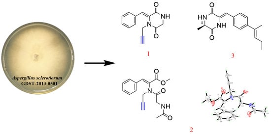
Alkaloids, as one of the largest classes of natural products with diverse structures, are an important source of innovative medicines. Filamentous fungi, especially those derived from the marine environment, are one of the major producers of alkaloids. In this study, three new alkaloids, sclerotioloids A–C (13), along with six known analogs (49), were obtained under the guidance of the MS/MS-based molecular networking from the marine-derived fungus, Aspergillus sclerotiorum ST0501, collected from the South China Sea. Their chemical structures were elucidated by comprehensive analysis of the spectroscopic data, including 1D and 2D NMR and HRESIMS. Additionally, the configuration of compound 2 was unambiguously determined by X-ray single crystal diffraction, and that of compound 3 was determined by the TDDFT-ECD approach. Sclerotioloid A (1) represents the first example of 2,5-diketopiperazine alkaloid with a rare terminal alkyne. Sclerotioloid B (2) showed the inhibition of NO production induced by lipopolysaccharide (LPS), with an inhibition rate of 28.92% higher than that of dexamethasone (25.87%). These results expanded the library of fungal-derived alkaloids and further prove the potential of marine fungi in the generation of alkaloids with new scaffolds.

Graphical Abstract

1. Introduction

In the past few years, natural products isolated from marine-derived fungi have aroused great interest due to their unique structures, interesting the pharmacological and biological properties [1]. Among the marine-derived fungi, Aspergillus is the largest source of fungal natural products. The growth of marine-derived fungi research rose from 2015 to 2019, with natural products from marine-derived fungi sources already accounting for nearly half (47%) of the total number of new marine natural products reported by 2019 [2]. Additionally, a large part of the compounds isolated from Aspergillus show anti-bacterial, anti-cancer, anti-inflammatory, anti-tuberculosis, and cytotoxic activities [3]. Metabolites of marine-derived fungi, such as polyketides, alkaloids, terpenes, lactones and peptides, are rich sources of biologically active natural products [4]. It was reported that alkaloids represent about 18% of all the marine natural products in 2019 [2]. Recent studies of marine fungal metabolites looking for bioactive compounds suggest that they have the potential to become a source of new drugs [5,6]. For example, plinabulin is a 2,5-diketopiperazine alkaloid isolated from the marine-derived fungus Aspergillus sp., which was first used in clinical trials for the treatment of non-small-cell lung cancer (NSCLC) [7]. Ecteinascidin-743 (ET743) is a novel anti-tumor drug on the market [8]. Aspergillus sclerotiorum have potential to biosynthesize alkaloids, though alkaloids are the most bioactive metabolites that possess marked antimicrobial and cytotoxic activities [9]. Sclerotiamides C and F, which are isolated from the A. sclerotiorum LZDX-33-4, a fungus derived from a gorgonian coral (LZDX-33), showed a significant inhibitory effect against a panel of tumor cell lines with IC50 values ranging from 1.6 to 7.9 μM [10]. A new cytotoxic indole-3-ethenamide isolated from A. sclerotium PT06-1 showed moderate (3.0 mM) and weak (27 mM) cytotoxicity toward A-549 and HL-60 cells, respectively [11].
During our ongoing research for new bioactive secondary metabolites from marine-derived fungi in the South China Sea [12,13,14,15], plenty of marine-derived alkaloids have been isolated. For example, emestrins L and M were obtained from the marine-derived fungus A. terreus RA2905. Emestrin M displayed antibacterial activity against Pseudomonas aeruginosa ATCC 27,853, with a minimum inhibitory concentration (MIC) value of 64 µg/mL [16]. Three new indole-diketopiperazine alkaloids, spirotryprostatin G and cyclotryprostatins F and G, were obtained from fungal strain HBU-136, and among them, spirotryprostatin G exhibited cytotoxicity against the HL-60 cell line, with an IC50 value of 6.0 μM, while cyclotryprostatins F and G exhibited cytotoxicity against the MCF-7 cell line, with IC50 values of 7.6 and 10.8 μM, respectively [17].
The fungal strain A. sclerotiorum ST0501 was isolated from the inner part of an unidentified sponge, GDST-2013-05, collected from the South China Sea. In the previous work, three new 2,5-diketopiperazine alkaloids, speramide C, 3,21-epi-taichunamide F and 2-epi-amoenamide C, were obtained from the same fungus strain. Speramide C represents the first prenylated indole alkaloid with an ethylene oxide ring on the isopentenyl side chain [18]. However, in this paper, we discovered three new alkaloids (13) and six known analogs (49) from ethyl acetate extract of A. sclerotiorum ST0501 based on the MS/MS molecular network. Herein, we report the isolation, structure elucidation and biological activities of these isolated compounds.

2. Results and Discussion

2.1. Elucidation of Chemical Structures

In order to fully explore the alkaloids of ST0501, molecular networking analysis was performed on the crude ethyl acetate extract (EtOAc) of solid rice and wheat cultures of A. sclerotiorum ST0501. The results show that there are signals associated with alkaloids. The relationship between compound 2 and compound 6 is close. Based on the indication of molecular networking (Figure 1), three unreported (13) and six previously described alkaloids (49) (Figure 2) were isolated by using a combination of column chromatography including silica gel, octadecylsilyl, Sephadex LH-20 columns and semi-preparative HPLC. The structures of gartryprostatin C (4) [19], stephacidin A (5) [20], sclerotiamide (6) [21], notoamide B (7) [22], speramide C (8) [18] and stephacidin B (9) [23] were elucidated by comparison of their spectroscopic data with those previously reported in the literature.
Sclerotioloid A (1) was isolated as a yellow powder. Its molecular formula C14H12N2O2 was deduced from its HRESIMS m/z 263.0794 [M+Na]+ (calcd for C14H12N2O2Na, 263.0797) and 1D NMR data analysis, suggesting ten degrees of unsaturation. The IR spectrum of 1 featured typical absorption bands for alkyne (2361 cm−1) and conjugated ketone (1698 cm−1). The 1H NMR spectrum of 1 (Table 1) exhibited one amine proton (δH 8.42, s, 1-NH), five aromatic protons (δH 7.35–7.40, m, 1-substituted benzene ring), one olefinic proton (δH 7.10, s, H-10), two methylene protons (δH 4.13 d, J = 2.4 Hz, H-7; δH 4.01 d, J = 2.3 Hz, H-6) and one acetylenic proton (δH 3.10, t, J = 2.4 Hz, H-9). The 13C NMR spectrum, in combination with HSQC spectra, revealed the presence of fourteen carbons, including two amide carbonyl (δC 165.7, C-5 and δC 163.4, C-2), six aromatic carbons (δC 133.4, C-11; δC 129.4, C-13 and C-15; δC 128.8, C-14; δC 128.6, C-12 and C-16), two olefinic carbons (δC 129.9, C-3 and δC 120.8, C-10), two alkynyl carbons (δC 78.2, C-8 and δC 74.5, C-9) and two methylene sp3 (δC 44.5, C-6 and δC 32.9, C-7). The correlations between H-1 and H-6 in the 1H-1H COSY spectrum and the HMBC correlations between NH-1 and C-3 and between H-6 and C-2/C-5 revealed the existence of a 2,5-diketopiperazine ring. Furthermore, the HMBC correlations between H-10 and C-2/C-3/C-16 indicated that the benzene ring was connected to C-3 of the 2,5-diketopiperazine ring through C-10. Based on the HMBC correlations between H-7 and C-3/C-5/C-9, the alkynyl group was connected to N-4 of the 2,5-diketopiperazine ring (Figure 3). According to the (Z)-vinyl proton chemical shifts of H-7 (δH 7.03) in nocazine C [24] and H-7 (δH 6.54) in (3S,6E)-3-benzyl-6-benzylidenepiperazine-2,5-dione [25], the Δ3,10 double bond in 1 was deduced to have a Z configuration due to the relative downfield shift of H-10 (δH 7.10), where the (Z)-vinyl proton had a larger downfield shift than that of the (E)-vinyl proton because of the deshielding effect of the carbonyl in 2,5-diketopiperazine [24,25,26,27]. Therefore, the structure of 1 was elucidated.
Sclerotioloid B (2) was also isolated as a yellow powder. The HRESIMS spectrum of 2 indicates a molecular formula of C17H18N2O4 based on the prominent peak [M+Na]+ at m/z 337.1160 (calcd for C17H18N2O4Na, 337.1164), as well as 1D NMR data analysis, requiring ten degrees of unsaturation. The IR spectrum of 2 featured typical absorption bands for alkyne (2361 cm−1) and conjugated ketone (1557 cm−1). The 1H NMR and 13C NMR spectroscopic data of 2 (Table 2) showed high similarity with 1, except for the appearance of one methoxyl protons resonating at δH 3.81 (H-12) and one methyl protons resonating at δH 1.77 (H-1), as well as the additional carbonyl carbon resonating at δC 169.0 (C-2). The existence of an acetamide group was suggested by the HMBC correlations between NH-3 and C-2, between H-1 and C-2 and between H-4 and C-5, as well as the COSY correlation between NH-3 and H-4. The additional HMBC correlations between H-12 and C-11 and between H-13 and C-11/C-15 proved that the benzene ring is connected to the methyl acrylate portion at C-13 (Figure 4). Finally, a single crystal of 2 was obtained after one week of slow crystallization in 95% MeOH (H2O) at 4 °C by optimizing the conditions. Therefore, the structure of 2 was confirmed by X-ray crystal diffraction analysis, and this further proved the Z configuration of the Δ10,13 double bond.
Sclerotioloid C (3) was isolated as a white powder. Its molecular formula, C17H20N2O2, is based on its HRESIMS m/z 323.1370 [M+K]+ (calcd for C17H20N2O2K, 323.1162) and 1D NMR data analysis, suggesting nine degrees of unsaturation. The IR spectrum of 3 featured typical absorption bands for olefin (2923 cm−1) and conjugated ketone (1512 cm−1). The 1H NMR spectra of 3 (Table 3) displayed two amine protons (δH 9.79, s, 4-NH; 8.35-8.37, m, 1-NH), four aromatic protons (δH 7.44, d, J = 8.8 Hz, H-10 and H-14; δH 6.95, d, J = 8.8 Hz, H-11 and H-13), two olefinic protons (δH 6.64, s, H-8; δH 5.43, tt, J = 6.7, 1.4 Hz, H-16), one methylene (δH 4.55, d, J = 6.7 Hz, H-17), one methine (δH 4.11, d, J = 6.8 Hz, H-6), and the protons of three methyl groups (δH 1.74, s, H-19; δH 1.71, d, J = 1.3 Hz, H-18; δH 1.33, d, J = 7.0 Hz, H-7). 13C NMR and HSQC data revealed the presence of seventeen carbons, including two amide carbonyl (δC 167.6, C-2 and δC 160.7, C-5), ten sp2 resonated between δC 158.2 and δC 114.3, one methylene sp3 (δC 64.3, C-17), one methine sp3 (δC 50.3, C-6) and three methyl (δC 25.4, C-19; δC 19.2, C-7; δC 18.0, C-18) carbons. The COSY correlations between NH-1 and H-6 and between H-6 and H-7 and the HMBC correlations between NH-4 and C-2/C-5/C-6, between H-6 and C-2/C-5/C-7 and between H-7 and C-2/C-6 revealed the existence of the 2,5-diketopiperazine ring. The COSY correlations between H-6 and H-7 and the HMBC correlations between H-7 and C-6 revealed that the methyl group is connected to C-6 of the 2,5-diketopiperazine ring. The COSY correlations between H-13 and H-14 and the HMBC correlations between H-12 and C-13/C-14 and between H-14 and C-10 revealed the existence of 1,4-disubstituted benzene ring. The HMBC correlations between H-8 and C-2/C-10 and between H-10 and C-3 indicate that the 2,5-diketopiperazine ring is connected to C-9 of the benzene ring through C-8. The COSY correlations between H-16 and H-17 and between H-and to H-18 and the HMBC correlations between H-17 and C-12/C-16, between H-18 and C-15/C-16 and between H-19 and C-15/C-16, indicate that the cis-2-pentene is connected to C-12 of the benzene ring (Figure 5). Therefore, the planar structure of 3 was elucidated as a new diketopiperazine derivative. The configuration of the double bond in 3 was established by the analysis of its NOESY spectrum (Figure 5). The NOESY spectrum did not show any correlation between H-16 and H-19, confirming the E configuration of the double bond between C-15 and C-16. In addition, there was no NOESY correlation between H-8 and 4-NH, suggesting Z configuration of the double bond between C-3 and C-8. The absolute configuration of C-6 in 3 was determined by the TDDFT-ECD approach. The experimental ECD spectrum of 3 matched well with the calculated ECD spectrum of S-3 (Figure 6) and further supported its absolute configuration.

2.2. Bioassays of Compounds

2.2.1. Cytotoxicity Assay

Compound 3 was screened for inhibitory activity on 20 human tumor cell lines at a concentration of 20 μM. However, it showed no observable activities against the abovementioned tumor cell lines (Table S1).

2.2.2. Anti-Microbial Activity Assay

Compounds 19 were tested for the anti-microbial activity against 20 pathogenic bacteria, including Enterobacter cloacae, E. hormaechei, Aeromonas salmonicida, Escherichia coli, Pseudomonas fulva, Photobacterium halotolerans, P. aeruginosa, Staphylococcus aureus, Bacillus subtilis, Vibrio Parahemolyticus HUB183, V. alginolyticus HUB184, V. anguillarum HUB185, Staphylococcus aureus ATCC 43300, Enterococcus faecalis ATCC 51299, E. faecium ATCC 35667, S. aureus ATCC 33591, S. aureus ATCC 29213, S. aureus ATCC 25923, V. vulnificus MCCC E1758, V. campbellii MCCC E333 and two pathogenic fungi, Candida albicans ATCC 24,433 and C. parapsilosis ATCC 22019. The initial screening concentration was 50 μg/mL, and none of them showed observable activities against abovementioned pathogens.

2.2.3. Anti-Oxidant Activity Assays

To test the anti-oxidant activity, 1,1-diphenyl-2-picrylhydrazyl (DPPH) assay was used. The initial screening concentration was 50 μg/mL, but none of them showed anti-oxidant activity.

2.2.4. Anti-Inflammatory Activity Assays

Compounds 13 were screened for NO production inhibitory activity. Compound 2 showed inhibition of NO production induced by lipopolysaccharide (LPS), with an inhibition rate of 28.92 ± 3.49% (Table S2). Compound 2 has a higher inhibition rate than dexamethasone does, which makes it promising as a good anti-inflammatory drug.
Compared with other alkaloid compounds, the activity of compounds 13 is not good, and other activity models need to be further screened.

3. Materials and Methods

3.1. General Experimental Procedure

Optical rotations were measured by using a JASCO P-1020 digital polarimeter (589 nm, 20 °C) (JASCO Ltd., Tokyo, Japan). UV spectra were recorded using an HITACHI UH 5300 UV spectrophotometer (Hitachi, Tokyo, Japan). IR spectra were acquired using a Nicolet-Nexus-470 spectrometer in KBr discs (400–4000 cm−1) (PerkinElmer Ltd., Boston, MA, USA). NMR spectra were recorded using a JEOL JEM-ECP NMR spectrometer (JEOL Ltd., Tokyo, Japan) with 500 MHz for 1H NMR and 125 MHz for 13C NMR and using TMS as an internal standard. HRESIMS were analyzed using a Thermo MAT95XP high resolution mass spectrometer (Thermo Fisher Scientific, Bremen, Germany), and ESIMS spectra were analyzed using a Thermo DSQ EImass spectrometer (Thermo Fisher Scientific, Bremen, Germany). Single-crystal X-ray crystallographic analysis was performed using a Bruker D8 venture X-ray single crystal diffractometer (Bruker, Karlsruhe, Germany). HPLC analysis was performed using an HITACHI L-2000 HPLC system coupled with a L-2455 photodiode array detector and using a semi-prepared C18 column (Kromasil 250 mm × 10 mm, 5 µm). For HPLC analysis, the mobile phase consisted of methanol (A) and water (B) with a flowrate of 2 mL/min with 10 µL injection volume (10 mg/mL) and monitored at UV length of 190–400 nm. The elution gradient was as follows: 0–40 min, 40–100% A; 40–45 min, 100% A; 45–50 min, 100–40% A; 50–55 min, 40% A. Sephadex LH-20 (Amersham Biosciences Co., Buckinghamshire, UK) and octadecylsilyl silica gel (Unicorn; 45–60 µm) were used for column chromatography (CC). Precoated silica gel plates (Yan Tai Zi Fu Chemical Group Co.; G60, F-254) were used for thin-layer chromatography analysis.

3.2. Fungal Material

The fungal strain, A. sclerotiorum ST0501, was isolated from the inner part of an unidentified sponge, GDST-2013-05, collected from the South China Sea (Guangdong, China) in May 2013. The fungal identification was performed by analysis of its morphological characteristics and ITS region of the rDNA [19]. The sequence data were submitted to Genbank with accession number MT534582. The strain was deposited in a −80 °C refrigerator in the laboratory.

3.3. Fermentation, Extraction and Isolation

The fungal strain was cultivated on a solid rice and wheat medium (3.6 g of natural sea salt from Yangkou saltern, China; 80 g of rice; 20 g wheat; 100 mL of H2O in 1000-mL Erlenmeyer flask) for 30 d at room temperature. Total fermentation of 45 flasks was extracted repeatedly with equal volume of ethyl acetate (EtOAc) two times, and the organic solvent was evaporated to dryness under vacuum to give a crude extract of 24.0 g. The crude extract was first fractionated by silica gel column chromatography (CC) using a step gradient elution with EtOAc-petroleum ether (0–100%), then with methanol-EtOAc (0–100%) to provide eleven fractions (Fr.1–Fr.11). Fr.3 was separated into three subfractions (M1–M3) by silica gel CC eluted with a step gradient of petroleum ether-EtOAc (from 8/1 to 1/5, v/v). M1 was placed in a Sephadex LH-20 column and eluted with dichloromethane-methanol (1/1, v/v) to generate fourteen subfractions (N1–N14). N14 was purified by HPLC (50% methanol-H2O) to yield 1 (1.0 mg) and 2 (6.4 mg). Fr.6 was separated into eight subfractions (O1–O8) by silica gel CC eluting with a step gradient of methanol–dichloromethane (0–100%). O4 was further separated by the silica gel CC eluted with a step gradient of dichloromethane–methanol (from 100/1 to 5/1, v/v) to produce 3 (8.8 mg). Fr.7 was separated into eighteen subfractions (P1–P18) in an ODS column eluted with 30–100% methanol–H2O to give 4 (4.5 mg). P6 was separated by the silica gel CC eluted with dichloromethane–methanol (30/1, v/v) to produce 9 (4.8 mg). P12 was further separated by the silica gel CC eluted with dichloromethane-methanol (60/1, v/v) to generate 5 (2.3 mg). P13 was separated by the silica gel CC eluted with dichloromethane-methanol (20/1, v/v) to produce 8 (1 mg). Fr.8 was separated into five subfractions (Q1–Q5) by the silica gel CC eluted with a step gradient of dichloromethane-methanol (60/1 to 5/1, v/v) to produce 6 (116.5 mg). Q4 was separated into five subfractions (R1–R5) in the Sephadex LH-20 column and eluted with dichloromethane–methanol (1/1, v/v). R2 was further purified by HPLC (80% methanol–H2O) to produce 7 (1.9 mg).

3.4. LC-MS/MS and Molecular Networking Analysis

LC-MS/MS was performed using a Waters series 2695 HPLC instrument coupled with an amaZon SL ion trap Mass spectrometer (Bruker, Karlsruhe, Germany), with a Xchange C18 column (Acchrom Co., CO, USA) 250 mm × 4.6 mm, 5 μm, 0.5 mL/min). The organic portion was dissolved in MeOH at 10 mg/mL, filtered through a Gracepure C18 SPE cartridge and analyzed by LC–MS/MS. Ten μL aliquot of each sample was injected and eluted with a gradient program of MeOH-H2O (0.1% formic acid) (0–20 min 10–100%, 21–25 min 100%; 1.0 mL/min). Mass spectra were obtained in positive ESI mode and with an automated fully dependent MS/MS scan from 100 to 1000 Da. MS/MS data were converted digitally to mzXML files using Filezilla software. The molecular networking was performed using the GNPS data analysis workflow using the spectral clustering algorithm [28]. The spectral networks were imported into Cytoscape 3.9.1 and visualized using the force-directed layout.

3.5. Spectroscopic and Spectrometric Data

Sclerotioloid A (1): yellow powder; UV (MeOH) λmax (log ε) 221 (0.13), 288 (0.16) nm; HRESIMS (m/z 263.0794 [M+ Na]+, calcd for 263.0797); IR (KBr) νmax 3749, 2926, 2361, 1557, 1398, 1053 cm−1; 1H and 13C NMR data, see Table 1.
Sclerotioloid B (2): yellow powder; UV (MeOH) λmax (log ε) 219 (0.03), 288 (0.05) nm; HRESIMS (m/z 337.1160 [M+ Na]+, calcd for 337.1164); IR (KBr) νmax 2361, 1796, 1701, 1522, 1418, 1264 cm−1; 1H and 13C NMR data, see Table 2.
X-ray Crystallographic Analysis of Compounds 2. Colorless crystals of 2 suitable for X-ray diffraction were obtained from 95% MeOH (H2O) by slow evaporation. The crystal data were collected at 293 K using an Agilent Gemini Ultra (Agilent, PA, USA) diffractometer with Cu Kα radiation (λ = 1.54184 Å). The structure was solved using CrysAlisPro version 1.171.41.121 (Rigaku Oxford Diffraction, 2021). Empirical absorption correction using spherical harmonics implemented in SCALE3 ABSPACK scaling algorithm.
Crystal Data for 2. C17H19N2O4, Mr = 314.33, triclinic, space group P − 1 with a = 8.8982(11) Å, b = 9.6133(10) Å, c = 10.9564(14) Å, α = 67.791(11), β = 71.857(11), γ = 82.76(1), V = 824.51(19) Å3, Z = 2, Dx = 1.266 g/cm3, μ (Cu Kα) = 0.752 mm−1, and F (000) = 332. Crystal dimensions: 0.12 × 0.12 × 0.11 mm3. Independent reflections: 1915 (Rint = 0.0576). The final R1 value was 0.0576.
Sclerotioloid C (3): white powder; [α]20D = −11.3° (c 0.01, MeOH); UV (MeOH) λmax (log ε) 226 (0.01), 318 (0.01) nm; HRESIMS (m/z 323.1370 [M+K]+, calcd for 323.1162); IR (KBr) νmax 2923, 2824, 1735, 1512, 1455, 1253, 1179 cm−1; 1H and 13C NMR data, see Table 3.

3.6. Biological Assay

Cytotoxic activity against human cancer cell lines was evaluated following the CCK-8 assay [29]. Twenty cell lines were used, including human lung cancer cells (CCL-185), human breast cancer cells (HTB-22), human gastric cancer cells (TCP-1008), human colon cancer cells (CCL-247EMT), human hepatocellular carcinoma cells (HB-8065), human cervical cancer cells (CCL-2), human chronic myeloid leukemia cells (CRL-3344), human brain tumor cells (CRL-2020), normal human liver cells (CRL-2254), human renal clear cell adenocarcinoma cells (CRL-1932), human esophageal carcinoma cells (PDM-246), human bladder cancer cells (HTB-9), human prostate cancer cells (HTB-81), human thyroid cancer cells (CRL-3354), human pancreatic cancer cells (CRL-2151), human osteosarcoma cells (CRL-1543), human malignant melanoma cells (CRL-1619), human rhabdomyosarcoma cells (CRL-1598), human embryonic kidney cells (CRL-3216) and human gallbladder carcinoma cells (PDM-273). Adriamycin hydrochloride was used as a positive control.
Antibacterial activity was evaluated following the standards recommended by Pierce [30], with ampicillin sodium as a positive control. The antifungal bioassay was conducted following the standards recommended by the Clinical and Laboratory Standards Institute [30], with amphotericin B used as a positive control.
Compounds 19 were tested against 20 pathogenic bacteria, including Enterobacter cloacae, E. hormaechei, Aeromonas salmonicida, Escherichia coli, Pseudomonas fulva, Photobacterium halotolerans, P. aeruginosa, Staphylococcus aureus, Bacillus subtilis, Vibrio Parahemolyticus HUB183, V. alginolyticus HUB184, V. anguillarum HUB185, S. aureus ATCC 43300, Enterococcus faecalis ATCC 51299, E. faecium ATCC 35667, S. aureus ATCC 33591, S. aureus ATCC 29213, S. aureus ATCC 25923, V. vulnificus MCCC E1758, V. campbellii MCCC E333, and two pathogenic fungi Candida albicans ATCC 24,433 and C. parapsilosis ATCC 22019.
The DPPH scavenging assay was performed using the method described by Aquino et al. [31]. The reaction mixture consisted of freshly prepared DPPH dissolved in ethanol (100 µmol/L) mixed with different concentrations of the tested compound. The reaction mixture was incubated for 20 min at room temperature in the dark, and the optical density was recorded at 517 nm.
The bioassay for NO production inhibitory activity was conducted as described by Xia et al. [32]. The mouse macrophages were seeded in 96-well plates. In each well, LPS (1 μg/mL) was added after treating with or without the tested compound for 24 h. The NO production in the supernatant was detected by the Griess reaction. The absorbance at 540 nm was measured with a microplate reader. The NO concentration and the inhibitory rate were calculated as a calibration curve. Dexamethasone was used as the positive control. Experiments were performed in triplicate, and the data are described as mean ± SD of three independent experiments.

4. Conclusions

In conclusion, nine alkaloids, including three new ones, were obtained by using MS/MS-based molecular networking for continuous investigation of the marine-derived fungus, A. sclerotiorum ST0501. Sclerotioloid A (1) represents the first example of 2,5-diketopiperazine alkaloid with a rare terminal alkyne. Additionally, the absolute configuration of compound 2 was unambiguously determined by single crystal X-ray analysis. Compound 2 showed the inhibition of NO production induced by lipopolysaccharide (LPS). Compound 3 displayed weak proliferation inhibitory activity against human chronic myeloid leukemia cells K-562 and human renal clear cell adenocarcinoma cell 786-O. Therefore, the potential of the marine-derived fungus, A. sclerotiorum ST0501, to produce novel bioactive secondary metabolites is worthy of further exploration.

Supplementary Materials

The following supporting information can be downloaded at: https://www.mdpi.com/article/10.3390/md21040219/s1, Figures S1–S19: 1H NMR, 13C NMR, 1H-1H COSY, HSQC, HMBC, HRESIMS, UV and IR spectra of compounds 1 and 2. Figure S20–S28: 1H NMR, 13C NMR, 1H-1H COSY, HSQC, HMBC, NOESY, HRESIMS, UV and IR spectra of compound 3. Table S1: The activity results about anti-tumor of compound 3. Table S2: The activity results about anti-inflammatory of compounds 13.

Author Contributions

Conceptualization, Y.L. and G.-S.Y.; methodology, J.-Q.M.; data analysis, J.-Q.M., Y.-Y.Z.; bioassays, J.-Q.M.; writing—original draft preparation, J.-Q.M.; writing—review and editing, C.-Y.W., Y.L. and G.-S.Y. All authors have read and agreed to the published version of the manuscript.

Funding

This work was supported by grants from the National Natural Science Foundations of China (NOs. 81874300; 41830535), Shandong Provincial Natural Science Foundation (Major Basic Research Projects) (NO. ZR2019ZD18); Natural Science Foundation of Fujian Province (NO. 2021J011043) and Minjiang University (NOs. MYK19011; JAT190622); the Program of Open Studio for Druggability Research of Marine Natural Products, National Laboratory for Marine Science and Technology (Qingdao, China) Directed by Kai-Xian Chen and Yue-Wei Guo; the Taishan Scholars Program, China.

Institutional Review Board Statement

This article does not contain any studies with animals performed by any of the authors.

Acknowledgments

We appreciate Xiaomei Teng and Ping Zhang of Qingdao marine biopharmaceutical research institute for HRESIMS testing. We are grateful to Houting Liu of Liaocheng university for X-ray crystallographic analysis.

Conflicts of Interest

The authors declare no conflict of interest.

References

  1. Carroll, A.R.; Carroll, A.R.; Davis, R.A.; Copp, B.R.; Davis, R.A.; Keyzers, R.A.; Prinsep, M.R. Marine natural products. Nat. Prod. Rep. 2022, 39, 1122–1171. [Google Scholar] [CrossRef]
  2. Carroll, A.R.; Copp, B.R.; Davis, R.A.; Keyzers, R.A.; Prinsep, M.R. Marine natural products. Nat. Prod. Rep. 2021, 38, 362–413. [Google Scholar] [CrossRef] [PubMed]
  3. Carroll, A.R.; Copp, B.R.; Davis, R.A.; Keyzers, R.A.; Prinsep, M.R. Marine natural products. Nat. Prod. Rep. 2020, 37, 175–223. [Google Scholar] [CrossRef] [PubMed]
  4. Rateb, M.E.; Ebel, R. Secondary metabolites of fungi from marine habitats. Nat. Prod. Rep. 2011, 28, 290–344. [Google Scholar] [CrossRef] [PubMed]
  5. Jin, L.; Quan, C.; Hou, X.; Fan, S. Potential pharmacological resources: Natural bioactive compounds from marine-derived fungi. Mar. Drugs 2016, 14, 76. [Google Scholar] [CrossRef] [PubMed] [Green Version]
  6. Youssef, F.S.; Ashour, M.L.; Singab, A.N.B.; Wink, M. A comprehensive review of bioactive peptides from marine fungi and their biological significance. Mar. Drugs 2019, 17, 559. [Google Scholar] [CrossRef] [PubMed] [Green Version]
  7. Natoli, M.; Herzig, P.; Pishali, B.E.; Buchi, M.; Ritschard, R.; Heinzelmann, V.; Trub, M.; Zippelius, A.; Kashyap, A.S.; Natoli, M. Plinabulin, a Distinct Microtubule-Targeting Chemotherapy, Promotes M1-Like Macrophage Polarization and Anti-tumor Immunity. Front. Oncol. 2021, 11, 644608. [Google Scholar] [CrossRef] [PubMed]
  8. Hendriks, H.R.; Fiebig, H.H.; Giavazzi, R.; Langdon, S.P.; Jimeno, J.M.; Faircloth, G.T. High antitumour activity of ET743 against human tumour xenografts from melanoma, non-small-cell lung and ovarian cancer. Ann. Oncol. 1999, 10, 1233–1240. [Google Scholar] [CrossRef] [PubMed]
  9. Ibrahim, S.R.M.; Abdallah, H.M.; Mohamed, G.A.; Deshmukh, S.K. Exploring Potential of Aspergillus sclerotiorum: Secondary Metabolites and Biotechnological Relevance. Mycol. Prog. 2022, 22, 8. [Google Scholar] [CrossRef]
  10. Guo, X.; Meng, Q.; Liu, J.; Wu, J.; Jia, H.; Liu, D.; Gu, Y.; Liu, J.; Huang, J.; Fan, A. Sclerotiamides C-H, Notoamides from a Marine Gorgonian-Derived Fungus with Cytotoxic Activities. J. Nat. Prod. 2022, 85, 1067–1078. [Google Scholar] [CrossRef]
  11. Wang, H.; Zheng, J.-K.; Qu, H.-J.; Liu, P.-P.; Wang, Y.; Zhu, W.-M. A new cytotoxic indole-3-ethenamide from the halotolerant fungus Aspergillus sclerotiorum PT06-1. J. Antibiot. 2011, 64, 679–681. [Google Scholar] [CrossRef] [PubMed] [Green Version]
  12. Chao, R.; Hou, X.M.; Xu, W.F.; Hai, Y.; Wei, M.Y.; Wang, C.Y.; Gu, Y.C.; Shao, C.L. Targeted Isolation of Asperheptatides from a Coral-Derived Fungus Using LC-MS/MS-Based Molecular Networking and Antitubercular Activities of Modified Cinnamate Derivatives. J. Nat. Prod. 2021, 84, 11–19. [Google Scholar] [CrossRef] [PubMed]
  13. Chen, M.; Shao, C.-L.; Fu, X.-M.; Xu, R.-F.; Zheng, J.-J.; Zhao, D.-L.; She, Z.-G.; Wang, C.-Y. Bioactive indole alkaloids and phenyl ether derivatives from a marine-derived Aspergillus sp. Fungus. J. Nat. Prod. 2013, 76, 547–553. [Google Scholar] [CrossRef]
  14. Liu, L.; Zheng, Y.-Y.; Shao, C.-L.; Wang, C.-Y. Metabolites from marine invertebrates and their symbiotic microorganisms: Molecular diversity discovery, mining, and application. Mar. Life Sci. Technol. 2019, 1, 60–94. [Google Scholar] [CrossRef] [Green Version]
  15. Wu, J.-S.; Yao, G.-S.; Shi, X.-H.; Rehman, S.U.; Fu, X.-M.; Zhang, X.-L.; Wang, C.-Y. Epigenetic Agents Trigger the Production of Bioactive Nucleoside Derivatives and Bisabolane Sesquiterpenes from the Marine-Derived Fungus Aspergillus versicolor. Front. Microbiol. 2020, 11, 85. [Google Scholar] [CrossRef] [PubMed] [Green Version]
  16. Wu, J.-S.; Shi, X.-H.; Yao, G.-S.; Shao, C.-L.; Fu, X.-M.; Zhang, X.-L.; Guan, H.-S.; Wang, C.-Y. New thiodiketopiperazine and 3,4-dihydroisocoumarin derivatives from the marine-derived fungus Aspergillus Terreus. Mar. Drugs 2020, 18, 132. [Google Scholar] [CrossRef] [PubMed] [Green Version]
  17. Zhang, Y.-H.; Geng, C.; Zhang, X.-W.; Zhu, H.-J.; Shao, C.-L.; Cao, F.; Wang, C.-Y. Discovery of bioactive indole-diketopiperazines from the marine-derived fungus Penicillium brasilianum aided by genomic information. Mar. Drugs 2019, 17, 514. [Google Scholar] [CrossRef] [Green Version]
  18. Wang, C.-Y.; Liu, X.-H.; Zheng, Y.-Y.; Ning, X.-Y.; Zhang, Y.-H.; Fu, X.-M.; Li, X.; Shao, C.-L.; Wang, C.-Y. 2,5-Diketopiperazines from a Sponge-Derived Fungus Aspergillus sclerotiorum. Front. Microbiol. 2022, 13, 808532. [Google Scholar] [CrossRef]
  19. Wang, B.; Park, E.M.; King, J.B.; Mattes, A.O.; Nimmo, S.L.; Clendinen, C.; Edison, A.S.; Anklin, C.; Cichewicz, R.H. Transferring Fungi to a Deuterium-Enriched Medium Results in Assorted, Conditional Changes in Secondary Metabolite Production. J. Nat. Prod. 2015, 78, 1415–1421. [Google Scholar] [CrossRef]
  20. Whyte, A.C.; Gloer, J.B.; Wicklow, D.T.; Dowd, P.F. Sclerotiamide: A new member of the paraherquamide class with potent antiinsectan activity from the sclerotia of Aspergillus sclerotiorum. J. Nat. Prod. 1996, 59, 1093–1095. [Google Scholar] [CrossRef]
  21. He, W.; Xu, Y.; Fu, P.; Zuo, M.; Liu, W.; Jiang, Y.; Wang, L.; Zhu, W. Cytotoxic Indolyl Diketopiperazines from the Aspergillus sp. GZWMJZ-258, Endophytic with the Medicinal and Edible Plant Garcinia multiflora. J. Agric. Food Chem. 2019, 67, 10660–10666. [Google Scholar] [CrossRef]
  22. Qian-Cutrone, J.; Huang, S.; Shu, Y.-Z.; Vyas, D.; Fairchild, C.; Menendez, A.; Krampitz, K.; Dalterio, R.; Klohr, S.E.; Gao, Q. Stephacidin A and B: Two structurally novel, selective inhibitors of the testosterone-dependent prostate LNCaP cells. J. Am. Chem. Soc. 2002, 124, 14556–14557. [Google Scholar] [CrossRef]
  23. Kato, H.; Yoshida, T.; Tokue, T.; Nojiri, Y.; Hirota, H.; Ohta, T.; Williams, R.M.; Tsukamoto, S. Notoamides A-D: Prenylated indole alkaloids isolated from a marine-derived fungus, Aspergillus sp. Angew. Chem. Int. Ed. 2007, 46, 2254–2256. [Google Scholar] [CrossRef]
  24. Fu, P.; Liu, P.; Qu, H.; Wang, Y.; Chen, D.; Wang, H.; Li, J.; Zhu, W. α-Pyrones and diketopiperazine derivatives from the marine-derived actinomycete Nocardiopsis dassonvillei HR10-5. J. Nat. Prod. 2011, 74, 2219–2223. [Google Scholar] [CrossRef] [PubMed]
  25. Shin, C.-G.; Kato, H.; Yonezawa, Y.; Hayakawa, M.; Yoshimura, J. Synthesis and structural assignment of naturally occurring 3-benzyl-6-benzylidene-2,5-piperazinedione. Heterocycles 1980, 14, 1767–1770. [Google Scholar] [CrossRef]
  26. Marcuccio, S.M.; Elix, J.A. Pyrazine chemistry. V. Synthesis of methylanhydropicroroccellin and dimethylpicroroccellin. Aust. J. Chem. 1985, 38, 1785–1796. [Google Scholar] [CrossRef]
  27. Sterns, M.; Patrick, J.M.; Patrick, V.A.; White, A.H. Conformational studies of some piperazine-2,5-diones: Crystal structures of three isomeric forms of methylanhydropicroroccellin (derivative of a lichen diketopiperazine) and a derivative bromohydrin. Aust. J. Chem. 1989, 42, 349–364. [Google Scholar] [CrossRef]
  28. Wang, M.; Carver, J.J.; Phelan, V.V.; Sanchez, L.M.; Garg, N.; Peng, Y.; Nguyen, D.D.; Watrous, J.; Kapono, C.A.; Luzzatto-Knaan, T. Sharing and community curation of mass spectrometry data with Global Natural Products Social Molecular Networking. Nat. Biotechnol. 2016, 34, 828–837. [Google Scholar] [CrossRef] [Green Version]
  29. Kunwar, A.; Barik, A.; Mishra, B.; Rathinasamy, K.; Pandey, R.; Priyadarsini, K.I. Quantitative cellular uptake, localization and cytotoxicity of curcumin in normal and tumor cells. Biochim. Biophys. Acta Gen. Subj. 2008, 1780, 673–679. [Google Scholar] [CrossRef]
  30. Pierce, C.G.; Uppuluri, P.; Tristan, A.R.; Wormley, F.L., Jr.; Mowat, E.; Ramage, G.; Lopez-Ribot, J.L. A simple and reproducible 96-well plate-based method for the formation of fungal biofilms and its application to antifungal susceptibility testing. Nat. Protoc. 2008, 3, 1494–1500. [Google Scholar] [CrossRef]
  31. Aquino, R.; Morelli, S.; Lauro, M.R.; Abdo, S.; Saija, A.; Tomaino, A. Phenolic constituents and antioxidant activity of an extract of Anthurium versicolor leaves. J. Nat. Prod. 2001, 64, 1019–1023. [Google Scholar] [CrossRef] [PubMed]
  32. Xia, W.; Luo, P.; Hua, P.; Ding, P.; Li, C.; Xu, J.; Zhou, H.; Gu, Q. Discovery of a New Pterocarpan-Type Antineuroinflammatory Compound from Sophora tonkinensis through Suppression of the TLR4/NFκB/MAPK Signaling Pathway with PU.1 as a Potential Target. ACS Chem. Neurosci. 2019, 10, 295–303. [Google Scholar] [CrossRef] [PubMed]
Figure 1. LC-MS/MS-based molecular networking of Aspergillus sclerotiorum ST0501.
Marinedrugs 21 00219 g001
Figure 2. The structure of 19.
Marinedrugs 21 00219 g002
Figure 3. The key 1H-1H COSY and HMBC correlations of compound 1.
Marinedrugs 21 00219 g003
Figure 4. The key 1H-1H COSY and HMBC correlations of 2 (left) and the ORTEP diagram of 2 (right).
Marinedrugs 21 00219 g004
Figure 5. Key 1H-1H COSY, HMBC and NOESY correlations of 3.
Marinedrugs 21 00219 g005
Figure 6. Experimental and calculated ECD spectra of 3.
Marinedrugs 21 00219 g006
Table 1. NMR data for compound 1 (DMSO-d6, 400 MHz/100 MHz).
PositionδC, mult.ΔH, mult. (J in Hz)COSYHMBC
1NH8.42 (s)H-6C-3
2163.4, C C-6, C-10
3129.9, C 1-NH, C-7
4N
5165.7, C C-6, C-7
644.5, CH24.01, d, (2.4)1-NHC-2, C-5
732.9, CH24.13, d, (2.4) C-3, C-5, C-9
878.2, C
974.5, CH3.10, t, (2.4) C-7
10120.8, CH7.10 (s) C-2, C-16
11133.4, C C-16
12128.6, CH7.35–7.40 (m) H-13
13129.4, CH7.35–7.40 (m) H-12, H-14
14128.8, CH7.35–7.40 (m) H-13, H-15
15129.4, CH7.35–7.40 (m) H-14, H-16
16128.6, CH7.35–7.40 (m) H-15C-10, C-11
Table 2. NMR data for compound 2 (DMSO-d6, 600 MHz/150 MHz).
PositionδC, mult.ΔH, mult. (J in Hz)COSYHMBC
121.9, CH31.77 (s) C-2
2169.0, C 1-NH
3NH 8.06 (s)H-4C-2
440.4, CH23.56, dd, (17.0, 5.7)
3.66, dd, (17.1, 5.7)
1-NHC-5
5168.4, C C-4
6N
735.5, CH24.24, dd, (17.6, 2.5)
4.38, dd, (17.5, 2.5)
C-8, C-9
877.5, C C-7
9 75.8, CH3.15 (s) C-7
10126.6, C
11164.5, C C-12, C-13
1252.5, OCH33.81 (s) C-11
13139.5 CH7.84 (s) C-11, C-14 C-15
14131.5, C C-13
15130.2, CH7.75 (s)H-16C-13
16128.8, CH7.46, d, (7.6)H-15, H-17
17131.0, CH7.49, d, (6.9)H-16, H-18
18128.8, CH7.46, d, (7.6)H-17, H-19
19130.2, CH7.73 (s)H-18
Table 3. NMR data for compound 3 (DMSO-d6, 500 MHz/125 MHz).
PositionδC, mult.ΔH, mult. (J in Hz)COSYHMBCNOESY
1NH8.35–8.37, mH-6C-3, C-5H-6, H-7
2160.7, C 4-NH, C-6, C-8
3125.3, C 1-NH, C-10, C-14
4NH9.79, s C-2, C-5, C-6H-12, H-13
5167.6, C 1-NH, 4-NH, C-6, C-7
650.3, CH4.11, d, (6.8)1-NH, H-7C-2, C-5, 4-NH, C-71-NH, H-7
719.2, CH31.33, d, (7.0)H-6C-5, C-61-NH, H-6
8114.3, CH6.64 (s) C-2, C-10
9125.7, C
10130.8, CH7.44, d, (8.8)H-14C-8, C-12, C-14
11114.8, CH6.95, d, (8.8)H-13C-3, C-12, C-13H-17
12158.2, C C-10, C-11, C-13, C-14, C-174-NH
13114.8, CH 6.95, d, (8.8)H-11C-3, C-11, C-12H-17
14130.8, CH 7.44, d, (8.8)H-10C-10, C-12
15137.3, C C-17, C-18, C-19
16119.8, CH5.43, tt, (6.7, 1.4)H-17C-17, C-18, C-19H-17, H-18
1764.3, CH24.55, d, (6.7)H-16, H-18C-12, C-15, C-16H-11, H-13, H-16
1818.0, CH31.71, d, (1.3)H-17C-15, C-16H-16
1925.4, CH31.74 (s) C-15, C-16
Disclaimer/Publisher’s Note: The statements, opinions and data contained in all publications are solely those of the individual author(s) and contributor(s) and not of MDPI and/or the editor(s). MDPI and/or the editor(s) disclaim responsibility for any injury to people or property resulting from any ideas, methods, instructions or products referred to in the content.