Article

Determination of the Biological Activity and Structure Activity Relationships of Drugs Based on the Highly Cytotoxic Duocarmycins and CC-1065

by
Lutz F. Tietze
*,
Birgit Krewer
,
J. Marian Von Hof
,
Holm Frauendorf
and
Ingrid Schuberth
Institute of Organic and Biomolecular Chemistry, Georg-August-University Göttingen, Tammannstr. 2, 37077 Göttingen, Germany
*
Author to whom correspondence should be addressed.
Toxins 2009, 1(2), 134-150; https://doi.org/10.3390/toxins1020134
Submission received: 3 November 2009 / Revised: 28 November 2009 / Accepted: 1 December 2009 / Published: 2 December 2009
(This article belongs to the Special Issue  Feature Papers)

Abstract

:
The natural antibiotics CC‑1065 and the duocarmycins are highly cytotoxic compounds which however are not suitable for cancer therapy due to their general toxicity. We have developed glycosidic prodrugs of seco-analogues of these antibiotics for a selective cancer therapy using conjugates of glycohydrolases and tumour-selective monoclonal antibodies for the liberation of the drugs from the prodrugs predominantly at the tumour site. For the determination of structure activity relationships of the different seco-drugs, experiments addressing their interaction with synthetic DNA were performed. Using electro­spray mass spectrometry and high performance liquid chromatography, the experiments revealed a correlation of the stability of these drugs with their cytotoxicity in cell culture investigations. Furthermore, it was shown that the drugs bind to AT-rich regions of double-stranded DNA and the more cytotoxic drugs induce DNA fragmentation at room temperature in several of the selected DNA double-strands. Finally, an explanation for the very high cytotoxicity of CC-1065, the duocarmycins and analogous drugs is given.

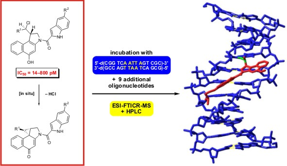
Graphical Abstract

1. Introduction

The natural antibiotics CC‑1065 (1, Figure 1) and the duocarmycins such as duocarmycin SA (2) are highly cytotoxic compounds and several derivatives of these natural products have already entered clinical trials [1,2,3,4]. Unfortunately, however, the general toxicity of these drugs is very high since their selectivity for tumour cells is rather low. The mode of action of the natural products as well as of related toxins is supposed to involve a sequence selective alkylation of cellular DNA in AT-rich regions that induces cell death [3,4,5].
Figure 1. (+)-CC-1065 (1) and (+)-duocarmycin SA (2) with highlighted pharmacophoric units.
Toxins 01 00134 g001
To overcome the insufficient selectivity of these compounds, we have developed glycosidic prodrugs such as 3a, 3b and 3c (Figure 2) [6,7,8,9]. The glycosidic bonds in 3a-c can be cleaved selectively at the tumour site by using a suitable monoclonal-antibody-glycohydrolase-conjugate (here β-d-galactosidase) that removes the sugar moiety to give the corresponding seco-drugs 4a-c. Due to the monoclonal-antibody, the conjugate binds to tumour-associated antigens, thus limiting the activation of the prodrugs to the tumour tissue [10,11]. The seco-drugs 4a-c as well as 4d cyclise rapidly with loss of HCl to give the corresponding drugs 5a-d which contain a spirocyclopropyl-cyclo­hexa­dienone moiety similar to the one that can be found in the natural products 1 and 2 (Figure 1). The alkylating moiety of 5c and 5d (R1 = H), which is formed in situ, is the 1,2,9,9a-tetra­hydro­cyclo­propa[1,2‑c]benz[1,2-e]indol-4-one (CBI) subunit developed by Boger et al. [12,13,14,15], whereas 5a and 5b (R1 = Me) bear the methylated CBI subunit developed in our group [6,7].
Whereas prodrugs 3a, 3b and 3c showed an excellent selectivity in cell culture experiments with QIC50 (QIC50 = IC50 (prodrug)/IC50 (prodrug + enzyme)) values of 4800 (3a), 1100 (3b) and 3500 (3c) [6,7,8], compounds 3a and 3b exhibited a reduced stability when being incubated in cell culture media or phosphate buffer [16]. The obviously relatively easy replacement of the secondary chloro atom in 3a and 3b by a hydroxyl group, which renders the prodrugs inactive, resulted in a half life of approximately 14 h for these prodrugs. In contrast, 3c showed less then 1% of hydrolysis after incubation for 24 h at 37 °C in cell culture medium [8,17].
Figure 2. Glycosidic prodrugs 3a-c, seco-drugs 4a-d and cyclisation of the latter under loss of HCl to give the drugs 5a-d as analogues of the natural products 1 and 2.
Toxins 01 00134 g002
Investigations of the reactivity of 4a against synthetic models of potential cellular target molecules using electrospray ionisation Fourier transform ion cyclotron resonance mass spectrometry (ESI-FTICR MS) had shown a highly effective alkylation of double-stranded DNA by the drug 5a formed in situ from 4a [16,18]. In contrast, the reactivity of 5a against single-stranded DNA, double-stranded RNA and the tripeptide glutathione was only low, suggesting that alkylation of double-stranded DNA is the predominant mode of action of this kind of drugs [19]. Since the DNA alkylation of analogue drugs did not correlate quantitatively with their relative cytotoxicity, we assumed that parameters like differences in the sequence selectivity [20,21] or in the stability of the compounds under physiological conditions [22,23] might modulate their cytotoxicity.
Here we describe investigations aiming at a better understanding of structure-activity relationships regarding this class of compounds. For these studies, the reactivity of the four seco-drugs 4a-d against ten different DNA oligonucleotides was investigated using electrospray ionisation Fourier transform ion cyclotron resonance (ESI-FTICR) mass spectrometry and high performance liquid chromato­graphy (HPLC).

2. Results and Discussion

The cytotoxicity of the hydrochlorides of seco-drugs 4a-4d against human bronchial carcinoma cells of line A549 (Table 1) was determined using a modified tumour colony forming ability test as reported previously [6,7,8,9,24].
Table 1. In vitro cytotoxicity of the hydrochlorides of seco-drugs 4a-d against human bronchial carcinoma cells of line A549. Cells were exposed to various concentrations of the test substance for 24 h at 37 °C; after 12 days of subsequent incubation the clone formation was compared to an untreated control assay and the relative clone forming rate was determined. IC50: Concentration required for 50% growth inhibition of target cells.
seco-drugIC50 (pm)
4a·HCl750
4b·HCl800
4c·HCl26
4d·HCl14
The seco-drugs 4c and 4d containing a hydrogen atom at the pharmacophoric unit (R1 = H) were found to show a much higher cytotoxicity than their analogues 4a and 4b containing a methyl group (R1 = Me) at the same position. This is presumably due to the fact that an attack of the cellular target, namely N3 of the nucleobase adenine, at the electrophilic cyclopropane moiety of the corresponding drugs 5a-d resulting in a subsequent formation of the corresponding DNA adducts, is less hindered in the absence of the sterically more demanding methyl group. Additionally, the lower hydrolytic stability of the drugs 5a and 5b bearing a methyl group decreases their concentration in cell culture media [16,17], which would lower the cytotoxicity of these drugs determined in cell culture assays. The different side chains (R2) in 4a and 4b on the one hand and 4c and 4d on the other hand seem to have only a small influence on the drugs’ cytotoxicity since 4a and 4b as well as 4c and 4d, respectively, show cytotoxicities within the same order of magnitude. This is understandable because both side chains contain a tertiary amine and have approximately the same size. Denny et al. as well as Boger et al. have previously reported that substituents at C-5 of the DNA binding subunit, which are here represented by R2, have a pronounced effect on the rate and efficiency of the DNA alkylation and the resulting biological potency of CC‑1065 analogues [25,26,27,28], but the exact type of the substituent does not seem to be as important as its length.

2.1. Investigations on the Reactivity and Sequence Selectivity of 4a-d against DNA Oligomers

The reactivity and sequence selectivity of 4a-d against ten different synthetic double-stranded DNA oligomers (ds-1-ds‑10) and against the synthetic single-stranded DNA oligo­nucleo­tide ON-1 of ds‑1 (Figure 3) was investigated using ESI-FTICR MS in order to rationalise differences in the compounds’ cytotoxicities.
An alkylation of the DNA oligonucleotides ON-1 and ON-2 by the drugs 5a-d results in the form­ation of covalent adducts denoted by ON-1*a-d and ON-2*a-d, respectively (Figure 4). In the absence of suitable nucleophiles like DNA, the drugs are partially hydrolysed to give the inactive alcohols 6a-d.
Figure 3. Base sequences of the ten double-stranded DNA oligonucleotides used in the present study. Double-stranded DNA oligonucleotides are denoted with the praefix “ds-“. Oligonucleotides with lower molecular mass are denoted with the abbreviation “ON-1” and oligonucleotides with higher molecular mass with the abbreviation “ON-2”. A: Adenine, G: Guanine, C: Cytosine, T: Thymine.
Toxins 01 00134 g003
Figure 4. Reaction of the drugs 5a-d with the DNA oligonucleotides ON-1 or ON-2 under formation of the alkylated oligonucleotides ON-1*a-d and ON-2*a-d, respectively, or hydrolysis of the drugs to give the alcohols 6a-d.
Toxins 01 00134 g004
For the mass spectrometric investigations, the hydrochlorides of seco-drugs 4a-d were incubated with the DNA oligo­nucleotides in water for 24 h at 25 °C. The samples were then diluted with equivalent amounts of methanol and investigated directly by means of ESI-FTICR mass spectro­metry without preliminary purification or enrichment of the products as described previously [16,18,19]. Covalent adducts were identified by comparison of calculated and found masses and alkylation positions were determined by identification of the characteristic fragment ions formed from the alkylated oligonucleotides applying capillary skimmer dissociation (CSD) [16,18,19,29,30,31,32].
If only one of the two oligonucleotides of the double-stranded DNA was alkylated, the percentage of alkylation of ON-1 or ON-2 was calculated based on the relative peak intensities of the corresponding isotope peaks of the unreacted oligonucleotides of ON-1 and ON-2 after 24 h of incubation as compared to the same ratio after 0 h of incubation. In case both oligonucleotides were alkylated, the preferred alkylation of one of the two oligo­nucleo­tides was determined based on the relative peak intensities of the covalent adducts. Analogously, if an oligonucleotide was alkylated in more than one position, the preferred binding site was determined based on the relative peak intensities of the respective characteristic fragment ions. The percentage of alkylation of the single-stranded oligo­nucleotide ON-1 of ds-1 was determined by calculating the ratio of the peak intensity of the covalent adduct as compared to the unmodified oligonucleotide.
Figure 5. Base sequences of the ten double-stranded DNA oligonucleotides used in the present study with alkylation positions marked by underlines. Potential alkylation positions in the oligonucleotides are denoted with a superscript and differing nucleo­bases in the 5'‑direction of potential competing binding sites are marked in red.
Toxins 01 00134 g005
Figure 5 and Table 2 show the results of the mass spectrometric investigations (as described above) in terms of the main alkylation site and the percentage of alkylation. In Figure 5, alkylation positions are denoted by underlines and important differences in the base sequence in the 5'‑direction of potential competing binding sites of ON-1 as compared to ON-2 are marked in red. Notably, all drugs alkylated mainly the nucleobase adenine (A) in AT-rich DNA regions of at least four consecutive AT base pairs with the alkylated adenine situated at the 3'-end of this sequence. This sequence selectivity could also be observed for the natural products CC‑1065 and duocarmycin SA [5] and the alkylation of the oligonucleotide ON-1 of ds-4 by the drug 5d is the only exception hereof. Furthermore, all drugs prefer the nucleobase adenine (A) over thymine (T) in the first, second and third position in the 5'-direction of the binding site as is obvious from the preferred binding positions in the oligonucleotides ds-6, ds-7 and ds-8, respectively (Figure 5 and Table 3). Hence, the drugs preferably alkylate the oligo­nucleotides ON-2 of these double-stranded oligonucleotides instead of the oligonucleotides ON-1.
Table 2. Alkylation efficiency and alkylation position after incubation of the DNA oligo­nucleotides ds‑1-ds‑10 and ON-1 of ds-1 with the hydrochlorides of 4a-d in a 1:1 ratio for 24 h at 25 °C.
Alkylation [%]Alkylation position
DNA4a4b4c4d4a4b4c4d
ds-175534628A10 (ON-1)
ds-26066931A14 (ON-1)
ds-355672932A10 (ON-1)
ds-4- a- a- a32- bON-1 a
ds-5ON-1 and ON-2 c6A10 (ON-1) ≥ A10 (ON-2)A10 (ON-1)
ds-630201713A8 (ON-2) ≥ A9 (ON-2)
ds-7ON-1 and ON-2 cA10 (ON-2) ≥ A10 (ON-1)
ds-8ON-1 and ON-2 cA7 (ON-2) ≥ A7 (ON-1)
ds-977822130A10 (ON-2) ≥ A11 (ON-2)
ds-10- a- a- a- a- b
ON-1 of ds-12025- a- a- b
a No significant amount of alkylation could be detected. b The alkylation position could not be deter­mined due to the low abundance of the covalent alkylation product or of the specific fragments of the latter. c Since both oligonucleotides were alkylated, no percentage of alkylation could be determined.
In addition, all drugs show a preference for purine bases (A or G) over pyrimidine bases (C or T) in the first position in the 3'-direction of the binding site. This results for example in the alkylation of A8 of ON-2 in ds‑6 additionally to A9 of the same DNA oligonucleotide (Figure 5, Table 3).
Table 3. Prefered nucleobases in the vicinity of the alkylation position (0) as observed after incubation of the hydrochlorides of seco-drugs 4a-d with the DNA oligonucleotides ds‑1-ds‑10.
5'43210-13'
4a, 4c, 4b, 4d A/T > G/C, G/C > A/TA > TA > TA > TAA/G > C/T
Interestingly, the seco-drugs 4a and 4c containing a right hand dimethylamino-ethoxyindole subunit prefer AT base pairs in the fourth position in the 5'-direction of the binding site with the AT-rich sequence being located in the middle of the double strand. This is obvious due to the lower alkylation efficiencies of the double-strands ds-2 and ds-3 as compared to ds‑1. In contrast, the seco-drugs 4b and 4d containing a right hand morpholinoindole subunit prefer CG base pairs in the fourth position in the 5'-direction of the binding site and AT-rich sequences located at the end of the double strand. This can be seen by the higher alkylation efficiencies of the double-strands ds-6 and ds-7 in comparison to ds-1. Furthermore, all drugs showed a much higher reactivity against the double-stranded DNA oligo­nucleotide ds‑1 than against the respective single-stranded oligonucleotide ON-1 of ds-1 (Table 2).
In summary, all drugs show a high selectivity for adenines in AT-rich DNA regions and differ only slightly in the preferred base sequence. Thus, differences in cytotoxicity are presumably not due to differences in sequence selectivity. Unexpectedly, also the alkylation efficiency regarding the same DNA oligonucleotide does not seem to correlate with the cytotoxicity of the drugs because, with the exception of ds-4, 4a and 4b alkylated the DNA with a significantly higher efficiency than the seco-drugs 4c and 4d even though they show a lower cytotoxicity as observed in the cell culture assays than the latter (Table 2).

2.2. Investigations on the Reaction Kinetics of the Seco-Drugs

Consequently other factors seem to influence the biological activity of these alkylating agents and it could be argued that differences in the rate of the cyclisation reaction of the seco-drugs 4a-d to give the drugs 5a-d or maybe differences in the rate of formation of the DNA adducts might play a crucial role. Therefore, the hydrochlorides of seco-drugs 4a-d were incubated with the double-stranded DNA oligo­nucleo­tides in buffer or water for up to 24 h at 25 °C and the kinetics of the reactions were investigated using HPLC. Since covalent and non-covalent adducts of the DNA and the drugs elute with the same retention time under the conditions of these measurements [16], mixtures of both covalent and non-covalent adducts are denoted ds‑x(*)a-d in the following whereas purely covalently bound adducts as detected by means of mass spectrometry are denoted ds‑x*a-d. For evaluation, the area under the curve (AuC) in the HPLC chromatograms of the respective seco-drugs and their derivatives was determined based on their absorption of light at λ = 350 nm because at this wavelength the absorption of unmodified DNA can be neglected. It should be noted that the results obtained are only semi-quantitative, since we did not correct the calculated concentrations according to the molar extinction coefficients of the seco-drugs and their derivatives; however, it can be assumed that the extinction coefficients are approximately the same. Indeed, the results allow a good and straight­forward comparison of the reactivity of the seco-drugs 4a-d and their drugs 5a-d, respectively.
Figure 6 shows a series of HPLC chromatograms obtained at 0-4 h after starting the incubation of the hydrochloride of seco-drug 4a with ds-1 in phosphate buffer (pH 7). As can clearly be seen, the seco-drug cyclises to give the corresponding drug 5a very rapidly and the formation of the respective DNA-adduct ds-1(*)a has finished already after 2 h. Table 4 shows the resulting AuCs determined after up to 6 hours of incubation of a 1:1 mixture of the hydrochlorides of the seco-drugs 4a-d with the DNA in phosphate buffer (pH 7).
The seco-drugs 4a and 4b cyclise rapidly to give the corresponding drugs 5a and 5b which subsequently form the respective DNA adducts ds-1(*)a and ds-1(*)b within two hours. Hence, no free seco-drugs or drugs can be observed after two hours of incubation. In contrast, the more cytotoxic seco-drugs 4c and 4d cyclise to the corresponding drugs 5c and 5d with a much lower reaction rate and, moreover, the DNA adduct formation proceeds quite slowly. Thus, after six hours of incubation, DNA adduct formation is not completed and therefore, seco-drugs 4c and 4d can still be detected in considerable amounts.
Figure 6. HPLC chromatograms (λ = 350 nm)obtained 0-4 h after starting the incubation of the hydrochloride of seco-drug 4a with ds-1 in phosphate buffer (pH 7).
Toxins 01 00134 g006
Table 4. AuC after indicated times of incubation of the DNA oligonucleotide ds-1 with the the hydrochlorides of seco-drugs 4a-d in phosphate buffer (pH 7).
AuC [%] after the indicated time of incubation
Reaction mixtureSpecies0 h2 h4 h6 h
ds-1/4a4a78---
5a22---
ds-1(*)a-100100100
ds-1/4b4b89---
5b11---
ds-1(*)b-100100100
ds-1/4c4c74451912
5c262014-
ds-1(*)c-356788
ds-1/4d4d76513419
5d24201719
ds-1(*)d-294962
Since seco-drugs such as 4a-d cyclise to the corresponding drugs nearly quantitatively in less than 90 minutes in buffer without DNA [16,17], 4c and 4d obviously are stabilised by an interaction with the DNA. This interaction is relatively weak because it can be disrupted under conditions of chromato­graphy to give back the free seco-drugs 4c and 4d as well as unchanged DNA. In addition, the formation of stable non-covalent and covalent complexes of the respective drugs 5c and 5d with the DNA oligonucleotides seems to be disfavoured because the amounts of ds-1(*)c ds-1(*)d increase only slowly and after prolonged incubation times, free drugs 5c (up to 4 h) and 5d (up to 6 h) can still be detected. Interestingly, the products of hydrolysis 6a-d are not observed, indicating that the rate of nucleophilic attack by water is reduced in the presence of DNA.
Further, we investigated whether there might be differences in seco-drug stabilisation or DNA adduct formation correlating with the base sequence. Using again HPLC, the interaction of the four seco-drugs 4a-d with the DNA oligonucleotides ds-1-ds-10 was analysed. Table 5 and Table 6 show the respective results obtained after 24 hours of incubation of a 1:1 mixture of DNA with the hydrochlorides of 4a-d in water at 25 °C.
Table 5. AuC after 24 h of incubation of the DNA oligonucleotides ds-1-ds-10 with the hydrochlorides of seco-drugs 4a and 4b in water (pH 7).
ds-x/4ads-x/4b
ds-x4a [%]5a [%]ds-x(*)a [%]4b [%]5b [%]ds-x(*)b[%]
ds-1--1002197
ds-2-2989289
ds-3-199-496
ds-4-6436-6238
ds-5-1993196
ds-6-2773-4060
ds-7-397-397
ds-8--1002197
ds-9-199-199
ds-10-6337-4060
After 24 hours of incubation, only traces (≤9%) of the seco-drugs 4a and 4b were detected. Furthermore, with the exception of ds-4,ds-6 and ds-10, nearly all detectable amounts of the drugs 5a and 5b which had been formed during the time of incubation, were converted to the corresponding DNA adducts ds‑x(*)a and ds‑x(*)b, respectively. Additionally, even in those cases where only small amounts of DNA adduct were formed (ds-4,ds-6 undds-10), no hydrolysis of the drugs 5a and 5b to the hydroxylated derivatives 6a and 6b (Figure 4) occurred in contrast to a notable hydrolysis of these drugs in the absence of DNA [16]. This indicates a weak interaction of the drugs with the DNA oligo­nucleo­tides that stabilises the drugs against hydrolysis on the one hand, but disfavours the alkylation reaction on the other hand. Consistent with this observation, the formation of covalent adducts as determined by the mass spectrometric investigations is low regarding ds-4,ds-6 and ds-10.
Table 6. AuC after 24 h of incubation of the DNA oligonucleotides ds-1-ds-10 with the seco-drugs 4c and 4d in water (pH 7).
ds-x/4cds-x/4d
ds-x4c [%]5c [%]ds-x(*)c [%]DNA fragments [%]4d [%]5d [%]ds-x(*)d [%]DNA fragments [%]
ds-11-99-45352-
ds-213-87-17281-
ds-38-92-19378-
ds-4156817-196813-
ds-559-41-63334-
ds-641353302784817
ds-7311731372847
ds-85-95-30367-
ds-92-8315213706
ds-10295318-334126-
Notably, the effect of stabilisation of the seco-drugs and drugs by the DNA is much more pro­nounced in the case of 4c and 4d as well as 5c and 5d, in comparison to that of 4a and 4b as well as 5a and 5b. Though in the absence of DNA all seco-drugs 4a-d cyclise quantitatively within 90 min in buffer or cell culture media [16,17], in the presence of DNA, the seco-drugs 4c and 4d cyclise to give the corresponding drugs 5c and 5d to a much lower extent than their methylated analogues 4a and 4b. Since the formation of the drugs is the prerequisite for the alkylation reaction, this might explain why the DNA alkylation is lower in case of 4c and 4d as compared to 4a and 4b. Interestingly, the extent of drug formation depends on the kind of double-stranded DNA oligonucleotide used, indicating a specific interaction of the seco-drugs with the DNA. The most pronounced stabilisation of the seco-drugs can be observed using ds-5, in the presence of which only about 40% of 4c and 4d, respectively, are converted to the corresponding drugs in 24 hours. Nevertheless, with the exception of ds-4,ds-6, ds-10 and partially also ds‑7, most of 5c and 5d reacted with the DNA oligonucleotides under formation of the non-covalent and covalent DNA adducts ds-x(*)c and ds-x(*)d as was already found for 5a and 5b. However, additional DNA fragments indicative of strand cleavage could be observed in case of ds-6,ds-7 and ds-9 after incubation with 4c and 4d in contrast to the respective incubations with 4a and 4b. Figure 7 displays HPLC chromatograms showing the DNA fragmentation exemplarily for the reaction of seco-drugs 4c and 4d with the oligonucleotide ds-6. DNA was detected using a wavelength of λ = 260 nm and the seco-drugs 4c and 4d as well as all their derivatives including DNA adducts were detected using a wavelength of λ = 350 nm. As can clearly be seen, the drugs 5c and 5d induce cleavage of the intact DNA to form DNA fragments a part of which is still covalently bound to the drug.
Figure 7. HPLC chromatograms (λ = 260 nm and λ = 350 nm) of ds-6 in water (Α), a solution of DNA oligo­nucleotide ds-6 incubated for 24 h with 4c·HCl in water (pH 7; B and D) and a solution of DNA oligo­nucleo­tide ds-6 incubated for 24 h with 4d·HCl in water (pH 7; C and E).
Toxins 01 00134 g007
The high cytotoxicity of 4c and 4d might thus also be modulated by DNA strand cleavage. DNA lesions such as single- and double-strand breaks have been reported previously to be caused by analogues of CC-1065 and the duocarmycins such as adozelesin and bizelesin [33]. These damages can lead to cell death in case they cannot be repaired by the cellular repair machinery [34].

3. Experimental Section

3.1. Materials

The hydrochlorides of seco-drugs 4a-d were synthesised according to previously published pro­cedures and stock solutions in DMSO were prepared [6,7,8,9]. The synthetic duplex-DNA oligomers ds-1-ds-10 and the single-stranded oligonucleotide ON-1 of ds-1 were purchased from IBA (Göttingen, Ger­many) as aqueous solutions (0.1 mm) of the sodium and ammonium salts, respectively. The phosphate buffer (pH 7.0) was composed of Na2HPO4/NaH2PO4 (10 mm) and NaCl (0.1 m) in bidistilled water.

3.2. Incubation of 4a-d with Synthetic DNA Oligonucleotides

Incubations of the hydrochlorides of seco-drugs 4a-d with the double-stranded DNA oligo­nucleo­tides ds-1 -10 and the single-stranded DNA oligonucleotide ON-1 of ds-1 were carried out in a 1:1 ratio of seco-drug to DNA. For each experi­ment, an aliquot (1 µL, 5 nmol) of the stock solution of the respective seco-drug hydrochlorides in DMSO (5 mmol L-14a-d) was mixed with an aliquot of DNA in water (50 µL, 5 nmol, 0.1 mmol L-1 DNA). Either, the reaction mixture was incubated at 25 °C for 24 h as such or phosphate buffer (pH 7, 50 µL) was added before starting the incubation. Samples were taken at different incubation times and analysed by means of mass spectrometry and chromatography.

3.3. Electrospray Ionisation Fourier Transform Ion Cyclotron Resonance Mass Spectrometry (ESI-FTICR MS)

For mass spectrometric investigations of the reaction mixtures, samples were taken at 0 h and 24 h, diluted with an equivalent amount of methanol and the resulting solution introduced directly into the ion source of the ESI-FTICR mass spectrometer. High-resolution mass spectrometry was performed using a 7 T-FTICR-MS instrument (APEX IV, Bruker Daltonics, Billerica, USA) equipped with an APOLLO electrospray ion source and a syringe pump (74900 series, Cole-Parmer, Vernon Hills, USA) with a flow rate of 2 µL min‑1 for sample injection. The ions were accumulated in the hexapole region for 0.8 s and transferred subsequently into the ICR cell. For gentle desolvatisation the drying gas temperature was set to 100 °C and the capillary exit voltage to -100 V. Enhanced fragmentation of alkylated oligonucleotides was achieved by capillary-skimmer dissociation (CSD) with a capillary exit voltage of -150 V. Ions were generated in the negative ion mode.

3.4. High Performance Liquid Chromatography (HPLC)

HPLC separations were performed with an Agilent 1200 with DAD from Agilent Technologies, an Aquapore OD-300 Column (220 × 4.6 mm, 7 µm) from Perkin Elmer and a Bondapak® C18 Column (300 × 3.9 mm, particle size 10 µm, pore size 125 Å) from Waters. Samples were eluted within 45 min with a flow rate of 1 mL min-1 (Aquapore OD-300) or 2 mL min-1 (Bondapak® C18) at 28 °C by applying a two-stage gradient (0-2 min: 5% B, 2-22 min: 5→20% B, 55-45 min: 20→80% B, 45-50 min: 80% B, 50-60 min: 80→5% B). Eluent A: 0.1 mol L-1 triethylammonium acetate buffer (H2O, pH 7.0). Eluent B: 0.1 mol L-1 triethylammonium acetate buffer (80% acetonitrile and 20% water, pH 7.0). The absorption of the drugs and their derivatives at λ = 350 nm was used to calculate the AuCs.

4. Conclusions

In summary, compounds 4c and 4d which are seco-analogues of the natural products CC‑1065 and duocarmycin SA, show a much higher cytotoxicity in cell culture investigations using a human bronchial carcinoma cell line than the respective derivatives 4a and 4b containing a methyl group instead of hydrogen in the pharmacophoric unit. In order to understand the structure activity relationships underlying the differences in cyto­toxicity, the reactivity of 4a-d against DNA as the proposed cellular target molecule was investigated by means of mass spectrometry and high performance liquid chromatography.
The seco-drugs 4a-d form the respective drugs 5a-d in situ, and the latter act as DNA alkylating agents. The drugs show high affinity to AT-rich DNA sequences and alkylate adenines at the 3'‑end of such sequences with high selectivity. All seco-drugs 4a-d are stabilised by the interaction with double stranded DNA oligonucleotides. This stabilisation delays the cyclisation reaction that gives the respective drugs 5a-d, and the effect is much more pronounced for seco-drugs 4c and 4d as compared to 4a and 4b. As a consequence, 4c and 4d are protected against hydrolytic deactivation and cyclise to the respective drugs 5c and 5d with a much lower reaction rate in the presence of DNA. In addition, the drugs 5c and 5d are less reactive than 5a and 5b and therefore alkylate double-stranded as well as single-stranded DNA much more slowly. The stability of the derivatives in the presence of DNA oligonucleotides correlates very well with the cytotoxicity of the compounds in cell culture investigations: The stronger the stabilisation of the seco-drugs and drugs by interaction with DNA oligonucleotides, the higher is their cytotoxicity. This observation is similar to results previously obtained by Boger et al. [35], in which a parabolic relationship between the cytotoxic potency of derivatives related to 5a-d and their stability against solvolysis was demonstrated. Furthermore, 5c and 5d seem to cause DNA fragmentation at room temperature whereas 5a and 5b do not. Based on our investigations, we assume that the mechanism responsible for the very high cytotoxicity of CC‑1065, the duocarmycins and of related synthetic analogues is an over-stabilisation of the DNA double strand by non-covalent interactions, which prohibits the repair, transcription or replication of DNA and therefore induce apoptosis. In the literature, the cytotoxicity of CC-1065, the duocarmycins and related compounds is always explained by their alkylation properties. However, we think that the alkylation only serves for an irreversible fixation of the compounds in the minor groove of the DNA and the astounding base selectivity of the alkylation is only a question of proximity. Thus, the discussion that the adenine which is alkylated might be a so called hot spot explaining the very high cytotoxicity of these compounds compared to other alkylating drugs as cyclophosphamide is in our belief not meaningful anymore.

Acknowledgements

This research was supported by the Deutsche Forschungsgemeinschaft (Sonderforschungs­bereich 416) and the Fonds der Chemischen Industrie. B.K. thanks the Deutsche Telekom Foundation for a Ph.D. scholarship. The authors thank F. Major for providing 4a and 4b.

References and Notes

  1. Martin, D.G.; Biles, C.; Gerpheide, S.A.; Hanka, L.J.; Krueger, W.C.; McGovren, J.P.; Mizsak, S.A.; Neil, G.L.; Stewart, J.C.; Visser, J. CC-1065 (NSC 298223), a Potent New Antitumor Agent. Improved Production and Isolation, Characterization and Antitumor Activity. J. Antibiot. 1981, 34, 1119–1125. [Google Scholar] [PubMed]
  2. Ichimura, M.; Ogawa, T.; Takahashi, K.; Kobayashi, E.; Kawamoto, I.; Yasuzawa, T.; Takahashi, I.; Nakano, H. Duocarmycin SA, a New Antitumor Antibiotic from Streptomyces sp. J. Antibiot. 1990, 43, 1037–1038. [Google Scholar] [PubMed]
  3. Tietze, L.F.; Krewer, B. Novel Analogues of CC-1065 and the Duocarmycins for the Use in Targeted Tumour Therapies. Anti-Cancer Agents Med. Chem. 2009, 9, 304–325. [Google Scholar]
  4. Baraldi, P.G.; Bovero, A.; Fruttarolo, F.; Preti, D.; Tabrizi, M.A.; Pavani, M.G.; Romagnoli, R. DNA Minor Groove Binders as Potential Antitumor and Antimicrobial Agents. Med. Res. Rev. 2004, 24, 475–528. [Google Scholar] [PubMed]
  5. Boger, D.L.; Johnson, D.S. CC-1065 and the Duocarmycins: Understanding Their Biological Function through Mechanistic Studies. Angew. Chem. Int. Ed. Eng. 1996, 35, 1438–1474. [Google Scholar]
  6. Tietze, L.F.; Major, F.; Schuberth, I. Antitumor Agents: Development of Highly Potent Glycosidic Duocarmycin Analogues for Selective Cancer Therapy. Angew. Chem. Int. Ed. 2006, 45, 6574–6577. [Google Scholar]
  7. Tietze, L.F.; Major, F.; Schuberth, I.; Spiegl, D.A.; Krewer, B.; Maksimenka, K.; Bringmann, G.; Magull, J. Selective treatment of Cancer: Synthesis, Biological Evaluation and Structural Elucidation of Novel Analogues of the Antibiotic CC-1065 and the Duocarmycins. Chem. Eur. J. 2007, 13, 4396–4409. [Google Scholar]
  8. Tietze, L.F.; von Hof, J.M.; Krewer, B.; Müller, M.; Major, F.; Schuster, H.J.; Schuberth, I.; Alves, F. Asymmetric Synthesis and Biological Evaluation of New Glycosidic Prodrugs for a Selective Cancer Therapy. ChemMedChem 2008, 3, 1946–1955. [Google Scholar] [PubMed]
  9. Tietze, L.F.; Krewer, B.; Major, F.; Schuberth, I. CD-Spectroscopy as a Powerful Tool for Investigating the Mode of Action of Unmodified Drugs in Living Cells. J. Am. Chem. Soc. 2009, 131, 13031–13036. [Google Scholar] [PubMed]
  10. Tietze, L.F.; Feuerstein, T.; Fecher, A.; Haunert, F.; Panknin, O.; Borchers, U.; Schuberth, I.; Alves, F. Proof of Principle in the Selective Treatment of Cancer by Antibody Directed Enzyme Prodrug Therapy. The Development of a Highly Potent Prodrug. Angew. Chem. Int. Ed. 2002, 41, 759–761. [Google Scholar] [CrossRef]
  11. Tietze, L.F.; Feuerstein, T. Highly Selective Compounds for the Antibody-Directed Enzyme Prodrug Therapy of Cancer. Aust. J. Chem. 2003, 56, 841–854. [Google Scholar]
  12. Boger, D.L.; Ishizaki, T.; Wysocki, R.J., Jr.; Munk, S.A.; Kitos, P.A.; Suntornwat, O. Total Synthesis and Evaluation of (±)-N-(tert-Butyloxycarbonyl)-CBI, (±)-CBI-CDPI1, and (±)-CBI-CDPI2: CC-1065 Functional Agents Incorporating the Equivalent 1,2,9,9a-Tetra­hydro­cyclo­prop[1,2-cbenz[1,2-e]indol-4-one (CBI) Left-Hand Subunit. J. Am. Chem. Soc. 1989, 111, 6461–6463. [Google Scholar]
  13. Boger, D.L.; Ishizaki, T. Resolution of a CBI Precursor and Incorporation into the Synthesis of (+)-CBI, (+)-CBI-CDPI1, (+)-CBI-CDPI2: Enhanced Functional Analogs of (+)-CC-1065. A Criti­cal Appraisal of a Proposed Relationship Between Electrophile Reactivity, DNA Binding Proper­ties,and Cytotoxic Potency. Tetrahedron Lett. 1990, 31, 793–794. [Google Scholar] [CrossRef]
  14. Boger, D.L.; Ishizaki, T. Synthesis of N-(tert-Butyloxycarbonyl)-CBI, CBI, CBI-CDPI1, and CBI-CDPI2: Enhanced Functional Analogues of CC-1065 Incorporating the 1,2,9,9a-Tetra­hydro­cyclo­propa[1,2-c]benz[1,2-e]indol-4-one (CBI) Left-Hand Subunit. J. Org. Chem. 1990, 55, 5823–5832. [Google Scholar]
  15. Jin, W.; Trzupek, J.D.; Rayl, T.J.; Bro­ward, M.A.; Vielhauer, G.A.; Weir, S.J.; Hwang, I.; Boger, D.L. A Unique Class of Duocarmycin and CC-1065 Analogues Subject to Reductive Activation. J. Am. Chem. Soc. 2007, 129, 15391–15397. [Google Scholar] [PubMed]
  16. Tietze, L.F.; Krewer, B.; Frauendorf, H. Investigation of the transformations of a novel anticancer agent combining HPLC, HPLC-MS and direct ESI-HRMS analyses. Anal. Bioanal. Chem. 2009, 395, 437–448. [Google Scholar] [PubMed]
  17. Tietze, L.F.; von Hof, J.M.; Krewer, B. Unpublished Results.
  18. Tietze, L.F.; Krewer, B.; Frauendorf, H.; Major, F.; Schuberth, I. Investigation of Reactivity and Selectivity of DNA-Alkylating Duocarmycin Analogues by High-Resolution Mass Spectrometry. Angew. Chem. Int. Ed. 2006, 45, 6570–6574. [Google Scholar]
  19. Tietze, L.F.; Krewer, B.; Frauendorf, H. Probing the mechanism of action of potential anticancer agents at a molecular level using electrospray Fourier transform ion cyclotron resonance mass spectrometry. Eur. J. Mass. Spectrom. 2009, 15, 661–672. [Google Scholar]
  20. Shinohara, K.-I.; Bando, T.; Sasaki, S.; Sakakibara, Y.; Minoshima, M.; Sugiyama, H. Antitumor activity of sequence-specific alkylating agents: Pyrrole-imidazole CBI conjugates with indole linker. Cancer Sci. 2006, 97, 219–225. [Google Scholar] [CrossRef] [PubMed]
  21. Nelson, S.M.; Ferguson, L.R.; Denny, W.A. DNA and the chromosome-varied targets for chemotherapy. Cell Chromosome 2004, 3:2. [Google Scholar]
  22. Tichenor, M.S.; MacMillen, K.S.; Stover, J.S.; Wolkenberg, S.E.; Pavani, M.G.; Zanella, L.; Zaid, A.N.; Spalluto, G.; Rayl, T.J.; Hwang, I.; Baraldi, P.G.; Boger, D.L. Rational Design, Synthesis, and Evaluation of Key Analogues of CC-1065 and the Duocarmycins. J. Am Chem. Soc. 2007, 129, 14092–14099. [Google Scholar] [PubMed]
  23. Parrish, J.P.; Hughes, T.V.; Hwang, I.; Boger, D.L. Establishing the Parabolic Relationship between Reactivity and Activity for Derivatives and Analogues of the Duocarmycins and CC‑1065 Alkylation Subunits. J. Am. Chem. Soc. 2004, 126, 80–81. [Google Scholar] [PubMed]
  24. Tietze, L.F.; Hannemann, R.; Buhr, W.; Lögers, M.; Menningen, P.; Lieb, M.; Starck, D.; Grote, T.; Döring, A.; Schuberth, I. Prodrugs of the Cytostatic CC-1065 That Can Be Activated in a Tumor-Selective Manner. Angew. Chem. Int. Ed. Engl. 1996, 35, 2674–2677. [Google Scholar]
  25. Milbank, J.J.B.; Tercel, M.; Atwell, G.J.; Wilson, W.R.; Hogg, A.; Denny, W.A. Synthesis of 1-Substituted 3-(Chloromethyl)-6-aminoindoline (6-Amino-seco-CI) DNA Minor Groove Alkylating Agents and Structure-Activity Relationships for Their Cytotoxicity. J. Med. Chem. 1999, 42, 649–658. [Google Scholar] [PubMed]
  26. Atwell, G.J.; Milbank, J.J.B.; Wilson, W.R.; Hogg, A.; Denny, W.A. 5-Amino-1-(chloromethyl)-1,2-dihydro-3H-benz[e]indoles: Relationships between Structure and Cytotoxicity for Analogues Bearing Different DNA Minor Groove Binding Subunits. J. Med. Chem. 1999, 42, 3400–3411. [Google Scholar] [PubMed]
  27. Parrish, J.P.; Kastrinsky, D.B.; Stauffer, F.; Hedrick, M.P.; Hwang, I.; Boger, D.L. Establishment of Substituent Effects in the DNA Binding Subunit of CBI Analogues of the Duocarmycins and CC-1065. Bioorg. Med. Chem. 2003, 11, 3815–3838. [Google Scholar] [PubMed]
  28. Boger, D.L.; Bollinger, B.; Johnson, D.S. Examination of the role of the duocarmycin SA methoxy substituents: identification of the minimum, fully potent DNA binding subunit. Bioorg. Med. Chem. Lett. 1996, 6, 2207–2210. [Google Scholar]
  29. Hofstadler, S.A.; Sannes-Lowery, K.A.; Hannis, J.C. Analysis of Nucleic Acids by FTICR MS. Mass Spectrom. Rev. 2005, 24, 265–285. [Google Scholar] [CrossRef] [PubMed]
  30. Banoub, J.H.; Newton, R.P.; Esmans, E.; Ewing, D.F.; Mackenzie, G. Recent Developments in Mass Spectrometry for the Characterization of Nucleosides, Nucleotides, Oligonucleotides, and Nucleic Acids. Chem. Rev. 2005, 105, 1869–1915. [Google Scholar] [CrossRef] [PubMed]
  31. Beck, J.L.; Colgrave, M.L.; Stephen, F.R.; Sheil, M.M. Electrospray Ionization Mass Spectrometry of Oligonucleotide Complexes with Drugs, Metals, and Proteins. Mass Spectrom. Rev. 2001, 20, 61–87. [Google Scholar] [CrossRef] [PubMed]
  32. McLuckey, S.; Habibi-Goudarzi, S. Decompositions of Multiply Charged Oligonucleotide Anions. J. Am. Chem. Soc. 1993, 115, 12085–12095. [Google Scholar] [CrossRef]
  33. Cao, P.-R.; McHugh, M.M.; Melendy, T.; Beerman, T. The DNA Minor Groove-alkylating Cyclopopylpyrroloindole Drugs Adozelesin and Bizelesin Induce Different DNA Damage Response Pathways in Human Colon Carcinoma HCT116 Cells. Mol. Canc. Ther. 2003, 2, 651–659. [Google Scholar]
  34. Schärer, O.D. Chemistry and Biology of DNA Repair. Angew. Chem. Int. Ed. 2003, 42, 2946–2974. [Google Scholar] [CrossRef]
  35. Parrish, J.P.; Hughes, T.V.; Hwang, I.; Boger, D.L. Establishing the Parabolic Relationship between Reactivity and Activity for Derivatives and Analogues of Duocarmycin and CC-1065 Alkylation Subunits. J. Am. Chem. Soc. 2004, 126, 80–81. [Google Scholar] [PubMed]