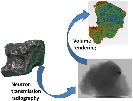
Structural Characterization of Iron Meteorites through Neutron Tomography
Abstract
:1. Introduction
2. Materials and Methods
2.1. Materials
2.2. Methods
2.2.1. ICON Experimental Set-Up
2.2.2. DINGO Experimental Set-Up
3. Results and Discussion
4. Conclusions
Supplementary Materials
Acknowledgments
Author Contributions
Conflicts of Interest
Abbreviations
| X-Ray CT | X-Ray Computed Tomography |
| NCT | Neutron Computed Tomography |
| BCC | Body centered cubic |
| FCC | Face centered cubic |
References
- Grady, M.M.; Pratesi, G.; Cecchi, V.M. Atlas of Meteorites, 1st ed.; Cambridge University Press: Cambridge, UK, 2014; pp. 322–329. [Google Scholar]
- Wasson, J.T. Meteorites: Classification and Properties, 1st ed.; Springer-Verlag: Berlin, Germany, 1974; p. 316. [Google Scholar]
- Frost, M.J. Kamacite plate width estimation in octahedrites. Mineral. Magazine 1965, 35, 640–642. [Google Scholar] [CrossRef]
- Buchwald, V.F.B. Handbook of Iron Meteorites; University of California Press: Berkeley, CA, USA, 1975. [Google Scholar]
- Hezel, D.C.; Friedrich, J.M.; Uesugi, M. Looking inside: 3D structures of meteorites. Geochim. Cosmochim. Acta 2013, 116, 1–4. [Google Scholar] [CrossRef]
- Friedrich, J.M. Quantitative methods for three-dimensional comparison and petrographic description of chondrites. Comput. Geosci. 2008, 34, 1926–1935. [Google Scholar] [CrossRef]
- Ebel, D.S.; Rivers, M.L. Meteorite 3-D synchrotron microtomography: Methods and applications. Meteorit. Planet. Sci. 2007, 42, 1627–1646. [Google Scholar] [CrossRef]
- Hezel, D.C.; Elangovan, P.; Viehmann, S.; Howard, L.; Abel, R.L.; Armstrong, R. Visualisation and quantification of CV chondrite petrography using micro-tomography. Geochim. Cosmochim. Acta 2012, 116, 33–40. [Google Scholar] [CrossRef]
- Uesugi, M.; Uesugi, K.; Takeuchi, A.; Suzuki, Y.; Hoshino, M.; Tsuchiyama, A. Three-dimensional observation of carbonaceous chondrites by synchrotron radiation X-ray CT—Quantitative analysis and developments for the future sample return missions. Geochim. Cosmochim. Acta 2013, 116, 17–32. [Google Scholar] [CrossRef]
- Ebel, D.S.; Weisberg, M.K.; Hertz, J.; Campbell, A.J. Shape, metal abundance, chemistry and origin of chondrules in the Renazzo (CR) chondrite. Meteorit. Planet. Sci. 2008, 43, 1725–1740. [Google Scholar] [CrossRef]
- Friedrich, J.M.; Macke, R.J.; Wignarajah, D.P.; Rivers, M.L.; Britt, D.T.; Ebel, D.S. Pore size distribution in an uncompacted equilibrated ordinary chondrite. Planet. Space Sci. 2008, 56, 895–900. [Google Scholar] [CrossRef]
- Friedrich, J.M.; Rivers, M.L. Three-dimensional imaging of ordinary chondrite microporosity at 2.6 μm resolution. Geochim. Cosmochim. Acta 2013, 116, 63–70. [Google Scholar] [CrossRef]
- Tsuchiyama, A.; Nakamura, T.; Okazaki, T.; Uesugi, K.; Nakano, T.; Sakamoto, K.; Akaki, T.; Iida, Y.; Kadono, T.; Jogo, K.; et al. Three-dimensional structures and elemental distributions of Stardust impact tracks using synchrotron microtomography and X-ray fluorescence analysis. Meteorit. Planet. Sci. 2009, 44, 1203–1224. [Google Scholar] [CrossRef]
- Ebel, D.S.; Greenberg, M.; Rivers, M.L.; Newville, M. Three-dimensional textural and compositional analysis of particle tracks and fragmentation history in aerogel. Meteorit. Planet. Sci. 2009, 44, 1445–1463. [Google Scholar] [CrossRef]
- Rantzsch, U.; Franz, A.; Kloess, G. Moldavite porosity: A 3-D X-ray micro-tomography study. Eur. J. Mineral. 2013, 25, 705–710. [Google Scholar] [CrossRef]
- Pratesi, G.; Caporali, S.; Loglio, F.; Giuli, G.; Dziková, L.; Skála, R. Quantitative Study of Porosity and Pore Features in Moldavites by Means of X-ray Micro-CT. Materials 2014, 7, 3319–3336. [Google Scholar] [CrossRef]
- Lehmann, E.; Deschler-Erb, E.; Ford, A. Neutron tomography as a valuable tool for the non-destructive analysis of historical bronze sculptures. Archaeometry 2010, 52, 272–285. [Google Scholar] [CrossRef] [Green Version]
- Barzagli, E.; Grazzi, F.; Salvemini, F.; Civita, F.; Scherillo, A.; Sato, H.; Shinohara, T.; Kamiyama, T.; Kiyanagi, Y.; Tremsin, A.; et al. Determination of the metallurgical properties of four ferrous Japanese arrow tips through Time of Flight Neutron Diffraction and Wavelength Resolved Neutron Transmission analysis: Identification of single crystal particles in historical metallurgy. Eur. Phys. J. Plus 2014, 7, 129–158. [Google Scholar]
- Fedrigo, A.; Grazzi, F.; Williams, A.; Scherillo, A.; Civita, F.; Zoppi, M. Neutron diffraction characterization of Japanese armour components. J. Anal. Atom. Spectrom. 2013, 286, 908–915. [Google Scholar] [CrossRef]
- Salvemini, F.; Grazzi, F.; Peetermans, S.; Civita, F.; Franci, R.; Hartmann, S.; Lehmann, E.; Zoppi, M. Quantitative characterization of Japanese ancient swords through energy-resolved neutron imaging. J. Anal. Atom. Spectrom. 2012, 27, 1494. [Google Scholar] [CrossRef]
- Vontobel, P.; Lehmann, E.; Carlson, W.D. Comparison of X-ray and Neutron Tomography Investigations of Geological Materials. IEEE Trans. Nucl. Sci. 2005, 52, 338–341. [Google Scholar] [CrossRef]
- Cnudde, V.; Dierick, M.; Vlassenbroeck, J.; Masschaele, B.; Lehmann, E.; Jacobs, P.; van Hoorebeke, L. High-speed neutron radiography for monitoring the water absorption by capillarity in porous materials. Nucl. Instrum. Meth. B 2008, 266, 155–163. [Google Scholar] [CrossRef]
- Dawson, M.; Francis, J.; Carpenter, R. New views of plant fossils from Antarctica: A comparison of X-ray and neutron imaging techniques. J. Paleontol. 2014, 88, 702–707. [Google Scholar] [CrossRef]
- Peetermans, S.; Grazzi, F.; Salvemini, F.; Lehmann, E.H.; Caporali, S.; Pratesi, G. Energy-selective neutron imaging for morphological and phase analysis of iron-nickel meteorites. Analyst 2013, 138, 5303–5308. [Google Scholar] [CrossRef] [PubMed]
- Neutron Scattering Lengths and Cross Sections. Available online: https://www.ncnr.nist.gov/resources/n-lengths/ (accessed on 28 December 2015).
- Kaestner, A.P.; Hartmann, S.; Kühne, G.; Frei, G.; Grünzweig, C.; Josic, L.; Schmid, F.; Lehmann, E.H. The ICON beamline—A facility for cold neutron imaging at SINQ. Nucl. Instr. Meth. A 2011, 659, 387–393. [Google Scholar] [CrossRef]
- Garbe, U.; Randall, T.; Hughes, C. The new neutron radiography/tomography/imaging station DINGO at OPAL. Nucl. Instr. Meth. A 2011, 651, 42–46. [Google Scholar] [CrossRef]
- Garbe, U.; Randall, T.; Hughes, C.; Davidson, G.; Pangelis, S.; Kennedy, S.J. A new Neutron Radiography/Tomography/Imaging Station DINGO at OPAL. Phys. Procedia 2015, 69, 27–32. [Google Scholar] [CrossRef]
- Dierick, M.; Masschaele, B.; van Hoorebeke, L. Octopus, a fast and user-friendly tomographic reconstruction package developed in LabView (R). Meas. Sci. Technol. 2004, 15, 1366–1370. [Google Scholar] [CrossRef]
- VGstudio. Available online: http://www.volumegraphics.com/en/products/vgstudio/basic-functionality/ (accessed on 28 December 2015).
- Van Niekerk, D.; Greenwood, R.C.; Franchi, I.A.; Scott, E.R.D.; Keil, K. Seymchan: A main group pallasite—Not an iron meteorite. Meteorit. Planet. Sci. Suppl. 2004, 42, 5196. [Google Scholar]
- Scott, E.R.D.; Wasson, J.T. Chemical classification of iron meteorites—VIII. Groups IC. IIE, IIIF and 97 other irons. Geochim. Cosmochim. Acta 1976, 40, 103–115. [Google Scholar] [CrossRef]


| Structural Class | Symbol | Kamacite Bandwidth (mm) |
|---|---|---|
| Hexahedrites | H | >50 |
| Coarsest octahedrites | Ogg | 3.3–50 |
| Coarse octahedrites | Og | 1.3–3.3 |
| Medium octahedrites | Om | 0.5–1.3 |
| Fine octahedrites | Of | 0.2–0.5 |
| Finest octahedrites | Off | <0.2 |
| Plessitic octahedrites | Opl | <0.2 spindles |
| Ataxites | D | <0.03 |
| Element | Scattering: Total Bound Cross Section (barn *) | Absorption: Cross Section for 2200 m/s Neutrons (barn *) |
|---|---|---|
| Ni | 18.5 | 4.49 |
| Fe | 11.62 | 2.56 |
| Catalogue Number | Meteorite Name | Chemical Group | Structural Group |
|---|---|---|---|
| MSN-RI3218 | Seymchan | Pallasite | Coarse octahedrite (Og) |
| MSN-RI3219 | Sikhote-Alin | IIAB | Coarsest octahedrite (Ogg) |
| MSN-RI3220 | Agoudal | IIAB | Coarse octahedrite (Og) |
| MSN-RI3221 | Campo del Cielo | IAB | Hexahedrite (H) |
| MSN-RI3222 | Muonionalusta | IVA | Fine octahedrite (Of) |
© 2016 by the authors; licensee MDPI, Basel, Switzerland. This article is an open access article distributed under the terms and conditions of the Creative Commons by Attribution (CC-BY) license (http://creativecommons.org/licenses/by/4.0/).
Share and Cite
Caporali, S.; Grazzi, F.; Salvemini, F.; Garbe, U.; Peetermans, S.; Pratesi, G. Structural Characterization of Iron Meteorites through Neutron Tomography. Minerals 2016, 6, 14. https://doi.org/10.3390/min6010014
Caporali S, Grazzi F, Salvemini F, Garbe U, Peetermans S, Pratesi G. Structural Characterization of Iron Meteorites through Neutron Tomography. Minerals. 2016; 6(1):14. https://doi.org/10.3390/min6010014
Chicago/Turabian StyleCaporali, Stefano, Francesco Grazzi, Filomena Salvemini, Ulf Garbe, Steven Peetermans, and Giovanni Pratesi. 2016. "Structural Characterization of Iron Meteorites through Neutron Tomography" Minerals 6, no. 1: 14. https://doi.org/10.3390/min6010014
APA StyleCaporali, S., Grazzi, F., Salvemini, F., Garbe, U., Peetermans, S., & Pratesi, G. (2016). Structural Characterization of Iron Meteorites through Neutron Tomography. Minerals, 6(1), 14. https://doi.org/10.3390/min6010014


