
Sonographic Assessment of Complex Ultrasound Morphology Adnexal Tumors in Pregnant Women with the Use of IOTA Simple Rules Risk and ADNEX Scoring Systems

 , , and
Abstract
:1. Introduction
2. Material and Methods
2.1. Study Design and Participants
2.2. Ultrasound Assessment
2.3. Tumor Markers
2.4. Statistical Analysis
3. Results
4. Discussion
5. Strengths and Limitations
Supplementary Materials
Author Contributions
Funding
Institutional Review Board Statement
Informed Consent Statement
Data Availability Statement
Conflicts of Interest
References
- Condous, G.; Khalid, A.; Okaro, E.; Bourne, T. Should we be examining the ovaries in pregnancy? Prevalence and natural history of adnexal pathology detected at first-trimester sonography. Ultrasound Obstet. Gynecol. 2004, 24, 62–66. [Google Scholar] [CrossRef]
- Yazbek, J.; Salim, R.; Woelfer, B.; Aslam, N.; Lee, C.T.; Jurkovic, D. The value of ultrasound visualization of the ovaries during the routine 11–14 weeks nuchal translucency scan. Eur. J. Obstet. Gynecol. Reprod. Biol. 2007, 132, 154–158. [Google Scholar] [CrossRef]
- Canavan, T.P. Sonographic Tips for Evaluation of Adnexal Masses in Pregnancy. Clin. Obstet. Gynecol. 2017, 60, 575–585. [Google Scholar] [CrossRef]
- Dłuski, D.F.; Mierzyński, R.; Czajkowska, P.E.; Gorzelak, L.B. Ovarian Cancer and Pregnancy—A Current Problem in Perinatal Medicine: A Comprehensive Review. Cancers 2020, 12, 3795. [Google Scholar] [CrossRef] [PubMed]
- Korenaga, T.R.K.; Tewari, K.S. Gynecologic cancer in pregnancy. Gynecol. Oncol. 2020, 157, 799–809. [Google Scholar] [CrossRef]
- Goh, W.; Bohrer, J.; Zalud, I. Management of the adnexal mass in pregnancy. Curr. Opin. Obstet. Gynecol. 2014, 26, 49–53. [Google Scholar] [CrossRef]
- Han, S.N.; Lotgerink, A.; Gziri, M.M.; Van Calsteren, K.; Hanssens, M.; Amant, F. Physiologic variations of serum tumor markers in gynecological malignancies during pregnancy: A systematic review. BMC Med. 2012, 10, 86. [Google Scholar] [CrossRef] [Green Version]
- Wang, Z.; Zhou, F.; Xiao, X.; Ying, C. Serum levels of human epididymis protein 4 are more stable than cancer antigen 125 in early and mid-term pregnancy. J. Obstet. Gynaecol. Res. 2018, 44, 2053–2058. [Google Scholar] [CrossRef]
- Gasiorowska, E.; Kluz, T.; Lipski, D.; Warchoł, W.; Tykarski, A.; Markwitz, N.E. Human Epididymis Protein 4 (HE4) Reference Limits in Polish Population of Healthy Women, Pregnant Women, and Women with Benign Ovarian Tumors. Dis. Markers 2019, 2019, 1–7. [Google Scholar] [CrossRef] [Green Version]
- Uslu, B.; Dogan, S.; Özdem, S.; Şimşek, T. Serum concentrations of HE4 and Ca125 in uncomplicated pregnancies: A longitudinal study. J. Obstet. Gynaecol. 2019, 40, 70–76. [Google Scholar] [CrossRef] [PubMed]
- Valentin, L. Pattern recognition of pelvic masses by gray-scale ultrasound imaging: The contribution of Doppler ultrasound. Ultrasound Obstet. Gynecol. 1999, 14, 338–347. [Google Scholar] [CrossRef] [PubMed]
- Koneczny, J.; Czekierdowski, A.; Florczak, M.; Poziemski, P.; Stachowicz, N.; Borowski, D. The use of sonographic subjective tumor assessment, IOTA logistic regression model 1, IOTA Simple Rules and GI-RADS system in the preoperative prediction of malignancy in women with adnexal masses. Ginekol. Polska 2017, 88, 647–653. [Google Scholar] [CrossRef] [PubMed] [Green Version]
- Froyman, W.; Timmerman, D. Methods of Assessing Ovarian Masses: International Ovarian Tumor Analysis Approach. Obstet. Gynecol. Clin. North Am. 2019, 46, 625–641. [Google Scholar] [CrossRef] [PubMed]
- Timmerman, D.; Testa, A.C.; Bourne, T.; Ameye, L.; Jurkovic, D.; Van Holsbeke, C.; Paladini, D.; Van Calster, B.; Vergote, I.; Van Huffel, S.; et al. Simple ultrasound-based rules for the diagnosis of ovarian cancer. Ultrasound Obstet. Gynecol. 2008, 31, 681–690. [Google Scholar] [CrossRef] [PubMed]
- Timmerman, D.; Ameye, L.; Fischerova, D.; Epstein, E.; Melis, G.B.; Guerriero, S.; Van Holsbeke, C.; Savelli, L.; Fruscio, R.; Lissoni, A.A.; et al. Simple ultrasound rules to distinguish between benign and malignant adnexal masses be-fore surgery: Prospective validation by IOTA group. BMJ 2010, 341, c6839. [Google Scholar] [CrossRef] [Green Version]
- Nunes, N.; Ambler, G.; Foo, X.; Naftalin, J.; Widschwendter, M.; Jurkovic, D. Use of IOTA simple rules for diagnosis of ovarian cancer: Meta-analysis. Ultrasound Obstet. Gynecol. 2014, 44, 503–514. [Google Scholar] [CrossRef] [Green Version]
- Van Calster, B.; Van Hoorde, K.; Valentin, L.; Testa, A.C.; Fischerova, D.; Van Holsbeke, C.; Savelli, L.; Franchi, D.; Epstein, E.; Kaijser, J.; et al. Evaluating the risk of ovarian cancer before surgery using The ADNEX model to differentiate between benign, borderline, early and advanced stage invasive, and secondary metastatic tumours: Prospective multicentre diagnostic study. BMJ 2014, 349, 5920. [Google Scholar] [CrossRef] [Green Version]
- Timmerman, D.; Van Calster, B.; Testa, A.; Savelli, L.; Fischerova, D.; Froyman, W.; Wynants, L.; Van Holsbeke, C.; Epstein, E.; Franchi, D.; et al. Predicting the risk of malignancy in adnexal masses based on the Simple Rules from the International Ovarian Tumor Analysis group. Am. J. Obstet. Gynecol. 2016, 214, 424–437. [Google Scholar] [CrossRef] [Green Version]
- Abramowicz, J.S.; Timmerman, D. Ovarian mass–differentiating benign from malignant: The value of the International Ovarian Tumor Analysis ultrasound rules. Am. J. Obstet. Gynecol. 2017, 217, 652–660. [Google Scholar] [CrossRef]
- Froyman, W.; Wynants, L.; Landolfo, C.; Bourne, T.; Valentin, L.; Testa, A.; Sladkevicius, P.; Franchi, D.; Fischerova, D.; Savelli, L.; et al. Validation of the Performance of International Ovarian Tumor Analysis (IOTA) Methods in the Diagnosis of Early Stage Ovarian Cancer in a Non-Screening Population. Diagnostics 2017, 7, 32. [Google Scholar] [CrossRef]
- Viora, E.; Piovano, E.; Poma, C.B.; Cotrino, I.; Castiglione, A.; Cavallero, C.; Sciarrone, A.; Bastonero, S.; Iskra, L.; Zola, P. The ADNEX model to triage adnexal masses: An external validation study and comparison with the IOTA two-step strategy and subjective assessment by an experienced ultrasound operator. Eur. J. Obstet. Gynecol. Reprod. Biol. 2020, 247, 207–211. [Google Scholar] [CrossRef] [PubMed]
- Education and Practical Standards Committee. European Federation of Societies for. Ultrasound in Medicine and Biology. Minimum training recommendations for the practice of medical ultrasound. Ultraschall Med. 2006, 27, 79–105. [Google Scholar] [CrossRef] [PubMed] [Green Version]
- Timmerman, D.; Valentin, L.; Bourne, T.H.; Collins, W.P.; Verrelst, H.; Vergote, I. Terms, definitions and measurements to describe the sonographic features of adnexal tumors: A consensus opinion from the International Ovarian Tumor Analysis (IOTA) group. Ultrasound Obstet. Gynecol. 2000, 16, 500–505. [Google Scholar] [CrossRef] [PubMed]
- SRR calculation tool. Available online: https://www.iotagroup.org/research/iota-models-software/iota-simple-rules-and-srrisk-calculator-diagnose-ovarian-cancer (accessed on 12 December 2020).
- Van Calster, B.; Van Hoorde, K.; Froyman, W.; Kaijser, J.; Wynants, L.; Landolfo, C.; Anthoulakis, C.; Vergote, I.; Bourne, T.; Timmerman, D. Practical guidance for applying the ADNEX model from the IOTA group to discriminate between different subtypes of adnexal tumors. Facts Views Vis. ObGyn 2015, 7, 32–41. [Google Scholar]
- ADNEX model website. Available online: https://www.iotagroup.org/sites/default/files/adnexmodel/IOTA%20-%20ADNEX%20model.html (accessed on 14 December 2020).
- ROMA model calculation tool. Available online: https://diagnostics.roche.com/global/en/article-listing/roma-calculator.html (accessed on 16 December 2020).
- Moore, R.G.; McMeekin, D.S.; Brown, A.K.; DiSilvestro, P.; Miller, M.C.; Allard, W.J.; Gajewski, W.; Kurman, R.; Bast, R.C.; Skates, S.J. A novel multiple marker bioassay utilizing HE4 and CA125 for the prediction of ovarian cancer in patients with a pelvic mass. Gynecol. Oncol. 2009, 112, 40–46. [Google Scholar] [CrossRef] [Green Version]
- Meys, E.; Rutten, I.; Kruitwagen, R.; Slangen, B.; Lambrechts, S.; Mertens, H.; Nolting, E.; Boskamp, D.; Van Gorp, T. Simple Rules, Not So Simple: The Use of International Ovarian Tumor Analysis (IOTA) Terminology and Simple Rules in Inexperienced Hands in a Prospective Multicenter Cohort Study. Ultraschall Med. Eur. J. Ultrasound 2017, 38, 633–641. [Google Scholar] [CrossRef]
- Landolfo, C.; Valentin, L.; Franchi, D.; Van Holsbeke, C.; Fruscio, R.; Froyman, W.; Sladkevicius, P.; Kaijser, J.; Ameye, L.; Bourne, T.; et al. Differences in ultrasound features of papillations in unilocular-solid adnexal cysts: A retrospective international multicenter study. Ultrasound Obstet. Gynecol. 2018, 52, 269–278. [Google Scholar] [CrossRef]
- Tritsch, T.I.E.; Foley, C.E.; Brandon, C.; Yoon, E.; Ciaffarrano, J.; Monteagudo, A.; Mittal, K.; Boyd, L. New sonographic marker of borderline ovarian tumor: Microcystic pattern of papillae and solid components. Ultrasound Obstet. Gynecol. 2019, 54, 395–402. [Google Scholar] [CrossRef]
- Li, Y.A.; Qiang, J.W.; Ma, F.H.; Li, H.M.; Zhao, S.H. MRI features and score for differentiating borderline from malignant epithelial ovarian tumors. Eur. J. Radiol. 2018, 98, 136–142. [Google Scholar] [CrossRef]
- Moro, F.; Mascilini, F.; Pasciuto, T.; Leombroni, M.; Destri, M.L.; De Blasis, I.; Garofalo, S.; Scambia, G.; Testa, A.C. Ultrasound features and clinical outcome of patients with malignant ovarian masses diagnosed during pregnancy: Experience of a gynecological oncology ultrasound center. Int. J. Gynecol. Cancer 2019, 29, 1182–1194. [Google Scholar] [CrossRef]
- Webb, K.; Sakhel, K.; Chauhan, S.; Abuhamad, A. Adnexal Mass during Pregnancy: A Review. Am. J. Perinatol. 2015, 32, 1010–1016. [Google Scholar] [CrossRef]
- Testa, A.C.; Mascilini, F.; Quagliozzi, L.; Moro, F.; Bolomini, G.; Mirandola, M.T.; Moruzzi, M.C.; Scambia, G.; Fagotti, A. Management of ovarian masses in pregnancy: Patient selection for interventional treatment. Int. J. Gynecol. Cancer 2020, 37, 516–523. [Google Scholar] [CrossRef]
- Sayasneh, A.; Wynants, L.; Preisler, J.; Kaijser, J.; Johnson, S.; Stalder, C.; Husicka, R.; Abdallah, Y.; Raslan, F.; Drought, A.; et al. Multicentre external validation of IOTA prediction models and RMI by operators with varied training. Br. J. Cancer 2013, 108, 2448–2454. [Google Scholar] [CrossRef] [PubMed]
- Knafel, A.; Banas, T.; Nocun, A.; Wiechec, M.; Jach, R.; Ludwin, A.; Turek, K.M.; Pietrus, M.; Pitynski, K. The Prospective External Validation of International Ovarian Tumor Analysis (IOTA) Simple Rules in the Hands of Level I and II Examiners. Ultraschall Med. Eur. J. Ultrasound 2015, 37, 516–523. [Google Scholar] [CrossRef]
- Navarro, R.; Poder, L.; Sun, D.; Jha, P. Endometriosis in pregnancy. Abdom. Radiol. 2020, 45, 1741–1753. [Google Scholar] [CrossRef]
- Mascilini, F.; Moruzzi, C.; Giansiracusa, C.; Guastafierro, F.; Savelli, L.; De Meis, L.; Epstein, E.; Timor-Tritsch, I.E.; Mailath-Pokorny, M.; Ercoli, A.; et al. Imaging in gynecological disease (10): Clinical and ultrasound characteristics of decidualized endometriomas surgically removed during pregnancy. Ultrasound Obstet. Gynecol. 2014, 44, 354–360. [Google Scholar] [CrossRef]
- Mascilini, F.; Savelli, L.; Scifo, M.C.; Exacoustos, C.; Timor-Tritsch, I.E.; De Blasis, I.; Moruzzi, M.C.; Pasciuto, T.; Scambia, G.; Valentin, L.; et al. Ovarian masses with papillary projections diagnosed and removed during pregnancy: Ultrasound features and histological diagnosis. Ultrasound Obstet. Gynecol. 2017, 50, 116–123. [Google Scholar] [CrossRef] [PubMed] [Green Version]
- Frühauf, F.; Fanta, M.; Burgetová, A.; Fischerová, D. Endometriosis in pregnancy-diagnostics and management. Ceska Gynekol. 2019, 84, 61–67. [Google Scholar] [PubMed]
- Takeuchi, M.; Matsuzaki, K.; Harada, M. Computed diffusion-weighted imaging for differentiating decidualized endometrioma from ovarian cancer. Eur. J. Radiol. 2016, 85, 1016–1019. [Google Scholar] [CrossRef] [PubMed]
- Hidalgo, J.J.; Ros, F.; Aubá, M.; Errasti, T.; Olartecoechea, B.; Zambrana, R.Á.; Alcázar, J.L. Prospective external validation of IOTA three-step strategy for characterizing and classifying adnexal masses and retrospective assessment of alternative two-step strategy using simple-rules risk. Ultrasound Obstet. Gynecol. 2019, 53, 693–700. [Google Scholar] [CrossRef] [PubMed]
- Chen, H.; Qian, L.; Jiang, M.; Du, Q.; Yuan, F.; Feng, W. Performance of IOTA ADNEX model in evaluating adnexal masses in a gynecological oncology center in China. Ultrasound Obstet. Gynecol. 2019, 54, 815–822. [Google Scholar] [CrossRef]
- Gaurilcikas, A.; Gedgaudaite, M.; Cizauskas, A.; Atstupenaite, V.; Paskauskas, S.; Gaurilcikiene, D.; Birzietis, T.; Vaitkiene, D.; Nadisauskiene, R.J. Performance of the IOTA ADNEX Model on Selected Group of Patients with Borderline Ovarian Tumours. Medicine 2020, 56, 690. [Google Scholar] [CrossRef]
- Ferraro, S.; Schiumarini, D.; Panteghini, M. Human epididymis protein 4: Factors of variation. Clin. Chim. Acta 2015, 438, 171–177. [Google Scholar] [CrossRef]
- Lu, J.; Zheng, Z.; Zhang, Q.; Li, G.; Li, F.; Le, Z.; Huang, J.; Xie, X.; Zhang, J. Measurement of HE4 and CA125 and establishment of reference intervals for the ROMA index in the sera of pregnant women. J. Clin. Lab. Anal. 2018, 32, 2368. [Google Scholar] [CrossRef] [Green Version]
- Valentin, L.; Jurkovic, D.; Van Calster, B.; Testa, A.; Van Holsbeke, C.; Bourne, T.; Vergote, I.; Van Huffel, S.; Timmerman, D. Adding a single CA 125 measurement to ultrasound imaging performed by an experienced examiner does not improve preoperative discrimination between benign and malignant adnexal masses. Ultrasound Obstet. Gynecol. 2009, 34, 345–354. [Google Scholar] [CrossRef] [PubMed]
- Dochez, V.; Randet, M.; Renaudeau, C.; Dimet, J.; Le Thuaut, A.; Winer, N.; Thubert, T.; Vaucel, E.; Caillon, H.; Ducarme, G. Efficacy of HE4, CA125, Risk of Malignancy Index and Risk of Ovarian Malignancy Index to Detect Ovarian Cancer in Women with Presumed Benign Ovarian Tumours: A Prospective, Multicentre Trial. J. Clin. Med. 2019, 8, 1784. [Google Scholar] [CrossRef] [PubMed] [Green Version]
- Chen, Y.N.; Ma, F.; Zhang, Y.D.; Chen, L.; Li, C.Y.; Gong, S.P. Ultrasound Features Improve Diagnostic Performance of Ovarian Cancer Predictors in Distinguishing Benign and Malignant Ovarian Tumors. Curr. Med. Sci. 2020, 40, 184–191. [Google Scholar] [CrossRef] [PubMed]


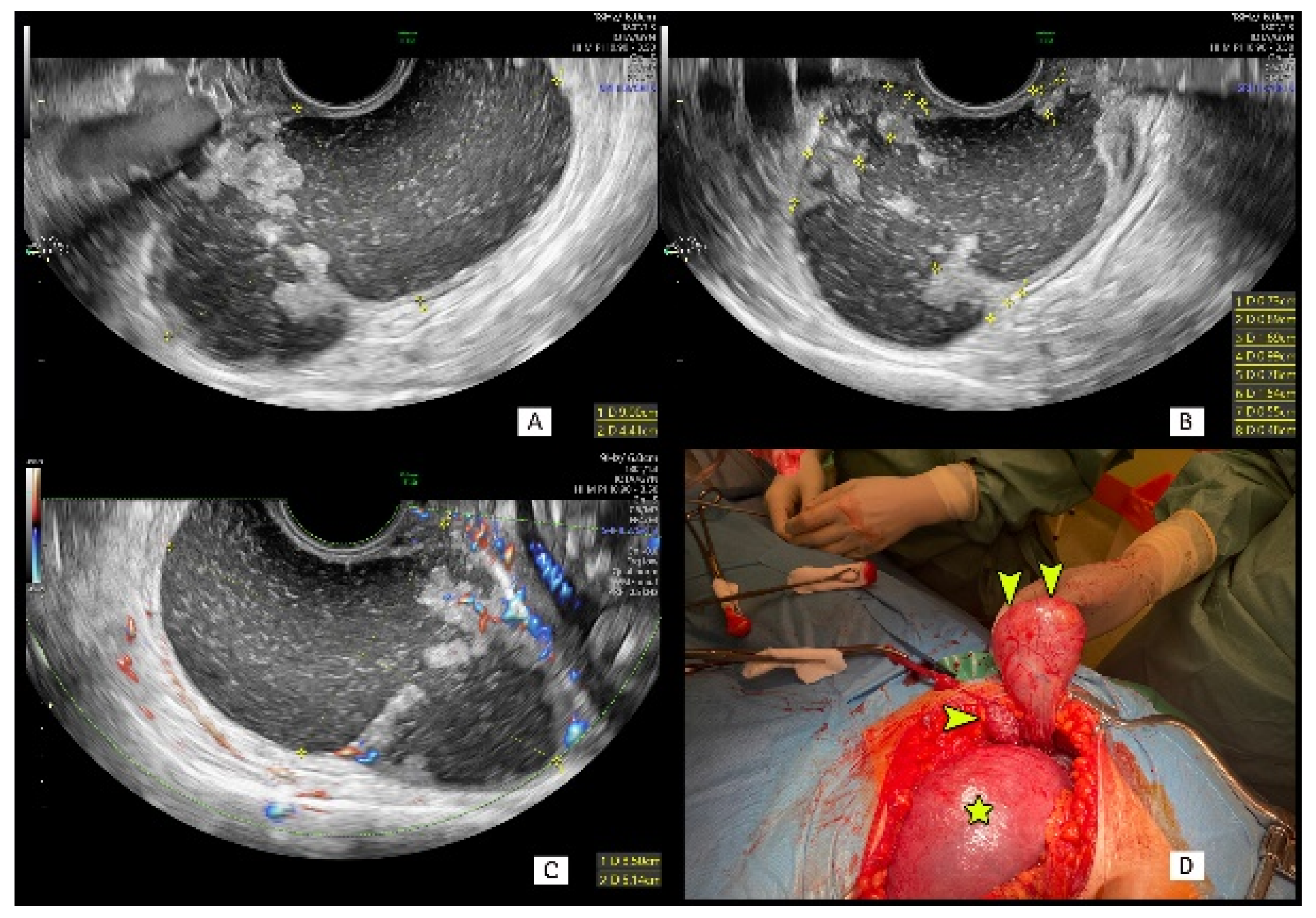
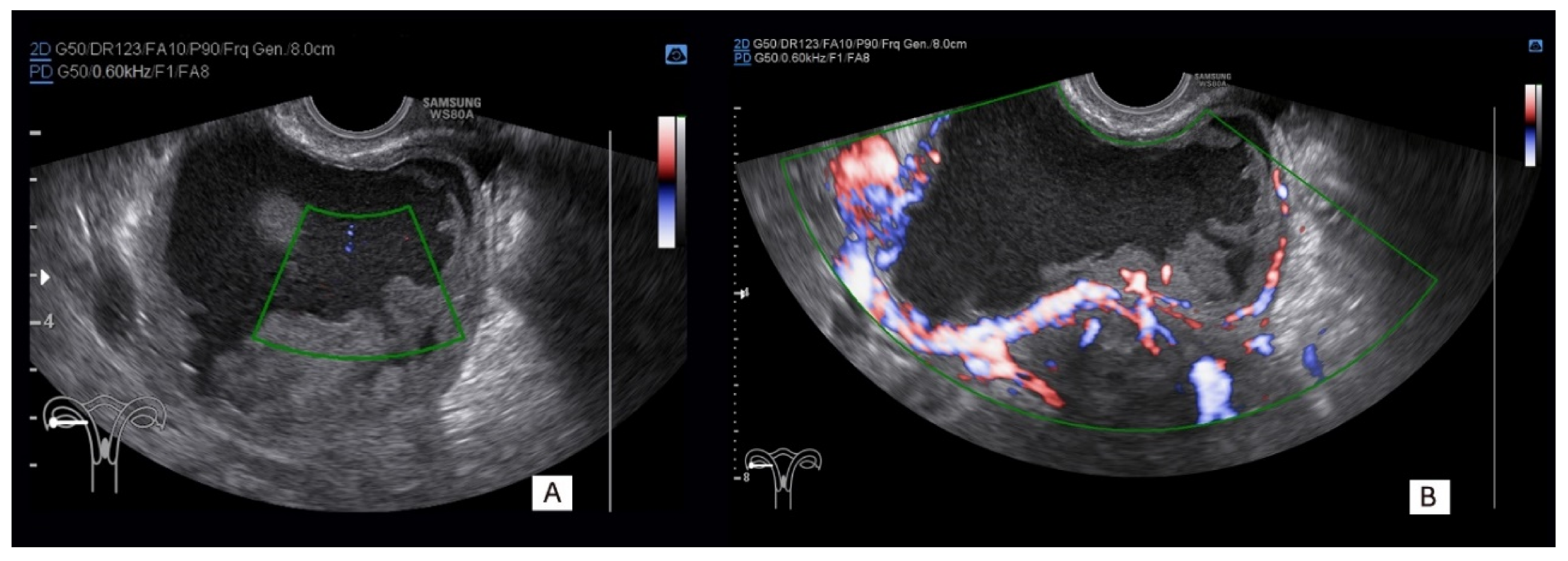
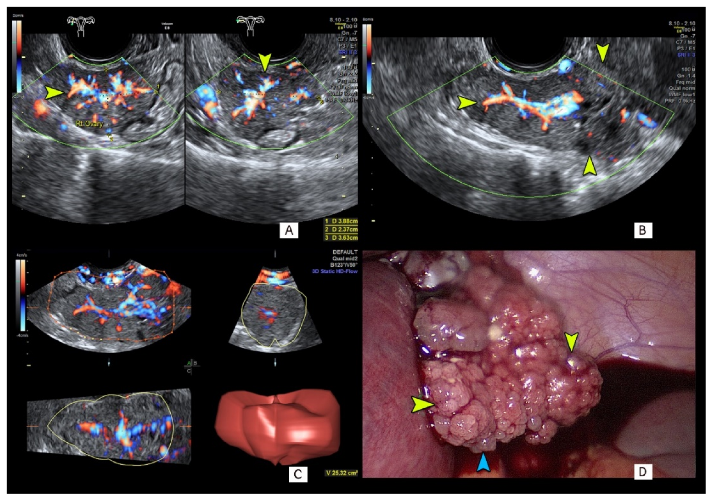
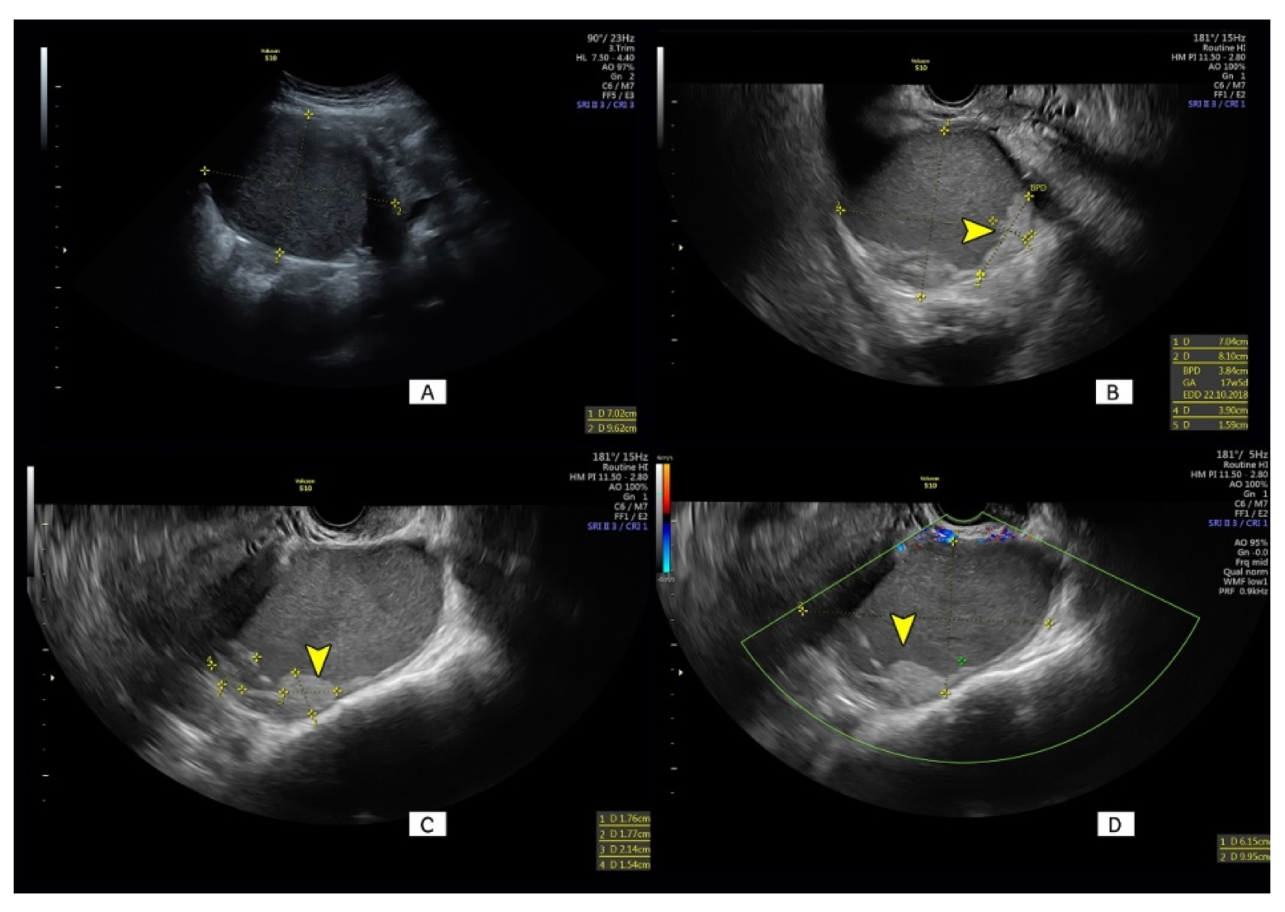
| Tumor Histology | (n = 36) |
|---|---|
| Benign tumors | 27 |
| Dermoid cyst | 11 |
| Decidualized endometrioma | 10 |
| Endometrioma | 1 |
| Fibrothecoma | 2 |
| Serous cystadenoma | 2 |
| Fibroma | 1 |
| Borderline | |
| Serous | 2 |
| Invasive ovarian tumors | |
| Serous ovarian cancer | 3 |
| Endometrioid ovarian cancer | 1 |
| Other | |
| Colorectal cancer (metastatic) | 2 |
| Dysgerminoma | 1 |
| Characteristic | All n = 36 (100%) | Benign n = 27 (75%) | Malignant n = 9 (25%) | p Value |
|---|---|---|---|---|
| Age at diagnosis (years) | 28.5 (20–42) | 28 (20–42) | 29 (24–41) | 0.57 |
| Nulliparous (%) | 22 (61.1) | 17 (63) | 5 (55.6) | 0.69 |
| Gestational age at diagnosis (weeks) | 13.5 (8–31) | 14 (8–31) | 12 (9–28) | 0.8 |
| Gestational age at surgery (weeks) | 21.5 (12–40) | 22 (14–40) | 21 (12–37) | 0.42 |
| Gestational age at delivery (weeks) | 39 (25–41) | 39.5 (38–41) | 38.5 (25–40) | 0.06 |
| Mode of delivery | 0.67 | |||
| Vaginal (n,%) | 12 (33.3) | 8 (29.6) | 4 (44.4) | |
| Cesarean section (n,%) | 20 (55.6) | 16 (59.3) | 4 (44.4) | |
| Ongoing Pregnancy (n, %) | 4 (11.1) | 3 (75) | 1 (25) |
| Sonographic Characteristics of Studied Tumors | All n = 36 (100%) | Benign n = 27 (75%) | Malignant n = 9 (25%) | p-Value * |
|---|---|---|---|---|
| Bilateral masses | 8 (22.2) | 6 (22.2) | 2 (22.2) | 0.64 |
| Maximum diameter of the lesion (mm) | 71.5 (39–206) | 71 (39–206) | 90 (45–135) | 0.14 |
| Type of tumor | 0.01 | |||
| Unilocular α | 8 (22.2) | 8 (29.6) | 0 (0) | |
| Multilocular | 2 (5.5) | 2 (7.4) | 0 (0) | |
| Unilocular–solid | 15 (41.7) | 13 (48.2) | 2 (22.2) | |
| Multilocular–solid | 6 (16.7) | 2 (7.4) | 4 (44.5) | |
| Solid | 5 (13.9) | 2 (7.4) | 3 (33.3) | |
| Echogenicity of cyst fluid | 0.06 | |||
| Anechoic | 12 (33.3) | 7 (25.9) | 5 (55.6) | |
| Low level | 1 (2.8) | 1 (3.7) | 0 (0) | |
| Ground glass | 6 (16.7) | 6 (22.2) | 0 (0) | |
| Mixed | 12 (33.3) | 11 (40.8) | 1 (11.1) | |
| Not relevant (solid mass) | 5 (13.9) | 2 (7.4) | 3 (33.3) | |
| Largest solid component (mm) | 21.5 (2–89) | 18 (2–77) | 39 (17–89) | 0.01 |
| Papillary projections | 16 (44.4) | 11 (40.7) | 5 (55.6) | |
| Number of papillary projections | 0.64 | |||
| 0 | 20 (55.6) | 16 (59.3) | 4 (44.5) | |
| 1 | 4 (11.1) | 3 (11.1) | 1 (11.1) | |
| 2 | 2 (5.5) | 2 (7.4) | 0 (0) | |
| 3 | 3 (8.3) | 2 (7.4) | 1 (11.1) | |
| 4 | 1 (2.8) | 1 (3.7) | 0 (0) | |
| >4 | 6 (16.7) | 3 (11.1) | 3 (33.3) | |
| Height of the largest papillary projection (mm) | 14.5 (3–42) | 12 (3–42) | 15 (11–17) | 0.65 |
| Papillation contour | 0.22 | |||
| Smooth | 13 (36.1) | 10 (37) | 3 (33.3) | |
| Irregegular | 3 (8.3) | 1 (3.7) | 2 (22.2) | |
| Papillation flow if papillary projections were present | 0.53 | |||
| NO | 25 (69.4) | 20 (74.1) | 5 (55.6) | |
| YES | 11 (30.6) | 7 (25.9) | 4 (44.4) | |
| Shadows behind papillae | 0.73 | |||
| NO | 33 (91.7) | 24 (88.9) | 9 (100) | |
| YES | 3 (8.3) | 3 (11.1) | 0 (0) | |
| Microcystic pattern of solid parts | 0.43 | |||
| NO | 34 (94.4) | 26 (96.3) | 8 (88.9) | |
| YES | 2 (5.6) | 1 (3.7) | 1 (11.1) | |
| Color Score | 0.07 | |||
| 1 | 13 (36.1) | 13 (48.2) | 0 (0) | |
| 2 | 5 (13.9) | 3 (11.1) | 2 (22.2) | |
| 3 | 14 (38.9) | 9 (33.3) | 5 (55.6) | |
| 4 | 4 (11.1) | 2 (7.4) | 2 (22.2) | |
| Ascites | 1 (2.8) | 0 (0) | 1 (11.1) | 0.56 |
| Free fluid in the pouch of Douglas | 0.09 | |||
| <5 mm | 34 (94.4) | 27 (100) | 7 (77.8) | |
| >5 mm | 2 (5.6) | 0 (0) | 2 (22.2) | |
| Metastatatic | 2 (5.6) | 0 (0) | 2 (22.2) | 0.09 |
| Ovarian crescent sign | 1 (2.8) | 1 (3.7) | 0 (0) | 0.56 |
| Diagnostic Test | All n = 36 (100%) | Benign n = 27 (75%) | Malignant n = 9 (25%) | p Value |
|---|---|---|---|---|
| CA 125 (U/mL) at diagnosis a | 27 (8.6–305) | 25.2 (8.6–126) | 63.1 (13.5–305) | 0.05 |
| HE4 at diagnosis b | 42.2 (28.9–75.9) | 41 (28.9–50.5) | 50.9 (39.6–75.9) | 0.03 |
| ROMA risk (%, range) b | 5.3 (2.2–18.7) | 5 (2.2–7.6) | 8.2 (4.5–18.7) | 0.04 |
| ROMA risk | 0.19 | |||
| Low (<11.4%) | 22 (91.7) | 16 (100) | 6 (75) | |
| High | 2 (8.3) | 0 (0) | 2 (25) | |
| Simple Rules Risk (SRR) | 0.03 | |||
| Low (<3%) | 10 (27.8) | 10 (37) | 0 (0) | |
| Intermediate (3–20%) | 7 (19.4) | 6 (22.2) | 1 (11.1) | |
| High (>20%) | 19 (52.8) | 11 (40.8) | 8 (88.9) | |
| ADNEX | 11.2 (0.4–91.3) | 7.6 (0.4–86.6) | 52 (4.4–91.3) | 0.02 |
| ADNEX Risk | 0.007 | |||
| Low (<3%) | 9 (25) | 9 (33.3) | 0 (0) | |
| Intermediate (3–20%) | 12 (33.3) | 10 (37) | 2 (22.2) | |
| High (>30%) | 15 (41.7) | 8 (29.7) | 7 (77.8) | |
| ADNEX highest risk | 0.002 | |||
| BENIGN n, % | 2 (5.6) | 2 (7.4) | 0 (0) | |
| Borderline tumors (BOT) n, % | 23 (63.9) | 20 (74.1) | 3 (33.3) | |
| FIGO stage I n, % | 7 (19.4) | 5 (18.5) | 2 (22.2) | |
| FIGO stage II–IV, n,% | 4 (11.1) | 0 (0) | 4 (44.5) | |
| Subjective Assessment (SA) | 0.0001 | |||
| BENIGN | 20 (55.6) | 19 (70.4) | 1 (11.1) | |
| BORDERLINE | 7 (19.4) | 6 (22.2) | 1 (11.1) | |
| MALIGNANT | 9 (25) | 2 (7.4) | 7 (77.8) |
| Test | TP | FP | FN | TN | SENS | SPEC | LR+ | LR− | PPV | NPV | ACC |
|---|---|---|---|---|---|---|---|---|---|---|---|
| CA125 a | 6 | 5 | 3 | 13 | 0.67 | 0.72 | 2.40 | 0.46 | 0.55 | 0.81 | 0.70 |
| HE4 b | 2 | 0 | 6 | 16 | 0.25 | 1.00 | - | 0.75 | 1.00 | 0.73 | 0.75 |
| ROM b | 2 | 0 | 6 | 16 | 0.25 | 1.00 | - | 0.75 | 1.00 | 0.73 | 0.75 |
| ADNEX | 7 | 8 | 2 | 19 | 0.78 | 0.70 | 2.63 | 0.32 | 0.47 | 0.90 | 0.72 |
| SA | 8 | 8 | 1 | 19 | 0.89 | 0.70 | 3.00 | 0.16 | 0.50 | 0.95 | 0.75 |
| SRR | 9 | 17 | 0 | 10 | 1.00 | 0.37 | 1.59 | 0.00 | 0.35 | 1.00 | 0.53 |
Publisher’s Note: MDPI stays neutral with regard to jurisdictional claims in published maps and institutional affiliations. |
© 2021 by the authors. Licensee MDPI, Basel, Switzerland. This article is an open access article distributed under the terms and conditions of the Creative Commons Attribution (CC BY) license (http://creativecommons.org/licenses/by/4.0/).
Share and Cite
Czekierdowski, A.; Stachowicz, N.; Smoleń, A.; Kluz, T.; Łoziński, T.; Miturski, A.; Kraczkowski, J. Sonographic Assessment of Complex Ultrasound Morphology Adnexal Tumors in Pregnant Women with the Use of IOTA Simple Rules Risk and ADNEX Scoring Systems. Diagnostics 2021, 11, 414. https://doi.org/10.3390/diagnostics11030414
Czekierdowski A, Stachowicz N, Smoleń A, Kluz T, Łoziński T, Miturski A, Kraczkowski J. Sonographic Assessment of Complex Ultrasound Morphology Adnexal Tumors in Pregnant Women with the Use of IOTA Simple Rules Risk and ADNEX Scoring Systems. Diagnostics. 2021; 11(3):414. https://doi.org/10.3390/diagnostics11030414
Chicago/Turabian StyleCzekierdowski, Artur, Norbert Stachowicz, Agata Smoleń, Tomasz Kluz, Tomasz Łoziński, Andrzej Miturski, and Janusz Kraczkowski. 2021. "Sonographic Assessment of Complex Ultrasound Morphology Adnexal Tumors in Pregnant Women with the Use of IOTA Simple Rules Risk and ADNEX Scoring Systems" Diagnostics 11, no. 3: 414. https://doi.org/10.3390/diagnostics11030414
APA StyleCzekierdowski, A., Stachowicz, N., Smoleń, A., Kluz, T., Łoziński, T., Miturski, A., & Kraczkowski, J. (2021). Sonographic Assessment of Complex Ultrasound Morphology Adnexal Tumors in Pregnant Women with the Use of IOTA Simple Rules Risk and ADNEX Scoring Systems. Diagnostics, 11(3), 414. https://doi.org/10.3390/diagnostics11030414