
Quantitative Assessment of Lung Volumes and Enhancement in Patients with COVID-19: Role of Dual-Energy CT
 , ,
, , 
Abstract
:1. Introduction
2. Materials and Methods
2.1. Setting and Participants
2.2. Imaging Acquisition
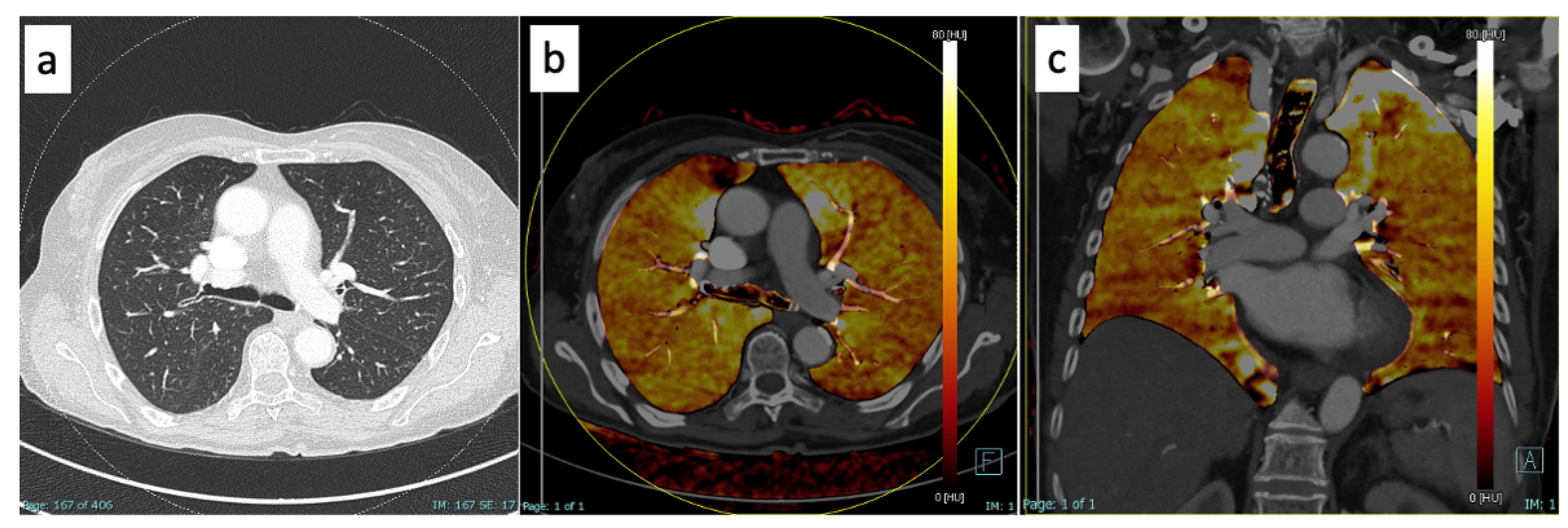
2.3. Imaging Post-Processing
2.4. Statistical Analysis
3. Results
4. Discussion
5. Conclusions
Author Contributions
Funding
Institutional Review Board Statement
Informed Consent Statement
Data Availability Statement
Conflicts of Interest
Abbreviations
| CT | Computed tomography |
| DECT | Dual-energy computed tomography |
| COVID-19 | Coronavirus disease 2019 |
References
- Kwee, T.C.; Kwee, R.M. Chest CT in COVID-19: What the Radiologist Needs to Know. RadioGraphics 2020, 40, 1848–1865. [Google Scholar] [CrossRef] [PubMed]
- Pontone, G.; Scafuri, S.; Mancini, M.E.; Agalbato, C.; Guglielmo, M.; Baggiano, A.; Muscogiuri, G.; Fusini, L.; Andreini, D.; Mushtaq, S.; et al. Role of computed tomography in COVID-19. J. Cardiovasc. Comput. Tomogr. 2021, 15, 27–36. [Google Scholar] [CrossRef] [PubMed]
- Li, X.; Zhao, Y.; Lu, Y.; Zheng, Y.; Mei, N.; Han, Q.; Ruan, Z.; Xiao, A.; Qiu, X.; Wang, D.; et al. Performances of clinical characteristics and radiological findings in identifying COVID-19 from suspected cases. BMC Med. Imaging 2022, 22, 55. [Google Scholar] [CrossRef]
- Axiaq, A.; Almohtadi, A.; Massias, S.A.; Ngemoh, D.; Harky, A. The role of computed tomography scan in the diagnosis of COVID-19 pneumonia. Curr. Opin. Pulm. Med. 2021, 27, 163–168. [Google Scholar] [CrossRef]
- Cellina, M.; Orsi, M.; Valenti Pittino, C.; Toluian, T.; Oliva, G. Chest computed tomography findings of COVID-19 pneumonia: Pictorial essay with literature review. Jpn J. Radiol. 2020, 38, 1012–1019. [Google Scholar] [CrossRef] [PubMed]
- Kato, S.; Ishiwata, Y.; Aoki, R.; Iwasawa, T.; Hagiwara, E.; Ogura, T.; Utsunomiya, D. Imaging of COVID-19: An update of current evidences. Diagn. Interv. Imaging 2021, 102, 493–500. [Google Scholar] [CrossRef]
- Idilman, I.S.; Telli Dizman, G.; Ardali Duzgun, S.; Irmak, I.; Karcaaltincaba, M.; Inkaya, A.C.; Demirkazik, F.; Durhan, G.; Akpinar, M.G.; Ariyurek, O.M.; et al. Lung and kidney perfusion deficits diagnosed by dual-energy computed tomography in patients with COVID-19-related systemic microangiopathy. Eur. Radiol. 2021, 31, 1090–1099. [Google Scholar] [CrossRef] [PubMed]
- Lang, M.; Som, A.; Mendoza, D.P.; Flores, E.; Reid, N.; Carey, D.; Li, M.; Witkin, A.; Rodriguez-Lopez, J.; Shepard, J.-A.; et al. Hypoxaemia related to COVID-19: Vascular and perfusion abnormalities on dual-energy CT. Lancet Infect. Dis. 2020, 20, 1365–1366. [Google Scholar] [CrossRef]
- Revzin, M.V.; Raza, S.; Warshawsky, R.; D’Agostino, C.; Srivastava, N.C.; Bader, A.S.; Malhotra, A.; Patel, R.D.; Chen, K.; Kyriakakos, C.; et al. Multisystem Imaging Manifestations of COVID-19, Part 1: Viral Pathogenesis and Pulmonary and Vascular System Complications. RadioGraphics 2020, 40, 1574–1599. [Google Scholar] [CrossRef]
- Olson, M.C.; Lubner, M.G.; Menias, C.O.; Mellnick, V.M.; Gettle, L.M.; Kim, D.H.; Elsayes, K.M.; Pickhardt, P.J. Update: Venous Thrombosis and Hypercoagulability in the Abdomen and Pelvis—Findings in COVID-19. RadioGraphics 2020, 40, E24–E28. [Google Scholar] [CrossRef]
- Perico, L.; Benigni, A.; Casiraghi, F.; Ng, L.F.; Renia, L.; Remuzzi, G. Immunity, endothelial injury and complement-induced coagulopathy in COVID-19. Nat. Rev. Nephrol. 2021, 17, 46–64. [Google Scholar] [CrossRef] [PubMed]
- McGonagle, D.; O’Donnell, J.S.; Sharif, K.; Emery, P.; Bridgewood, C. Immune mechanisms of pulmonary intravascular coagulopathy in COVID-19 pneumonia. Lancet Rheumatol. 2020, 2, e437–e445. [Google Scholar] [CrossRef] [PubMed]
- Le Berre, A.; Boeken, T.; Caramella, C.; Afonso, D.; Nhy, C.; Saccenti, L.; Tardivel, A.-M.; Gerber, S.; Roche, A.F.; Emmerich, J.; et al. Dual-energy CT angiography reveals high prevalence of perfusion defects unrelated to pulmonary embolism in COVID-19 lesions. Insights Imaging 2021, 12, 24. [Google Scholar] [CrossRef]
- Grillet, F.; Behr, J.; Calame, P.; Aubry, S.; Delabrousse, E. Acute Pulmonary Embolism Associated with COVID-19 Pneumonia Detected with Pulmonary CT Angiography. Radiology 2020, 296, E186–E188. [Google Scholar] [CrossRef] [Green Version]
- Léonard-Lorant, I.; Delabranche, X.; Séverac, F.; Helms, J.; Pauzet, C.; Collange, O.; Schneider, F.; Labani, A.; Bilbault, P.; Molière, S.; et al. Acute Pulmonary Embolism in Patients with COVID-19 at CT Angiography and Relationship to d-Dimer Levels. Radiology 2020, 296, E189–E191. [Google Scholar] [CrossRef] [Green Version]
- Poschenrieder, F.; Meiler, S.; Lubnow, M.; Zeman, F.; Rennert, J.; Scharf, G.; Schaible, J.; Stroszczynski, C.; Pfeifer, M.; Hamer, O.W. Severe COVID-19 pneumonia: Perfusion analysis in correlation with pulmonary embolism and vessel enlargement using dual-energy CT data. PLoS ONE 2021, 16, e0252478. [Google Scholar] [CrossRef] [PubMed]
- Rabiee, N.; Bagherzadeh, M.; Ghasemi, A.; Zare, H.; Ahmadi, S.; Fatahi, Y.; Dinarvand, R.; Rabiee, M.; Ramakrishna, S.; Shokouhimehr, M.; et al. Point-of-Use Rapid Detection of SARS-CoV-2: Nanotechnology-Enabled Solutions for the COVID-19 Pandemic. Int. J. Mol. Sci. 2020, 21, 5126. [Google Scholar] [CrossRef]
- Hong, Y.J.; Shim, J.; Lee, S.M.; Im, D.J.; Hur, J. Dual-Energy CT for Pulmonary Embolism: Current and Evolving Clinical Applications. Korean J. Radiol. 2021, 22, 1555. [Google Scholar] [CrossRef]
- Foti, G.; Silva, R.; Faccioli, N.; Fighera, A.; Menghini, R.; Campagnola, A.; Carbognin, G. Identification of pulmonary embolism: Diagnostic accuracy of venous-phase dual-energy CT in comparison to pulmonary arteries CT angiography. Eur. Radiol. 2021, 31, 1923–1931. [Google Scholar] [CrossRef]
- Monti, C.B.; Zanardo, M.; Cozzi, A.; Schiaffino, S.; Spagnolo, P.; Secchi, F.; De Cecco, C.N. Dual-energy CT performance in acute pulmonary embolism: A meta-analysis. Eur. Radiol. 2021, 31, 6248–6258. [Google Scholar] [CrossRef]
- Petritsch, B.; Pannenbecker, P.; Weng, A.M.; Veldhoen, S.; Grunz, J.P.; Bley, T.A.; Kosmala, A. Comparison of Dual- and Single-Source Dual-Energy CT for Diagnosis of Acute Pulmonary Artery Embolism. ROFO Geb. Rontgenstr. Nukl. 2021, 193, 427–436. [Google Scholar] [CrossRef] [PubMed]
- Jawad, S.; Ulriksen, P.S.; Kalhauge, A.; Hansen, K.L. Acute Pulmonary Embolism Severity Assessment Evaluated with Dual Energy CT Perfusion Compared to Conventional CT Angiographic Measurements. Diagn. Basel Switz. 2021, 11, 495. [Google Scholar] [CrossRef] [PubMed]
- Shi, F.; Wei, Y.; Xia, L.; Shan, F.; Mo, Z.; Yan, F.; Shen, D. Lung volume reduction and infection localization revealed in Big data CT imaging of COVID-19. Int. J. Infect. Dis. 2021, 102, 316–318. [Google Scholar] [CrossRef]
- Iwasawa, T.; Sato, M.; Yamaya, T.; Sato, Y.; Uchida, Y.; Kitamura, H.; Hagiwara, E.; Komatsu, S.; Utsunomiya, D. Ultra-high-resolution computed tomography can demonstrate alveolar collapse in novel coronavirus (COVID-19) pneumonia. Jpn J. Radiol. 2020, 38, 394–398. [Google Scholar] [CrossRef] [Green Version]
- Borghesi, A.; Maroldi, R. COVID-19 outbreak in Italy: Experimental chest X-ray scoring system for quantifying and monitoring disease progression. Radiol. Med. 2020, 125, 509–513. [Google Scholar] [CrossRef] [PubMed]
- Ridge, C.A.; Desai, S.R.; Jeyin, N.; Mahon, C.; Lother, D.L.; Mirsadraee, S.; Semple, T.; Price, S.; Bleakley, C.; Arachchillage, D.J.; et al. Dual-Energy CT Pulmonary Angiography Quantifies Vasculopathy in Severe COVID-19 Pneumonia. Radiol. Cardiothorac. Imaging 2020, 2, e200428. [Google Scholar] [CrossRef] [PubMed]
- Si-Mohamed, S.; Chebib, N.; Sigovan, M.; Zumbihl, L.; Turquier, S.; Boccalini, S.; Boussel, L.; Mornex, J.-F.; Cottin, V.; Douek, P. In vivo demonstration of pulmonary microvascular involvement in COVID-19 using dual-energy computed tomography. Eur. Respir. J. 2020, 56, 2002608. [Google Scholar] [CrossRef]
- Grillet, F.; Busse-Coté, A.; Calame, P.; Behr, J.; Delabrousse, E.; Aubry, S. COVID-19 pneumonia: Microvascular disease revealed on pulmonary dual-energy computed tomography angiography. Quant. Imaging Med. Surg. 2020, 10, 1852–1862. [Google Scholar] [CrossRef]
- Arru, C.D.; Digumarthy, S.R.; Hansen, J.V.; Lyhne, M.; Singh, R.; Rosovsky, R.; Nielsen-Kudsk, J.; Kabrhel, C.; Saba, L.; Kalra, M. Qualitative and quantitative DECT pulmonary angiography in COVID-19 pneumonia and pulmonary embolism. Clin. Radiol. 2021, 76, 392.e1–392.e9. [Google Scholar] [CrossRef]
- Brendlin, A.S.; Mader, M.; Faby, S.; Schmidt, B.; Othman, A.; Gassenmaier, S.; Nikolaou, K.; Afat, S. AI Lung Segmentation and Perfusion Analysis of Dual-Energy CT Can Help to Distinguish COVID-19 Infiltrates from Visually Similar Immunotherapy-Related Pneumonitis Findings and Can Optimize Radiological Workflows. Tomography 2021, 8, 22–32. [Google Scholar] [CrossRef]


| Parameter | Value |
|---|---|
| Patients (total) | 78 |
| Age (years) | 70 (24–97) [15] |
Good outcome
| 13/78 (16.7%) 9/78 (11.5%) 4/78 (5.2%) |
Bad outcome
| 65/78 (83.3%) 53/78 (68.0%) 8/78 (10.3%) 4/78 (5.1%) |
| Good Outcome | Bad Outcome | p Value | |
|---|---|---|---|
| Total Lung Volume (mL) | 4262.1 (±1295.5) (C.I.: 3795.1–4729.2) | 3577.8 (±1255.1) (C.I.: 3205.1–3950.5) | 0.0116 |
| Absolute Enhancement (hu) | 29.8 (±7.2) (C.I.: 27.2–32.5) | 31.2 (±8.1) (C.I.: 28.8–33.6) | 0.7824 |
| Relative Enhancement (%) | 124.2 (±65.0) (C.I.: 99.5–149.0) | 113.1 (±33.3) (C.I.: 103.2–123.0) | 0.1984 |
| N. | Mean | St. Dev. | Min | Max | |
|---|---|---|---|---|---|
| Total Lung Volume (mL) | 78 | 3858.5 | 1308.1 | 1197 | 6319 |
| Right | 78 | 2062.1 | 711.6 | 593 | 3422 |
| Left | 78 | 1801.4 | 645.2 | 596 | 3177 |
| Right Upper | 78 | 798.4 | 278.4 | 238 | 1430 |
| Right Middle | 78 | 495.1 | 242.8 | 118 | 1186 |
| Right Lower | 78 | 773.9 | 273.1 | 236 | 1419 |
| Left Upper | 78 | 820.8 | 309.1 | 265 | 1661 |
| Left Middle | 20 | 692.5 | 205.6 | 263 | 1066 |
| Left Lower | 78 | 808.2 | 304.5 | 261 | 1649 |
| N. | Mean | St. Dev. | Min | Max | |
|---|---|---|---|---|---|
| Tot Enhancement (hu) | 78 | 30.6 | 7.74 | 17 | 66 |
| Right | 78 | 30.2 | 8.04 | 4 | 66 |
| Left | 78 | 30.2 | 7.52 | 5 | 54 |
| Right Upper | 78 | 31.5 | 7.20 | 5 | 47 |
| Right Middle | 78 | 30.0 | 7.60 | 5 | 54 |
| Right Lower | 78 | 28.4 | 7.37 | 5 | 54 |
| Left Upper | 78 | 31.0 | 7.56 | 7 | 54 |
| Left Middle | 19 | 30.2 | 6.45 | 22 | 45 |
| Left Lower | 78 | 28.7 | 7.85 | 9 | 54 |
| Relative Enhancement (%) | 75 | 117.3 | 48.02 | 44 | 400 |
| Right | 75 | 116.4 | 49.79 | 12 | 412 |
| Left | 75 | 117.1 | 48.25 | 16 | 386 |
| Right Upper | 75 | 122.9 | 50.99 | 5 | 406 |
| Right Middle | 75 | 115.4 | 44.84 | 25 | 355 |
| Right Lower | 75 | 111.3 | 52.41 | 46 | 454 |
| Left Upper | 75 | 122.5 | 49.85 | 33 | 403 |
| Left Middle | 17 | 122.9 | 38.91 | 62 | 192 |
| Left Lower | 75 | 112.3 | 47.34 | 52 | 366 |
Disclaimer/Publisher’s Note: The statements, opinions and data contained in all publications are solely those of the individual author(s) and contributor(s) and not of MDPI and/or the editor(s). MDPI and/or the editor(s) disclaim responsibility for any injury to people or property resulting from any ideas, methods, instructions or products referred to in the content. |
© 2023 by the authors. Licensee MDPI, Basel, Switzerland. This article is an open access article distributed under the terms and conditions of the Creative Commons Attribution (CC BY) license (https://creativecommons.org/licenses/by/4.0/).
Share and Cite
Foti, G.; Longo, C.; Faccioli, N.; Guerriero, M.; Stefanini, F.; Buonfrate, D. Quantitative Assessment of Lung Volumes and Enhancement in Patients with COVID-19: Role of Dual-Energy CT. Diagnostics 2023, 13, 1201. https://doi.org/10.3390/diagnostics13061201
Foti G, Longo C, Faccioli N, Guerriero M, Stefanini F, Buonfrate D. Quantitative Assessment of Lung Volumes and Enhancement in Patients with COVID-19: Role of Dual-Energy CT. Diagnostics. 2023; 13(6):1201. https://doi.org/10.3390/diagnostics13061201
Chicago/Turabian StyleFoti, Giovanni, Chiara Longo, Niccolò Faccioli, Massimo Guerriero, Flavio Stefanini, and Dora Buonfrate. 2023. "Quantitative Assessment of Lung Volumes and Enhancement in Patients with COVID-19: Role of Dual-Energy CT" Diagnostics 13, no. 6: 1201. https://doi.org/10.3390/diagnostics13061201
APA StyleFoti, G., Longo, C., Faccioli, N., Guerriero, M., Stefanini, F., & Buonfrate, D. (2023). Quantitative Assessment of Lung Volumes and Enhancement in Patients with COVID-19: Role of Dual-Energy CT. Diagnostics, 13(6), 1201. https://doi.org/10.3390/diagnostics13061201