Multiparametric Ultrasound Approach Using a Tree-Based Decision Classifier for Inconclusive Focal Liver Lesions Evaluated by Contrast Enhanced Ultrasound

 ,
,  ,
,  , ,
, , 
Abstract
:1. Introduction
2. Materials and Methods
2.1. Study Design and Population
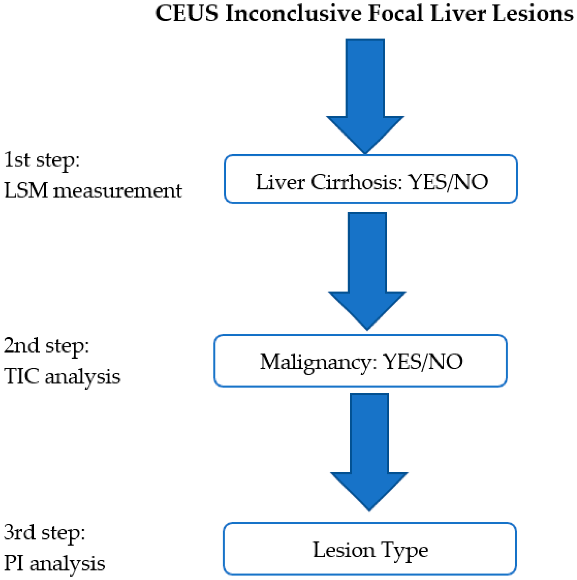
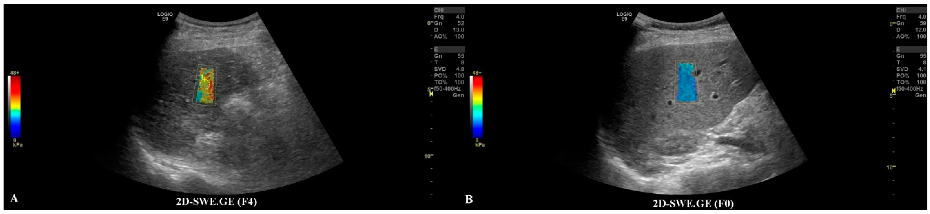
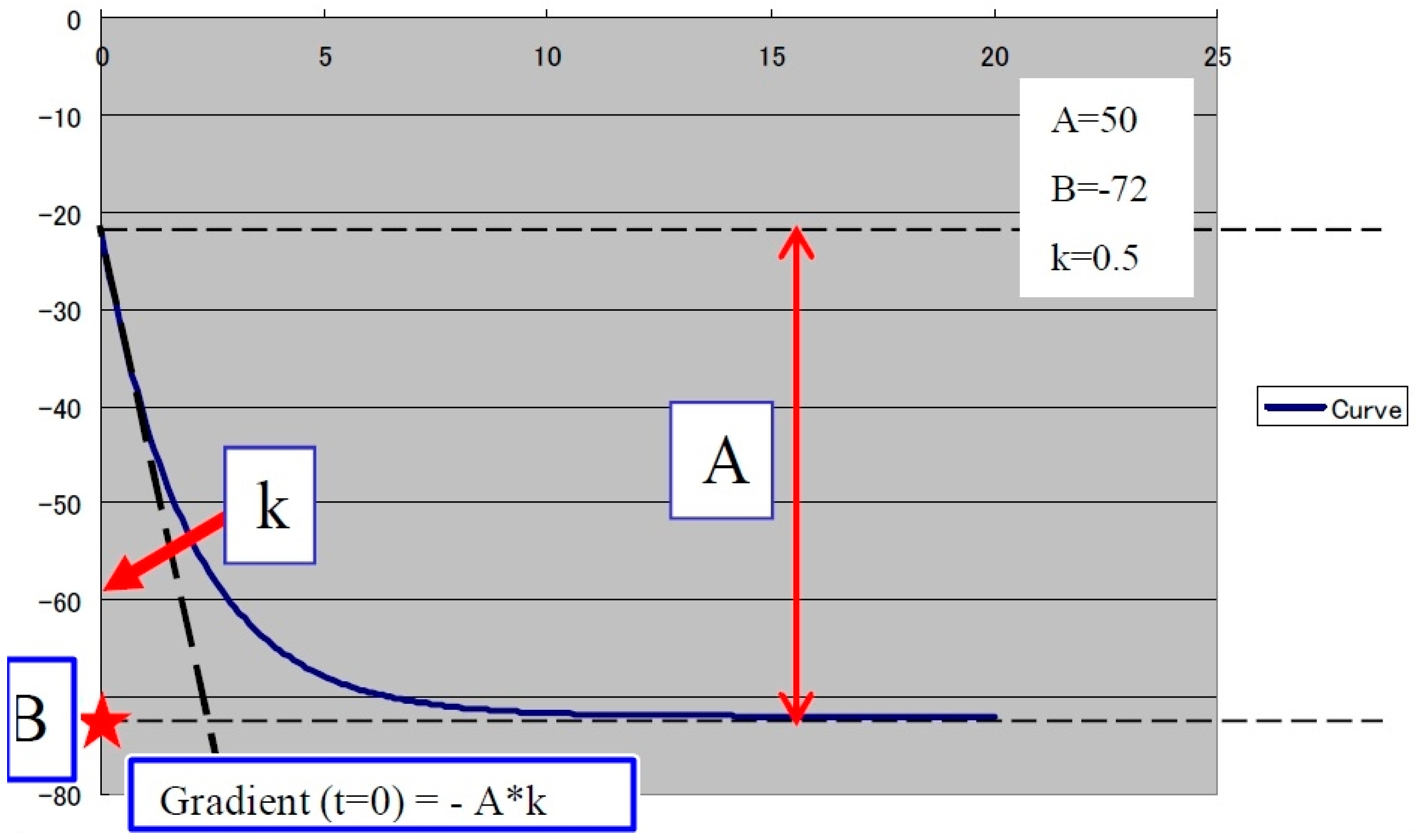
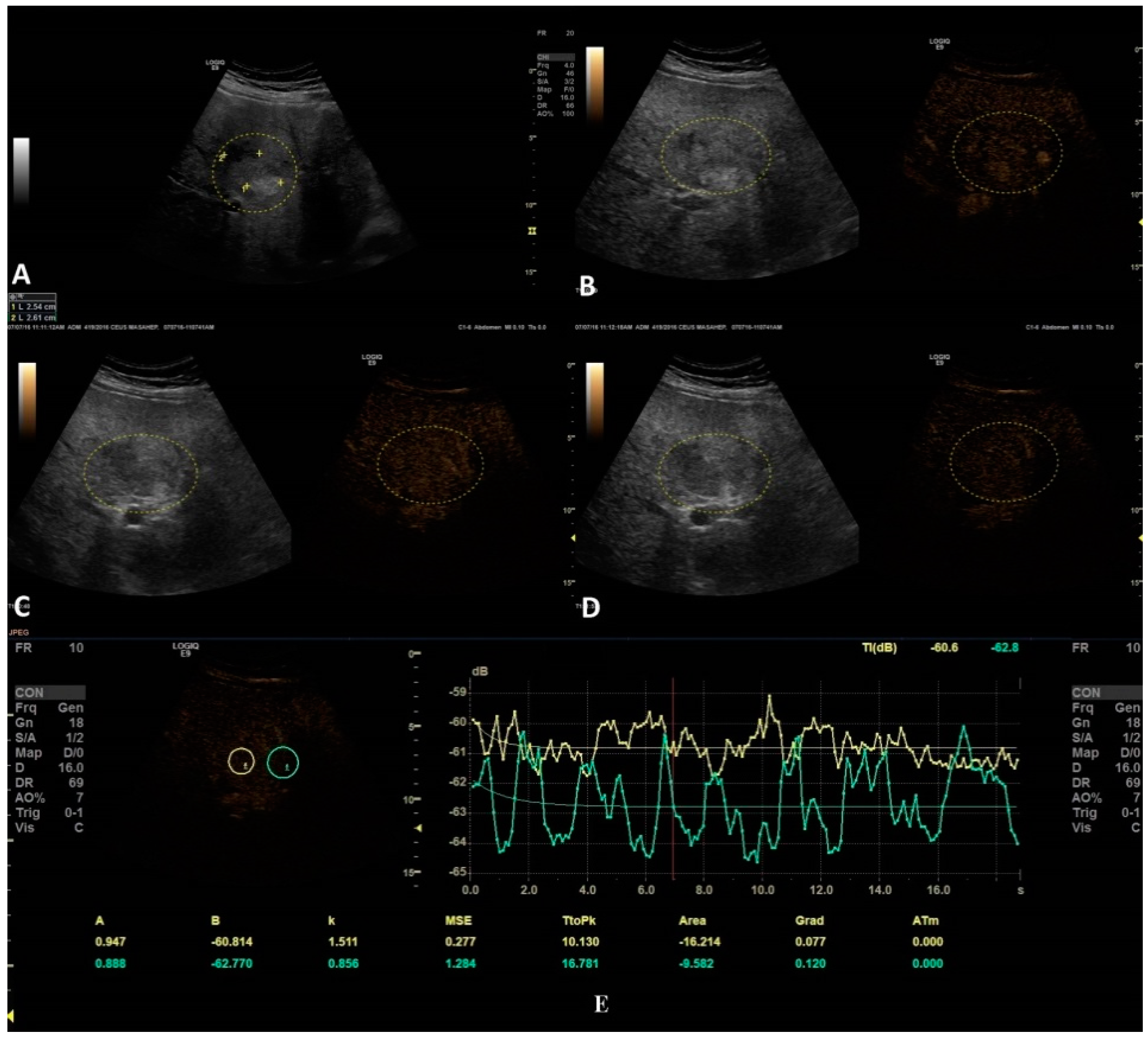
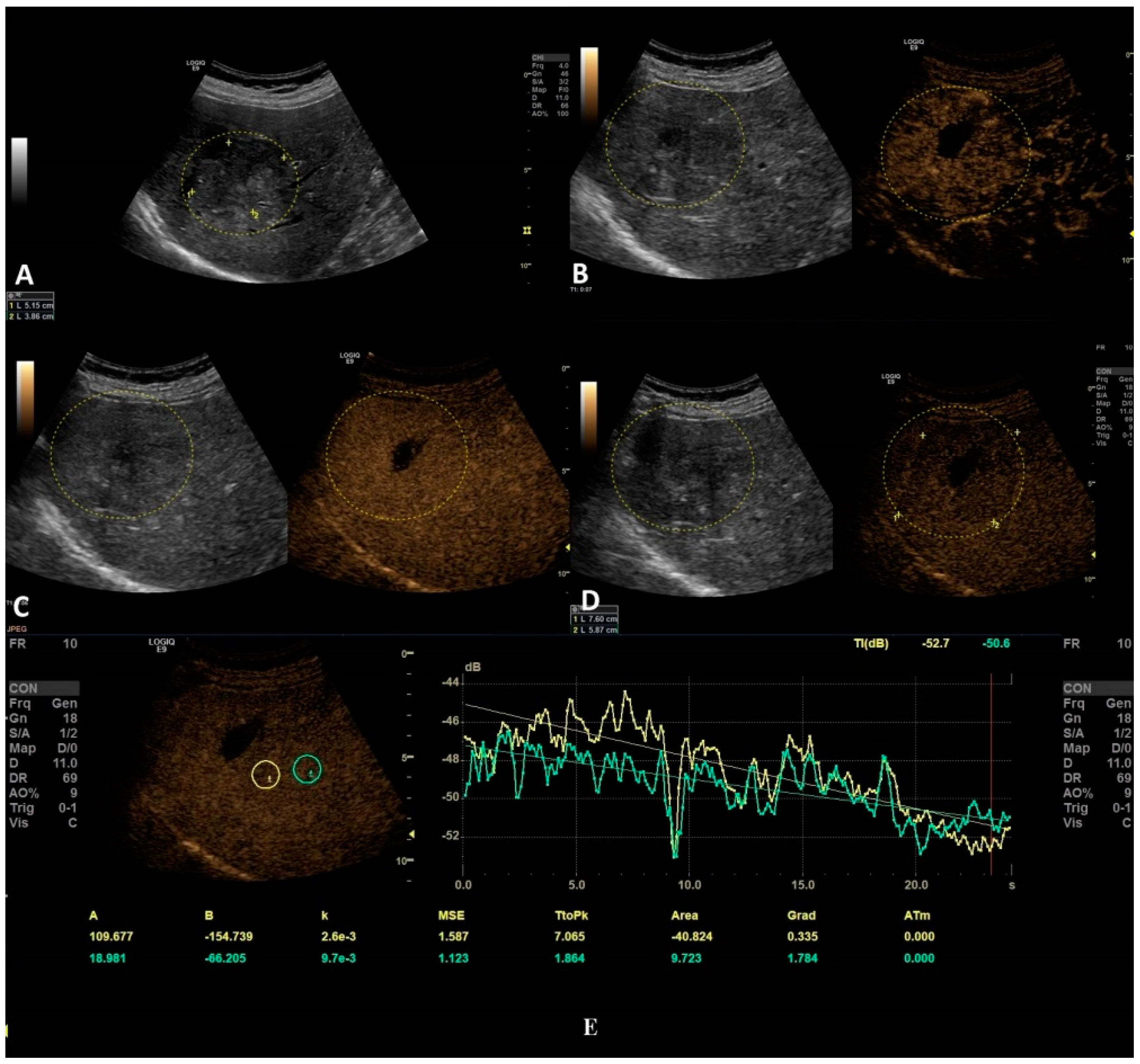
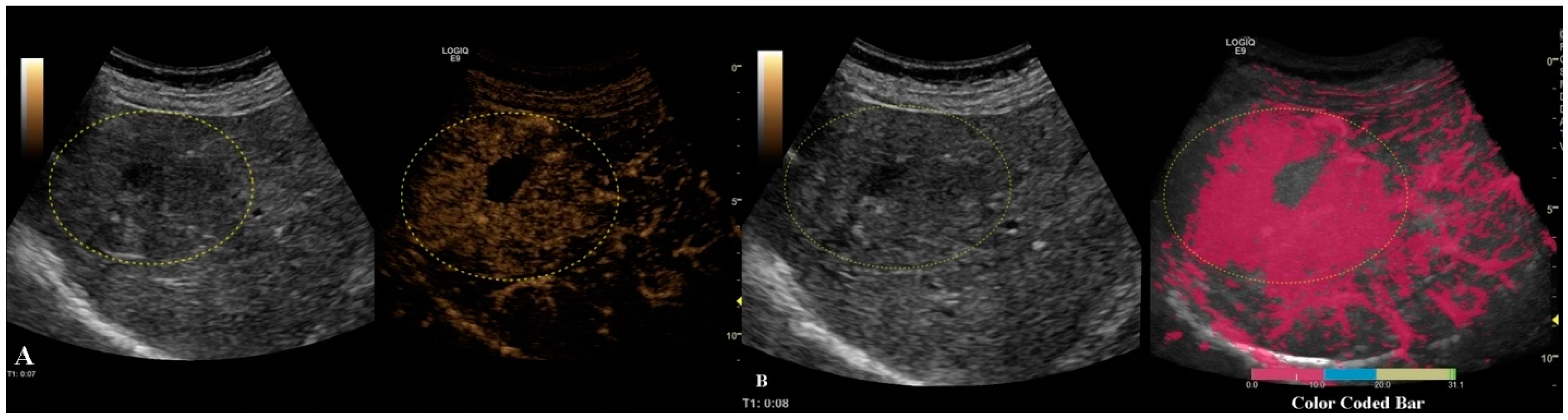
2.2. Multiparametric Ultrasound Steps
2.3. Statistical Analysis
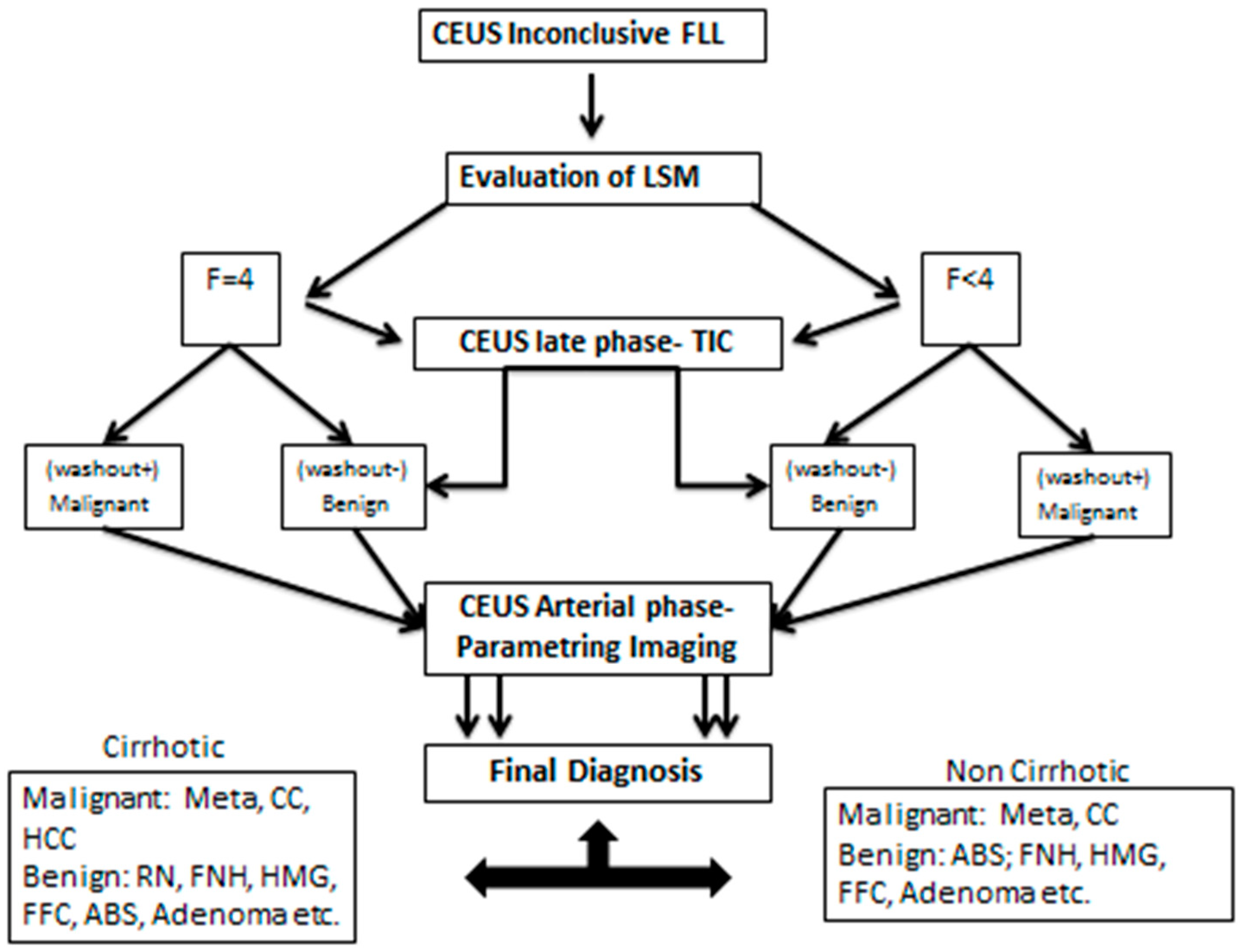
2.4. Binary Decision Tree Classifier
3. Results
BDTC Algorithm
4. Discussion
5. Conclusions
Author Contributions
Funding
Institutional Review Board Statement
Informed Consent Statement
Data Availability Statement
Conflicts of Interest
References
- Dietrich, C.F.; Nolsøe, C.P.; Barr, R.G.; Berzigotti, A.; Burns, P.N.; Cantisani, V.; Chammas, M.C.; Chaubal, N.; Choi, B.I.; Clevert, D.A.; et al. Guidelines and Good Clinical Practice Recommendations for Contrast-Enhanced Ultrasound (CEUS) in the Liver-Update 2020 WFUMB in Cooperation with EFSUMB, AFSUMB, AIUM, and FLAUS. Ultrasound Med. Biol. 2020, 46, 2579–2604. [Google Scholar] [CrossRef] [PubMed]
- Sporea, I.; Badea, R.; Brisc, C.; Ioanițescu, S.; Moga, T.; Popescu, A.; Săftoiu, A.; Săndulescu, L.; Spârchez, Z.; Șirli, R. Romanian National Guidelines on Contrast Enhanced Ultrasound in clinical practice. Med. Ultrason. 2017, 19, 401–415. [Google Scholar] [CrossRef] [PubMed] [Green Version]
- Moga, T.V.; Sirli, R.; Popescu, A.; Danila, M.; Ghiuchici, A.; Sporea, I. Misdiagnosis of focal liver lesions by means of CEUS. J. Hepatol. 2019, 70, E842. [Google Scholar] [CrossRef]
- Sidhu, P.S. Multiparametric Ultrasound (MPUS) Imaging: Terminology Describing the Many Aspects of Ultrasonography. Ultraschall Med. 2015, 36, 315–317. [Google Scholar] [CrossRef]
- Popescu, A. Multiparametric ultrasound (MPUS) or the many faces of ultrasonography. Med. Ultrason. 2019, 21, 369–370. [Google Scholar] [CrossRef]
- European Federation of Societies for Ultrasound in Medicine and Biology (EFSUMB). Minimum training requirements for the practice of Medical Ultrasound in Europe. Ultraschall Med. 2010, 31, 426–427. [Google Scholar] [CrossRef] [PubMed]
- Bende, F.; Sporea, I.; Sirli, R.; Popescu, A.; Mare, R.; Miutescu, B.; Lupusoru, R.; Moga, T.; Pienar, C. Performance of 2-D SWE-GEfor predicting different stages of liver fibrosis, using transient elastography as the reference method. Med. Ultrason. 2017, 19, 143. [Google Scholar] [CrossRef] [Green Version]
- De Lédinghen, V.; Wong, V.W.; Vergniol, J.; Wong, G.L.; Foucher, J.; Chu, S.H.; Le Bail, B.; Choi, P.C.; Chermak, F.; Yiu, K.K.; et al. Diagnosis of liver fibrosis and cirrhosis using liver stiffness measurement: Comparison between M and XL probe of FibroScan®. J. Hepatol. 2012, 56, 833–839. [Google Scholar] [CrossRef]
- Dietrich, C.F.; Bamber, J.; Berzigotti, A.; Bota, S.; Cantisani, V.; Castera, L.; Cosgrove, D.; Ferraioli, G.; Friedrich-Rust, M.; Gilja, O.H.; et al. EFSUMB guidelines and recommendations on the clinical use of liver ultrasound elastography, update 2017 (long version). Ultraschall Med. 2017, 38, e16–e47. [Google Scholar]
- Ultrasound Lab. GE Healthcare Time Intensity Curve Manual. Available online: https://www.logiqportal.net (accessed on 7 November 2005).
- LOGIQ™ XDclear™ Family Parametric Analysis. Available online: https://www.gehealthcare.com/products/logiq-xdclear-family (accessed on 15 July 2021).
- Friedrich-Rust, M.; Klopffleisch, T.; Nierhoff, J.; Herrmann, E.; Vermehren, J.; Schneider, M.D.; Zeuzem, S.; Bojunga, J. Contrast-Enhanced Ultrasound for the differentiation of benign and malignant focal liver lesions: A meta-analysis. Liver Int. 2013, 33, 739–755. [Google Scholar] [CrossRef]
- Niu, Y.; Huang, T.; Lian, F.; Li, F. Contrast-enhanced ultrasonography for the diagnosis of small hepatocellular carcinoma: A meta-analysis and meta-regression analysis. Tumour Biol. 2013, 34, 3667–3674. [Google Scholar] [CrossRef]
- Guang, Y.; Xie, M.; Ding, A.; Cai, L.; Huang, Y. Diagnosis value of focal liver lesions with SonoVue(R)-enhanced ultrasound compared with contrast-enhanced computed tomography and contrast-enhanced MRI: A meta-analysis. J. Cancer Res. Clin. Oncol. 2011, 137, 1595–1605. [Google Scholar] [CrossRef]
- Barr, R.G. Contrast enhanced ultrasound for focal liver lesions: How accurate is it? Abdom. Radiol. 2018, 43, 1128–1133. [Google Scholar] [CrossRef]
- Bernatik, T.; Seitz, K.; Blank, W.; Schuler, A.; Dietrich, C.F.; Strobel, D. Unclear focal liver lesions in contrast-enhanced ultrasonography-lessons to be learned from the DEGUM multicenter study for the characterization of liver tumors. Ultraschall Med. 2010, 31, 577–581. [Google Scholar] [CrossRef]
- Rafailidis, V.; Fang, C.; Leenknegt, B.; Ballal, K.; Deganello, A.; Sellars, M.E.; Yusuf, G.T.; Huang, D.Y.; Sidhu, P.S. Contrast-Enhanced Ultrasound Quantification Assessment of Focal Fatty Variations in Liver Parenchyma: Challenging the Traditional Qualitative Paradigm of Uniform Enhancement with Adjacent Parenchyma. J. Ultrasound Med. 2021, 40, 1137–1145. [Google Scholar] [CrossRef] [PubMed]
- Kierans, A.S.; Taneja, S.S.; Rosenkrantz, A.B. Implementation of Multi-parametric Prostate MRI in Clinical Practice. Curr. Urol. Rep. 2015, 16, 56. [Google Scholar] [CrossRef] [PubMed]
- Cantisani, V.; D’Andrea, V.; Biancari, F.; Medvedyeva, O.; Di Segni, M.; Olive, M.; Patrizi, G.; Redler, A.; De Antoni, E.E.; Patrizi, G.; et al. Prospective evaluation of multiparametric ultrasound and quantitative elastosonography in the differential diagnosis of benign and malignant thyroid nodules: Preliminary experience. Eur. J. Radiol. 2012, 81, 2678–2683. [Google Scholar] [CrossRef] [PubMed]
- Grgurevic, I.; Tjesic Drinkovic, I.; Pinzani, M. Multiparametric ultrasound in liver diseases: An overview for the practising clinician. Postgrad. Med. J. 2019, 95, 425–432. [Google Scholar] [CrossRef]
- Bartolotta, T.V.; Taibbi, A.; Midiri, M.; Matranga, D.; Solbiati, L.; Lagalla, R. Indeterminate focal liver lesions incidentally discovered at gray-scale US: Role of contrast-enhanced sonog-raphy. Investig. Radiol. 2011, 46, 106–115. [Google Scholar] [CrossRef] [Green Version]
- Ghiuchici, A.M.; Sporea, I.; Dănilă, M.; Șirli, R.; Moga, T.; Bende, F.; Popescu, A. Is There a Place for Elastography in the Diagnosis of Hepatocellular Carcinoma? J. Clin. Med. 2021, 10, 1710. [Google Scholar] [CrossRef] [PubMed]
- Schellhaas, B.; Goertz, R.S.; Pfeifer, L.; Kielisch, C.; Neurath, M.F.; Strobel, D. Diagnostic accuracy of contrastenhanced ultrasound for the differential diagnosis of hepatocellular carcinoma: ESCULAP versus CEUS-LI-RADS. Eur. J. Gastroenterol. Hepatol. 2017, 29, 1036–1044. [Google Scholar] [CrossRef] [PubMed]
- Terzi, E.; Iavarone, M.; Pompili, M.; Veronese, L.; Cabibbo, G.; Fraquelli, M.; Riccardi, L.; De Bonis, L.; Sangiovanni, A.; Leoni, S.; et al. Contrast ultrasound LI-RADS LR- identifies hepatocellular carcinoma in cirrhosis in a multicenter restropective study of 1006 nodules. J. Hepatol. 2018, 68, 485–492. [Google Scholar] [CrossRef]
- Ghiuchici, A.M.; Dănilă, M.; Popescu, A.; Șirli, R.; Moga, T.; Topan, M.; Bende, F.; Sporea, I. Contrast-enhanced ultrasound algorithm (ACR CEUS LI-RADSv 2017)—A valuable tool for the noninvasive diagnosis of hepatocellular carcinoma in patients with chronic liver disease. Med. Ultrason. 2021. [Google Scholar] [CrossRef] [PubMed]
- Schellhaas, B.; Bernatik, T.; Bohle, W.; Borowitzka, F.; Chang, J.; Dietrich, C.F.; Dirks, K.; Donoval, R.; Drube, K.; Friedrich-Rust, M.; et al. Strobel Contrast-Enhanced Ultrasound Algorithms (CEUS-LIRADS/ESCULAP) for the Noninvasive Diagnosis of Hepatocellular Carcinoma—A Prospective Multicenter DEGUM Study. Ultraschall Med. 2021, 42, 178–186. [Google Scholar] [CrossRef]
- Huang, Q.; Zhang, F.; Li, X. Machine Learning in Ultrasound Computer-Aided Diagnostic Systems: A Survey. BioMed Res. Int. 2018, 2018, 5137904. [Google Scholar] [CrossRef] [PubMed]
- Moga, T.V.; Popescu, A.; Sporea, I.; Danila, M.; David, C.; Gui, V.; Iacob, N.; Miclaus, G.; Sirli, R. Is Contrast Enhanced Ultrasonography a useful tool in a beginner’s hand? How much can a Computer Assisted Diagnosis prototype help in characterizing the malignancy of focal liver lesions? Med. Ultrason. 2017, 19, 252–258. [Google Scholar] [CrossRef] [PubMed] [Green Version]


| Lesion | Arterial Phase (10–20 to 30–45 s) |
|---|---|
| Hemangioma (HMG) | Peripheral nodular enhancement |
| FNH (focal nodular hyperplasia) | Centrifugal hyper-enhancement (possible with feeding artery) |
| Abscesses (ABS) | Peripheral enhancement possible with septa (no central enhancement—necrotic areas) |
| Metastasis (Meta) | Rim enhancement or hyper-enhancement |
| Hepatocellular carcinoma (HCC) | Hyper-enhancement |
| Cholangiocarcinoma (CC) | Rim-like hyper-enhancement with central hypo-enhancement |
| Focal fatty change (FFC) | Iso-enhancement |
| Parameter | Mean ± SD, n (%) |
|---|---|
| Age | 62.3 ± 6.4 |
| Gender (male) | 52.1% |
| Fibrosis stage | |
| Non-cirrhotic | 57 (62.6%) |
| Cirrhotic | 34 (37.4%) |
| Type of lesion | |
| Hepatocarcinoma | 34 (37.4%) |
| Metastasis | 13 (14.2%) |
| Haemangioma | 7 (7.7%) |
| Regenerative nodules | 7 (7.7%) |
| Focal liver alterations | 5 (5.5%) |
| Fatty free areas | 3 (3.3%) |
| Cholangiocarcinoma | 4 (4.4%) |
| Abscesses | 2 (2.2% |
| Adenomas | 5 (5.5%) |
| Complex biliary cysts | 4 (4.4%) |
| Parenchymal infarction areas | 5 (5.5%) |
| Vascular abnormalities | 2 (2.2%) |
| Parameter | Lesion | Parenchyma | p-Value |
|---|---|---|---|
| A | 5.5 ± 21.5 | −3.53 ± 61.8 | 0.98 |
| B | −57.3 ± 21.8 | −47.2 ± 59.7 | 0.72 |
| k | 1.5 ± 5.3 | 0.9 ± 1.3 | 0.50 |
| TtoPk | 7.3 ± 8.0 | 6.5 ± 7.7 | 0.89 |
| AREA | −25.0 ± 37.9 | −7.0 ± 42.6 | 0.04 |
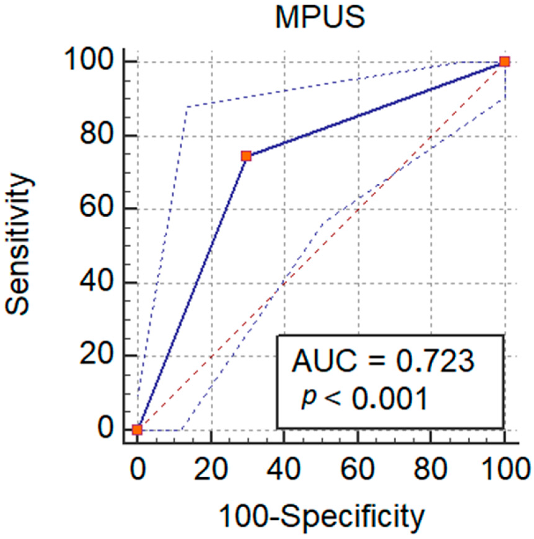
| Ultrasonography Techniques | AUROC | p-Value | Sensitivity | Specificity | PPV | NPV |
|---|---|---|---|---|---|---|
| B-mode | 0.52 | 0.96 | 15.6% | 84.6% | 57.1% | 43.4% |
| Elastography | 0.64 | <0.001 | 74.5% | 70% | 76% | 68.3% |
| CEUS-TIC | 0.58 | <0.001 | 74.0% | 45.7% | 72.5% | 64.2% |
| NEW model * | 0.72 | <0.001 | 90.2% | 22.5% | 59.7% | 64.3% |
Publisher’s Note: MDPI stays neutral with regard to jurisdictional claims in published maps and institutional affiliations. |
© 2021 by the authors. Licensee MDPI, Basel, Switzerland. This article is an open access article distributed under the terms and conditions of the Creative Commons Attribution (CC BY) license (https://creativecommons.org/licenses/by/4.0/).
Share and Cite
Moga, T.V.; David, C.; Popescu, A.; Lupusoru, R.; Heredea, D.; Ghiuchici, A.M.; Foncea, C.; Burdan, A.; Sirli, R.; Danilă, M.; et al. Multiparametric Ultrasound Approach Using a Tree-Based Decision Classifier for Inconclusive Focal Liver Lesions Evaluated by Contrast Enhanced Ultrasound. J. Pers. Med. 2021, 11, 1388. https://doi.org/10.3390/jpm11121388
Moga TV, David C, Popescu A, Lupusoru R, Heredea D, Ghiuchici AM, Foncea C, Burdan A, Sirli R, Danilă M, et al. Multiparametric Ultrasound Approach Using a Tree-Based Decision Classifier for Inconclusive Focal Liver Lesions Evaluated by Contrast Enhanced Ultrasound. Journal of Personalized Medicine. 2021; 11(12):1388. https://doi.org/10.3390/jpm11121388
Chicago/Turabian StyleMoga, Tudor Voicu, Ciprian David, Alina Popescu, Raluca Lupusoru, Darius Heredea, Ana M. Ghiuchici, Camelia Foncea, Adrian Burdan, Roxana Sirli, Mirela Danilă, and et al. 2021. "Multiparametric Ultrasound Approach Using a Tree-Based Decision Classifier for Inconclusive Focal Liver Lesions Evaluated by Contrast Enhanced Ultrasound" Journal of Personalized Medicine 11, no. 12: 1388. https://doi.org/10.3390/jpm11121388
APA StyleMoga, T. V., David, C., Popescu, A., Lupusoru, R., Heredea, D., Ghiuchici, A. M., Foncea, C., Burdan, A., Sirli, R., Danilă, M., Ratiu, I., Bizerea-Moga, T., & Sporea, I. (2021). Multiparametric Ultrasound Approach Using a Tree-Based Decision Classifier for Inconclusive Focal Liver Lesions Evaluated by Contrast Enhanced Ultrasound. Journal of Personalized Medicine, 11(12), 1388. https://doi.org/10.3390/jpm11121388