Article

Distribution and Excretion of Arsenic Metabolites after Oral Administration of Seafood-Related Organoarsenicals in Rats

Centre for Health and Environmental Risk Research, National Institute for Environmental Studies, 16-2 Onogawa, Tsukuba, Ibaraki 305-8506, Japan
*
Author to whom correspondence should be addressed.
Metals 2016, 6(10), 231; https://doi.org/10.3390/met6100231
Submission received: 29 June 2016 / Revised: 13 September 2016 / Accepted: 13 September 2016 / Published: 27 September 2016
(This article belongs to the Special Issue Metallomics)

Abstract

:
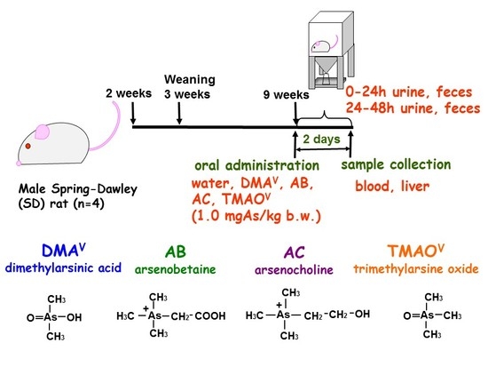
Less information is available on the metabolism of organic arsenicals compared to inorganic arsenic in mammals. In the present study, we investigated tissue distribution, metabolism and excretion in rats of organoarsenicals, dimethylarsinic acid (DMAV), arsenobetaine (AB), arsenocholine (AC) and trimethylarsine oxide (TMAOV). Among these animals, arsenic concentrations in red blood cells (RBCs) and spleen increased remarkably only in the DMAV group. Hepatic arsenic concentration increased significantly only in the AC group. Approximately 17%, 72% and 60% of the dose was excreted in urine in two days in the DMAV, AB and AC groups, respectively; virtually the entire dose was excreted in urine in one day in the TMAOV group. On the other hand, approximately 18%, 0.2%, 0.5% and 0.1% of the dose was excreted in feces in two days in the DMAV, AB, AC and TMAOV groups, respectively. A large amount of arsenic was accumulated in RBCs in the form of protein-bound dimethylarsinous acid (DMAIII), and dimethylmonothioarsinic acid (DMMTAV), a reportedly toxic thio-arsenical, was found in urine and fecal extract in the DMAV group. These results suggest that intake of DMAV is a potential health hazard, given that the metabolites of DMAV, such as DMAIII and DMMTAV, are known to be highly toxic.

Graphical Abstract

1. Introduction

Inorganic arsenite (iAsIII) and arsenate (iAsV) are well-known worldwide environmental contaminants. Inorganic arsenicals are absorbed by the gastrointestinal tract, methylated mainly in the liver and excreted mainly in urine as methylated pentavalent arsenicals, such as monomethylarsonic acid (MMAV) and dimethylarsinic acid (DMAV) [1,2]. Monomethylarsonous acid (MMAIII) and dimethylarsinous acid (DMAIII) also have been detected in human urine [3,4], and these trivalent arsenic metabolites are more toxic than iAsIII [5,6,7]. Recently, thiolated dimethylarsenic compounds, dimethylmonothioarsinic acid (DMMTAV) and dimethyldithioarsinic acid (DMDTAV), have been detected in urine [8,9,10,11]. Naranmandura et al. reported that DMMTAV was more cytotoxic than iAsIII in human bladder cancer cells [12].
Several organoarsenic compounds are present in seafood, including: arsenocholine (AC) in shrimps [13], seals and whales [14]; arsenobetaine (AB) in rock lobster tails [15], sharks [16,17], seals and whales [14]; trimethylarsine oxide (TMAOV) in fish [18]; arsenosugar in brown kelp [19]; and arsenolipid in demersal sharks [20] and tuna [21]. DMAV was reported as a major metabolite in human urine after ingestion of arsenosugar [22,23] and arsenolipid [24]. Orally-administered AC is almost completely absorbed from the gastrointestinal tract in mice and rats, and AB is the main urinary metabolite of AC in mice and rats [25]. Orally-administered AB is almost completely absorbed from the gastrointestinal tract and excreted as AB in mice, rats and rabbits [26]. Yoshida et al. [27] reported that a small portion of administered AB was converted to TMAOV, MMAV, DMAV, tetramethylarsonium (TeMA) and iAs after oral administration in rats. TMAOV was methylated to TeMA to some extent after one week and after seven months of oral administration of TMAOV, although most TMAOV was excreted in urine as TMAOV [28]. However, very little is known concerning fecal excretion of arsenic following the ingestion of those seafood-originating organoarsenicals, and data on the metabolic behavior of organoarsenicals are quite limited compared to those for iAs.
Rats are one of the most tolerant animal species to As, and As is known to accumulate in the red blood cells (RBCs) of rats following administration of iAs in the form of dimethylarsenical [29,30]. Lu et al. [31] reported that arsenic was bound to the α-chain of hemoglobin in the RBCs of rats that had been fed a DMAV-containing diet and that the main arsenical in RBCs was dimethylarsinous acid (DMAIII). The α-chain of rat hemoglobin includes a reactive cysteine 13 (Cys-13), and the lack of this cysteine in human hemoglobin may be responsible for the shorter retention of As in human blood compared to rat blood [32]. On the other hand, data on the arsenic speciation analysis of human RBCs are very limited. AB was detected in the RBCs of healthy male volunteers [33]. AB and dimethylarsenical were detected in the RBCs of humans who stopped drinking As-contaminated water two and five years before blood collection [34,35].
Standard rodent chow contains arsenic compounds derived from fish. Fish meal also contains a small amount of iAs [36], such that tissues obtained even from control rats maintained on standard commercial chow contain a measurable level of arsenic [37]. In the present study, we propose a method to reduce the background tissue and blood arsenic level. Using this method, we investigated the metabolism, distribution and excretion of seafood-derived organoarsenicals in rats.

2. Materials and Methods

2.1. Chemicals

Sodium arsenite (NaAsO2; iAsIII: 97.0%) and sodium arsenate (Na2HAsO4; iAsV: 99.0%) were purchased from Sigma (St. Louis, MO, USA). Monomethylarsonic acid ((CH3)AsO(OH)2; MMAV), dimethylarsinic acid ((CH3)2AsO(OH); DMAV), arsenobetaine ([(CH3)3As]+CH2COO; AB), arsenocholine ((CH3)3AsCH2CH2OH+; AC) and trimethylarsine oxide ((CH3)3AsO; TMAOV) were purchased from Tri Chemicals (Yamanashi, Japan). Stock solutions (containing the equivalent of 100 µg As/mL) of these compounds were stored at 4 °C in the dark. Working standard solutions (10 ng As/mL) were prepared daily using Milli-Q SP water (Yamato Millipore, Tokyo, Japan). Standard solutions containing arsenic (100 µg As/mL; Accustandard®, New Haven, CT, USA) and rhodium (internal standard, 1.01 mg Rh/mL; Wako Pure Chemical Industries, Osaka, Japan) were used for total arsenic analysis. Other chemicals were of analytical grade and were purchased from Wako Pure Chemical Industries.

2.2. Animals

Male Sprague-Dawley (SD) rats were obtained at two weeks of age with lactating female rats from Charles River Japan (Kanagawa, Japan). Following arrival, the lactating rats (with pups) were provided ad libitum with the commercial standard rodent chow (CE-2, Clea Japan Co., Tokyo, Japan) or the arsenic-depleted rodent chow (AIN-93G) [38], in which casein is used instead of fish as the protein source, purchased from Oriental Yeast Co. (Tokyo, Japan), and sterilized deionized-distilled water (DDW). Rats were maintained in an air-conditioned clean room at 22–25 °C, 50%–55% relative humidity and a 12-h light-dark cycle. After one week of lactation, the weanling male rats (three weeks old) were separated from the dams and were maintained on the same diet as the lactating rats.
To examine the effects of diet on tissue arsenic concentration, groups of four rats that had been maintained on either the standard (for up to ten weeks) or arsenic-depleted chow (for up to three weeks) were anesthetized with sodium pentobarbital (50 mg/kg body weight) and sacrificed by exsanguination through the abdominal aorta. Whole blood, liver and spleen samples were collected. In a separate experiment, additional groups of rats (nine weeks of age, n = 4) were fed the arsenic-depleted chow first for six weeks to reduce the basal level of arsenic in their body. These rats were orally administered with DMAV, AB, AC or TMAOV at a dose of 1.0 mg As/kg body weight and then placed for two days in metabolic cages designed for the separation of urine and feces. Control animals received the same volume of deionized water. These rats were sacrificed (as above) two days after administration, and whole blood, liver and spleen samples were collected. Plasma samples were prepared from heparinized blood and were separated from RBCs by centrifugation at 1600× g for 10 min. RBCs were washed three times with Tris-HCl-buffered saline, hemolyzed with four volumes of 50 mM ammonium acetate solution (pH approximately 6.5, 25 °C) and then centrifuged at 15,000× g for 30 min at 4 °C to obtain an RBC lysate. Urine samples were centrifuged at 1600× g for 10 min to remove suspended solids. The feces were dried with a lyophilizer (FZ-2.5CSCL; Laboconco, MO, USA) and powdered with a mortar. Arsenic compounds in feces samples were extracted with 50 mM ammonium acetate solution as described by Yoshida et al. [8] with a minor modification. In brief, the powder (25 mg) was soaked in 0.5 mL of 50 mM ammonium acetate solution in a 1.5-mL polypropylene microcentrifuge tube, and the contents were sonicated for 1 h (10 s sonicate-rest cycle, 25 °C) (Bioruptor, COSMO BIO Co., Ltd., Tokyo, Japan). The faces suspensions were centrifuged (15,000× g, 30 min), and the supernatants were filtered through a 0.45-µm syringe-driven filter before analysis.
All procedures were approved by the Animal Care and Use Committee of the National Institute for Environmental Studies (NIES).

2.3. Preparation of Rodent Chows and Tap Water for Analysis

The arsenic-depleted rodent chow and standard rodent chow were milled and sieved (250 µm) for ease of acid digestion and the extraction. The fine powder was dried in an oven at 50 °C for 3 h and then stored in a desiccator at room temperature. The methods for extraction of arsenic from rodent chow samples were performed using the same methods as described for extraction of the feces samples. Tap water was collected from three suction pipes of the animal room.

2.4. Synthesis of Dimethylthioarsenicals

Dimethylmonothioarsinic acid (DMMTAV) and dimethyldithioarsinic acid (DMDTAV) were synthesized according to the published procedure [39] with a minor modification. DMMTAV was prepared by stepwise addition of concentrated H2SO4 (2.4 mmol) to an aqueous solution containing 1.5 mmol DMAV and 2.4 mmol Na2S at the final molar ratio of DMAV/Na2S/H2SO4 = 1:1.6:1.6, and the reaction solution was allowed to stand for 1 h. DMMTAV was extracted with ethyl ether; the extract was evaporated under reduced pressure and blown to dryness with nitrogen gas. DMDTAV was prepared by stepwise addition of concentrated H2SO4 (3.75 mmol) to an aqueous solution containing 0.5 mmol DMAV and 3.75 mmol Na2S at the final molar ratio of DMAV/Na2S/H2SO4 = 1:7.5:7.5, and the reaction solution was allowed to stand for 24 h. Trimethylarsine sulfide (TMASV) was prepared by stepwise addition of concentrated H2SO4 (0.8 mmol) to an aqueous solution containing 0.5 mmol TMAOV and 0.8 mmol Na2S at the final molar ratio of DMAV/Na2S/H2SO4 = 1:1.6:1.6, and the reaction solution was allowed to stand for 24 h.

2.5. Procedure for Elemental Analyses

A 100-µL or 0.1-g portion of each sample was wet-digested with 0.6 mL nitric acid and 0.2 mL hydrogen peroxide at 90 °C overnight and then at 130 °C for 2–72 h in an aluminum block bath. The samples were diluted with Milli-Q water, and concentrations of total arsenic were determined with the internal standard (Rh; m/z 103) method by inductively-coupled argon plasma mass spectrometry (ICP-MS). We calculated the contents of arsenic in the organs and blood by multiplying the concentration of arsenic by the total weight of each sample. The weight of the whole blood was assumed to be 7% of the body weight [40]. The analytical method was validated by measuring arsenic concentrations in the reference samples; NIES CRM No. 18 human urine (NIES, Tsukuba, Japan) and NMIJ CRM 7403-a swordfish tissue (AIST, Tsukuba, Japan) (n = 3). The certified and the measured values for total arsenic were 0.137 ± 0.011 As mg/L and 0.144 ± 0.005 As mg/L for CRM No. 18 human urine and 6.62 ± 0.21 As mg/kg and 6.63 ± 0.22 As mg/kg for NMIJ CRM 7403-a, respectively.

2.6. HPLC-ICP-MS Analysis

The HPLC system (Shimadzu, Kyoto, Japan) consisted of a column oven (CTO-20AC), a solvent delivery module (LC-20AD), a degasser (DGU-20A3), a 7125 six-port injection valve with a 20-µL injection loop (Rheodyne, CA, USA) and a reversed phase C-18 column (ODS-3; 150 mm × 4.6 mm, 3-µm particle size; GL Science, Tokyo, Japan), an anion-exchange column (Shodex RSpak JJ50-4D; 150 mm × 4.6 mm i.d.; Showa Denko, Japan), a cation-exchange column (Shodex RSpak NN-614; 150 mm × 6.0 mm i.d.; Showa Denko) or a polymer-based gel filtration column (Shodex Asahipak GS-220 HQ; 300 mm × 7.6 mm i.d.; Showa Denko). The eluate was introduced directly into the nebulizer of ICP-MS (HP 7500c; Yokogawa Analytical Systems, Musashino, Japan) to detect arsenicals in samples. The signal intensity at m/z 75 was monitored for the measurement of arsenic, and the intensity at m/z 77 was also monitored for the detection of the molecular interference of argon chloride (ArCl+). The speciation of arsenic was carried out by HPLC-ICP-MS. An aliquot of the sample (20 µL) was applied to the column, and the columns were eluted with the pre-filtered (0.22 µm) mobile phase, constituted as follows. Reversed phase column: 5 mM tetrabutylammonium hydroxide (TBAH), 3 mM malonic acid and 5% (v/v) methanol, pH approximately 5.6, flow rate of 1.0 mL/min, at 50 °C. Anion-exchange column: 20 mM oxalic acid; pH was adjusted to 2.3 with ammonia solution, flow rate of 0.6 mL/min, at 25 °C. Cation-exchange column: 5 mM HNO3, 8 mM NH4NO3, pH approximately 2.4, flow rate of 0.8 mL/min, at 25 °C. Gel filtration column: 50 mM ammonium acetate, flow rate of 0.5 mL/min, at 25 °C. The analytical conditions for HPLC are summarized in Table 1.

2.7. Statistics

Values are presented as the means ± SD of three (rodent diet and tap water) or four samples (in vivo study). Statistical analysis of the data was carried out by one-way analysis of variance with Bonferroni’s post hoc comparison (Figure 1) or a t-test (Figure 2), and a probability value of less than 0.05 was accepted as indicative of a significant difference.

3. Results

3.1. Changes in the Concentration of Total Arsenic in Rats Fed Standard or Arsenic-Depleted Chow

The concentration of total arsenic in standard chow (CE-2) was 654 ± 13.6 ng/g; in arsenic-depleted chow (AIN-93G), the value was 7.65 ± 2.98 ng/g. The total arsenic concentration in tap water was below the limit of quantification (LOQ; <0.05 ng As/mL).
Concentrations of arsenic were determined to study the background arsenic levels in rats, as assayed in whole blood (Figure S1a), plasma (Figure S1b), liver (Figure S1c) and spleen (Figure S1d) from rats fed standard (open triangle) or arsenic-depleted diet (open circle). Small amounts of arsenic were found in the whole blood, plasma, liver and spleen in two-week-old rats (during lactation) (256 ± 26.6, 15.2 ± 3.72 ng As/mL, 17.4 ± 3.57 and 46.9 ± 6.47 ng As/g, respectively) (Figure S1). The concentrations of arsenic in whole blood in three-week-old rats (just before weaning) were 135 ± 27.0 ng As/mL for the arsenic-depleted diet group and 319 ± 60.5 ng As/mL for the standard diet group (Figure S1a). One week after weaning (at the age of four weeks), the concentration of arsenic in whole blood of the standard diet group was significantly increased compared to that of the arsenic-depleted diet group, and the concentration increased with age (Figure S1a). On the other hand, arsenic concentration in plasma was low even in the standard diet group (Figure S1b). Hepatic and splenic arsenic concentrations of rats fed the arsenic-depleted diet were 1.74 ± 0.37 and 28.9 ± 8.06 ng As/g, while those of rats fed the standard diet were 34.7 ± 9.40 and 80.6 ± 23.4 ng As/g at the age of three weeks, respectively (Figure S1c,d). The arsenic concentrations in liver and spleen after weaning increased with age, as observed in whole blood (Figure S1a,c,d).

3.2. Speciation of Arsenic in Rodent Chow

To elucidate the chemical forms of arsenical in standard rodent chow, an extract of the chow was prepared. The recovery of arsenic in the extract was approximately 50%. On the other hand, the concentration of As in the arsenic-depleted diet extract was below the quantification limit (<0.05 ng As/mL). The speciation of arsenicals in the chow was performed using both reversed phase (Figure S2a–c) and anion-exchange columns (Figure S2d–g).
Typical chromatograms were obtained for arsenic standards in deionized water (Figure S2a,d–f), in saline (Figure S2b) and in extracts of the basal diets (Figure S2c,g) by HPLC-ICP-MS. One major and four minor arsenic peaks were detected in the extract using the reversed phase column (Figure S2c). The major peak was eluted at a retention time corresponding to AB and/or iAsIII; high concentrations of chloride in the samples resulted in splitting and broadening of the iAsV and MMAV peaks (Figure S2b) [41]. To confirm whether the first peak shown in Figure S2c was AB or iAsIII and whether iAsV was contained in the basal diet or not, the samples were applied to an anion-exchange column (Figure S2g). AB could be separated from iAsIII using the anion-exchange column and iAsV was detected in the diet extract. These results indicate that AB was the major arsenic compound in the standard diet, which also contained small amounts of AC, iAsIII, DMAV and iAsV.

3.3. Tissue Distribution and Excretion of Metabolites Following Oral Administration of Organoarsenicals in Rats

The rats were fed on arsenic-depleted rodent chow to decrease the basal level of tissue arsenic contents.
The concentrations of arsenic were determined in whole blood (Figure 1a), plasma (Figure 1b), RBCs (Figure 1c), liver (Figure 1d) and spleen (Figure 1e) two days after the administration of As-containing compounds. In the control group, small amounts of arsenic were detected in whole blood, liver and spleen (98.1 ± 32.8, 4.09 ± 0.991 and 24.6 ± 1.15 ng As/mL, respectively) (Figure 1a,d,e), while the concentration of arsenic in plasma was below the LOQ (Figure 1b). Blood arsenic concentrations increased remarkably in the DMAV-treated group compared to the other groups (Figure 1a); however, the concentration of arsenic in plasma was not significantly increased compared to the control group (Figure 1b). Plasma arsenic concentrations were significantly increased in the AC-treated group (Figure 1b). Approximately 72%, 0.7% and 0.9% of the initial dose was present in the whole blood in the DMAV-, AB- and AC-treated groups, respectively (Figure 1a), and 0.07%, 0.6% and 0.7% in the plasma (Figure 1b). The arsenic concentration in RBCs was calculated based on the hematocrit value. Approximately 72%, 0.1% and 0.8% of the dose was present in the RBCs in the DMAV-, AB- and AC-treated groups, respectively (Figure 1c). On the other hand, the distribution of arsenic in the RBCs in the TMAOV-treated group was similar to that in the control group (Figure 1c). Hepatic arsenic concentration was increased significantly only in the AC-treated group (Figure 1d); splenic arsenic concentration was significantly increased only in the DMAV-treated group (Figure 1e). For the DMAV-, AB-, AC- and TMAOV-treated groups (respectively), the liver contained approximately 1.9%, 3.2%, 11% and 0.01% (Figure 1d) and the spleen contained approximately 0.6%, 0.1%, 0.1% and 0% of the administered dose (Figure 1e).
Excretion of arsenic was determined in urine (Figure 2a) and feces (Figure 2b). In the control group, small amounts of arsenic were excreted in the urine and feces (approximately 0.01–0.2 and 0.001–0.05 µg As/day, respectively) (Figure 2). Approximately 17%, 72% and 60% of the administered dose was excreted in the urine within two days in the DMAV-, AB- and AC-treated groups, respectively; in the TMAOV-treated group, most of the dose was excreted in the urine in the first day following administration (Figure 2a). On the other hand, significant levels were observed in the feces only for the DMAV-treated group compared to the control group, with recovery of approximately 18% of the administered dose within two days (Figure 2b). Approximately 0.2%, 0.5% and 0.1% of the dose was excreted in feces in two days in the AB-, AC- and TMAOV-treated groups, respectively (Figure 2b).
The recoveries of arsenic in two days after the administration in DMA-, AB-, AC- and TMAO-treated rats are approximately 110%, 76%, 73% and 110%, respectively. The lower values in the AB- and AC-treated groups suggested that the remaining AB and AC might have distributed in the other tissues (Table S1).

3.4. HPLC-ICP-MS Analysis of Arsenic Standards and Biological Samples

Equal volumes of the samples obtained from the individual rats of a given group were pooled, and a 20-μL aliquot was used for HPLC-ICP-MS. Figure 3 shows the elution profiles of arsenic standards (each at 20 ng As/mL) on gel filtration (Figure 3a) and cation-exchange columns (Figure 3b). The retention time of AB resembled that of TMAOV on the gel filtration column (Figure 3a), while the retention time of TMASV was closest to that of DMMTAV on the cation-exchange column (Figure 3b). Therefore, we used both columns for speciation of arsenicals in the various biological samples, except for the RBC lysate and plasma.
Figure 4 shows gel filtration column elution profiles of arsenic in RBC lysates (Figure 4a–e) and in plasma specimens (Figure 4f–j) derived from control rats (Figure 4a,f) and from rats treated with DMAV (Figure 4b,g), AB (Figure 4c,h), AC (Figure 4d,i) or TMAOV (Figure 4e,j). Two arsenic peaks were observed in the whole blood and plasma from DMAV-treated animals (Figure 4b,g). The major peak at the void volume appeared to correspond to protein-bound arsenicals, while the minor peak was eluted at the same retention time as that of authentic DMAV (Figure 3a and Figure 4b,g). The elution profiles of plasma in both AB- and AC-treated groups showed the major peak at the same retention time as that of AB; however, the minor peak at the void volume was observed only in the AC-treated group (Figure 4h,i). For the TMAOV-treated group, the elution profiles were similar to those observed in the control group (Figure 4a,e,f,j).
We used both gel filtration and cation-exchange columns for the speciation of arsenicals in the urine and fecal extract samples. Figure 5 shows the elution profiles of arsenic in urine with gel filtration (Figure 5a–e) and with cation exchange (Figure 5f–j). The arsenic compounds in control group urine were not detected as sharp peaks (Figure 5a,f). The results obtained by using the two different columns showed that chemical forms of arsenicals excreted in urine were DMAV (major peak), DMMTAV, DMDTAV, TMASV and TMAOV in the DMAV-treated group (Figure 5b,g); AB (major peak), TMASV and TMAOV in the AB-treated group (Figure 5c,h); AB (major peak), AC, TMASV and TMAOV in the AC-treated group (Figure 5d,i); and TMAOV (major peak) and TMASV in the TMAOV-treated group (Figure 5e,j). Furthermore, trace amounts of unknown arsenicals were observed in the elution profiles for the AB-, AC- and TMAOV-treated groups (Figure 5c–e,h–j). Additional experiments revealed that spiked AB in control rat urine (Figure S3b) eluted earlier than that in DDW (Figure S3a) and that AC and TMAOV could not be separated using the cation-exchange column (Figure S3b). Based on the results shown in Panels c, d, and e of Figure 5, we concluded that the arsenic peaks at 9.5 and 14.5 min (shown in Figure 5h–j) were AB and TMAOV, respectively. We also noted that the chemical forms of urine arsenicals recovered on the second day following administration were similar to those seen on the first day (Figure S4).
Figure 6 shows the elution profiles of arsenic in fecal extracts following separation by gel filtration (Figure 6a–e) or cation exchange (Figure 6f–j). The recovery of arsenic in fecal extract was approximately 65%. As observed with urine, arsenic compounds in control group feces were not detected as clear peaks (Figure 6a,f). The elution profile of feces in the DMAV-treated group was similar to that of urine from the same group (Figure 6b,g and Figure 5b,g). Although similar amounts of DMMTAV and DMDTAV were excreted in urine, the amount of DMMTAV exceeded that of DMDTAV in feces (Figure 5b and Figure 6b). Chemical forms of arsenicals excreted in feces were AB in the AB-treated group (Figure 6b,g); AB, AC and an unknown arsenical in the AC-treated group (Figure 6c,h); and TMASV, TMAOV and trace amounts of an unknown arsenical in the TMAOV-treated group (Figure 6e,j).
Those results of the HPLC-ICP-MS analyses are summarized in Table 2. The chemical forms of urinary and fecal extract arsenicals obtained from 24–48 h were similar to those obtained from 0–24 h (Figures S4 and S5).

4. Discussion

The U.S. National Research Council cautions against the use of rats as animal models in the study of metabolism [42] because the rat RBCs contain a larger amount of arsenic compared to those of the other animal species. Rat hemoglobin contains the reactive Cys-13α, and As is known to bind covalently to this site in the form of DMAIII [32]. DMAIII is unstable in biological fluids and is easily oxidized to DMAV, which makes the arsenic metabolic study difficult. Thus, in other words, rats are a suitable model animal for studying the conversion of DMAV to more toxic DMAIII in vivo. In the present study, we first depleted the blood arsenic level of rats using the formulated diet and investigated the differences in metabolites of the food-related organic arsenicals and also the involvement of the reduction of DMAV to DMAIII in the metabolic pathway by measuring the DMAIII level in RBCs semi-quantitatively.
The concentration of arsenic in whole blood and tissues in the standard diet group was significantly increased compared to that in the arsenic-depleted diet group (Figure S1). AB was the major chemical form of arsenic in the standard diet; however, small amounts of AC, iAsIII, DMAV and iAsV were also detected (Figure S2c,g). Using an arsenic-depleted diet, the background As level in blood and tissue is reduced. This is a better approach for the evaluation of organoarsenic metabolism and distribution.
In the present study, a large amount of arsenic was accumulated in RBCs following the administration of DMAV, but not following dosing with the rest of the organic species. For the AB- and AC-treated groups, approximately 0.1% and 0.8% (respectively) of the administered dose was present in RBCs, while the concentration of arsenic in RBCs from the TMAOV-treated group was similar to that of the control group. Presumably, the relatively large amount of arsenic detected in the spleen reflects the accumulation of arsenic in RBCs. The distribution of arsenic in blood, liver, kidneys and feces in the TMAOV-treated group was similar to that seen in the control group, and most of the dose was excreted in the urine during the first day of administration. These results suggest that TMAOV is absorbed easily and is excreted without tissue-interactive retention in the rat. Approximately 70% of AB and 60% of AC were excreted in urine and feces within two days of administration. On the other hand, concentrations of urinary and fecal arsenic were lower (35%) in the DMAV–treated group compared to the other As-treated groups, presumably because DMAV accumulated in the RBCs. The elution profiles of RBC lysate and plasma in the DMAV-treated group showed the presence of a major peak at the void volume; this peak appeared to correspond to protein-bound arsenicals. As DMAV is the major metabolite of inorganic arsenic [43], our current study suggests that both DMAV and iAs are incorporated into RBCs of rats when the animals are fed standard rodent chows. The major peak at the void volume appeared to correspond to protein-bound arsenicals in plasma from DMAV-treated animals. The ternary dimethylarsinous-hemoglobin-haptoglobin complex has been identified as the major arsenic-binding protein in rat plasma [44]. In the plasma of the AB- and AC-treated animals, the major arsenic peak was eluted at the retention time of AB; the minor peak at the void volume was detected in the AC-treated group, but not in the AB-treated rats. To clarify the chemical form of arsenic eluted at the void volume, the void volume fraction was collected and treated with H2O2. Though a small amount of DMAV was detected, another major arsenic peak was still observed in the void volume (Figure S6d). We used both gel filtration and cation exchange for speciation of arsenicals in the urine and feces samples. Urinary arsenicals in the DMAV-treated group were DMAV, DMMTAV, DMDTAV, TMASV and TMAOV. These arsenicals previously have been detected in the urine of rats following oral dosing with DMAV [8,9]. The observed chemical forms of As in urine were AB, TMAOV and TMASV in AB-treated rats; AB, AC, TMAOV and TMASV in AC-treated rats; and TMAOV and TMASV in TMAOV-treated rats; results that are compatible with previous reports [25,27,28]. The recovery of arsenic in fecal extract was approximately 65%. An unknown peak was detected in the fecal extract from the AC-treated group, and the ratio of unknown peak to AB varied among individuals (Figure S7). To partially characterize the unknown arsenic compound in the fecal extract of rats, the extract was treated with heat and H2O2. However, the arsenical elution profiles were not changed by these treatments (Figure S8), suggesting that the unknown arsenical is stable against heat and H2O2 treatment.
In summary, we investigated the differences in the metabolites of the food-related organic arsenicals in rats after reducing the arsenic level in blood and tissues. TMAOV hardly interacted with biological components and was excreted into urine within 24 h mostly in the authentic form (Table S2). AB and AC seem to be retained in the rat a little longer than TMAOV. AB was retained in plasma two days after the administration and excreted into urine and feces mainly in the authentic form (Table S2). AB, AC, TMAOV and TMASV were found in urine, and also, an unknown arsenical was found in feces in AC-treated rats (Table S2). The metabolic behavior of DMAV was different from that of the other examined organoarsenicals. The recovery of arsenic in RBCs was 72% of the dose in the DMAV-treated group, and DMAV was probably converted to DMAIII and was bound to cysteine residues in RBCs [32]. DMAV, DMMTAV, DMDTAV, TMAOV and TMASV were found in urine and feces in DMAV-treated rats (Table S2). The metabolites of DMAV, such as DMAIII and DMMTAV, should be the focus of future investigations of the health risks of seafood-derived arsenic.

Supplementary Materials

The following are available online at https://www.mdpi.com/2075-4701/6/10/231/s1.

Acknowledgments

We express our thanks to Ayano Mizumura, Ayaka Yamashiro (Graduate School of Pharmaceutical Sciences, Chiba University) for their assistance with the animal experiment. This work was partially supported by a Grant-in-Aid for Scientific Research (23790167) from The Japan Society for the Promotion of Science.

Author Contributions

Yayoi Kobayashi performed the experiment works, interpreted the results and prepared the manuscript. Seishiro Hirano helped with the planning of the experiment and interpreted the results. All co-authors contributed to manuscript proofing and submissions.

Conflicts of Interest

The authors state no conflict of interest.

Abbreviations

Asarsenic
ABarsenobetaine
ACarsenocholine
DDWdeionized-distilled water
DMAVdimethylarsinic acid
DMAIIIdimethylarsinous acid
DMDTAVdimethyldithioarsinic acid
DMMTAVdimethylmonothioarsinic acid
HPLChigh performance liquid chromatography
ICP-MSinductively-coupled argon plasma mass spectrometry
iAsVinorganic arsenate
iAsIIIinorganic arsenite
MMAVmonomethylarsonic acid
MMAIIImonomethylarsonous acid
RBCsred blood cells
TeMAtetramethylarsonium
TMAOVtrimethylarsine oxide
TMASVtrimethylarsine sulfide

References

  1. Crecelius, E.A. Changes in the chemical speciation of arsenic following ingestion by man. Environ. Health Perspect. 1977, 19, 147–150. [Google Scholar] [CrossRef] [PubMed]
  2. Smith, T.J.; Crecelius, E.A.; Reading, J.C. Airborne arsenic exposure and excretion of methylated arsenic compounds. Environ. Health Perspect. 1977, 19, 89–93. [Google Scholar] [CrossRef] [PubMed]
  3. Aposhian, H.V.; Zheng, B.S.; Aposhian, M.M.; Le, X.C.; Cebrian, M.E.; Cullen, W.; Zakharyan, R.A.; Ma, H.S.; Dart, R.C.; Cheng, Z.; et al. DMPS—Arsenic Challenge Test II. Modulation of arsenic species, including monomethylarsonous acid (MMA(III)), excreted in human urine. Toxicol. Appl. Pharmacol. 2000, 165, 74–83. [Google Scholar] [CrossRef] [PubMed]
  4. Mandal, B.K.; Ogra, Y.; Suzuki, K.T. Identification of dimethylarsinous and monomethylarsonous acids in human urine of the arsenic-affected areas in West Bengal, India. Chem. Res. Toxicol. 2001, 14, 371–378. [Google Scholar] [CrossRef] [PubMed]
  5. Styblo, M.; del Razo, L.M.; Vega, L.; Germolec, D.R.; LeCluyse, E.L.; Hamilton, G.A.; Reed, W.; Wang, C.; Cullen, W.R.; Thomas, D.J. Comparative toxicity of trivalent and pentavalent inorganic and methylated arsenicals in rat and human cells. Arch. Toxicol. 2000, 74, 289–299. [Google Scholar] [CrossRef] [PubMed]
  6. Hirano, S.; Kobayashi, Y.; Cui, X.; Kanno, S.; Hayakawa, T.; Shraim, A. The accumulation and toxicity of methylated arsenicals in endothelial cells: Important roles of thiol compounds. Toxicol. Appl. Pharmacol. 2004, 198, 458–467. [Google Scholar] [CrossRef] [PubMed]
  7. Hirano, S.; Kobayashi, Y. Cytotoxic effects of S-(dimethylarsino)-glutathione: A putative intermediate metabolite of inorganic arsenicals. Toxicology 2006, 227, 45–52. [Google Scholar] [CrossRef] [PubMed]
  8. Yoshida, K.; Kuroda, K.; Inoue, Y.; Chen, H.; Date, Y.; Wanibuchi, H.; Fukushima, S. Metabolism of dimethylarsinic acid in rats: Production of unidentified metabolites in vivo. Appl. Organomet. Chem. 2001, 15, 539–547. [Google Scholar] [CrossRef]
  9. Yoshida, K.; Kuroda, K.; Zhou, X.; Inoue, Y.; Date, Y.; Wanibuchi, H.; Fukushima, S.; Endo, G. Urinary sulfur-containing metabolite produced by intestinal bacteria following oral administration of dimethylarsinic acid to rats. Chem. Res. Toxicol. 2003, 16, 1124–1129. [Google Scholar] [CrossRef] [PubMed]
  10. Hansen, H.R.; Raab, A.; Jaspars, M.; Milne, B.F.; Feldmann, J. Sulfur-containing arsenical mistaken for dimethylarsinous acid DMA(III) and identified as a natural metabolite in urine: Major implications for studies on arsenic metabolism and toxicity. Chem. Res. Toxicol. 2004, 17, 1086–1091. [Google Scholar] [CrossRef] [PubMed]
  11. Raml, R.; Rumpler, A.; Goessler, W.; Vahter, M.; Li, L.; Ochi, T.; Francesconi, K.A. Thio-dimethylarsinate is a common metabolite in urine samples from arsenic-exposed women in Bangladesh. Toxicol. Appl. Pharmacol. 2007, 222, 374–380. [Google Scholar] [CrossRef] [PubMed]
  12. Naranmandura, H.; Ogra, Y.; Iwata, K.; Lee, J.; Suzuki, K.T.; Weinfeld, M.; Le, X.C. Evidence for toxicity differences between inorganic arsenite and thioarsenicals in human bladder cancer cells. Toxicol. Appl. Pharmacol. 2009, 238, 133–140. [Google Scholar] [CrossRef] [PubMed]
  13. Norin, H.; Ryhage, R.; Christakopoulos, A.; Sandstrom, M. New evidence for the presence of arsenocholine in shrimps (Pandalus borealis) by use of pyrolysis-gas chromatography-atomic-absorption spectrometry mass-spectrometry. Chemosphere 1983, 12, 299–315. [Google Scholar] [CrossRef]
  14. Goessler, W.; Rudorfer, A.; Mackey, E.A.; Becker, P.R.; Irgolic, K.J. Determination of arsenic compounds in marine mammals with high-performance liquid chromatography and an inductively coupled plasma mass spectrometer as element-specific detector. Appl. Organomet. Chem. 1998, 12, 491–501. [Google Scholar] [CrossRef]
  15. Edmonds, J.S.; Francesconi, K.A.; Cannon, J.R.; Raston, C.L.; Skelton, B.W.; White, A.H. Isolation, crystal structure and synthesis of arsenobetaine, the arsenical constituent of the western rock lobster Panulirus longipes cygnus George. Tetrahedron Lett. 1977, 18, 1543–1546. [Google Scholar] [CrossRef]
  16. Cannon, J.R.; Edmonds, J.S.; Francesconi, K.A.; Raston, C.L.; Saunders, J.B.; Skelton, B.W.; White, A.H. Isolation, crystal-structure and synthesis of arsenobetaine, a constituent of the western rock lobster, the dusky shark, and some samples of human-urine. Aust. J. Chem. 1981, 34, 787–798. [Google Scholar] [CrossRef]
  17. Hanaoka, K.; Tagawa, S. Isolation, and identification of arsenobetaine as a major water-soluble arsenic compound from muscle of blue pointer isurus-oxyrhincus and whitetip shark carcarhinus-longimanus. Bull. Jpn. Soc. Sci. Fish 1985, 51, 681–685. [Google Scholar] [CrossRef]
  18. Norin, H.; Christakopoulos, A.; Sandstrom, M.; Ryhage, R. Mass fragmentographic estimation of trimethylarsine oxide in aquatic organisms. Chemosphere 1985, 14, 313–323. [Google Scholar] [CrossRef]
  19. Edmonds, J.S.; Francesconi, K.A. Arseno-sugars from brown keip (Ecklonia radiata) as intermediats in cycling of arsenic in a marine ecosystem. Nature 1981, 289, 602–604. [Google Scholar] [CrossRef]
  20. Hanaoka, K.; Goessler, W.; Yoshida, K.; Fujitaka, Y.; Kaise, T.; Irgolic, K.J. Arsenocholine- and dimethylated arsenic-containing lipids in starspotted shark Mustelus manazo. Appl. Organomet. Chem. 1999, 13, 765–770. [Google Scholar] [CrossRef]
  21. Taleshi, M.S.; Edmonds, J.S.; Goessler, W.; Ruiz-Chancho, M.J.; Raber, G.; Jensen, K.B.; Francesconi, K.A. Arsenic-Containing lipids Are Natural Constituents of Sashimi Tuna. Environ. Sci. Technol. 2010, 44, 1478–1483. [Google Scholar] [CrossRef] [PubMed]
  22. Francesconi, K.A.; Tanggaard, R.; McKenzie, C.J.; Goessler, W. Arsenic metabolites in human urine after ingestion of an arsenosugar. Clin. Chem. 2002, 48, 92–101. [Google Scholar] [PubMed]
  23. Raml, R.; Goessler, W.; Traar, P.; Ochi, T.; Francesconi, K.A. Novel thioarsenic metabolites in human urine after ingestion of an arsenosugar, 2′,3′-dihydroxypropyl 5-deoxy-5-dimethylarsinoyl-β-d-riboside. Chem. Res. Toxicol. 2005, 18, 1444–1450. [Google Scholar] [CrossRef] [PubMed]
  24. Schmeisser, E.; Goessler, W.; Francesconi, K.A. Human metabolism of arsenolipids present in cod liver. Anal. Bioanal. Chem. 2006, 385, 367–376. [Google Scholar] [CrossRef] [PubMed]
  25. Marafante, E.; Vahter, M.; Dencker, L. Metabolism of arsenocholine in mice, rats and rabbits. Sci. Total. Environ. 1984, 34, 223–240. [Google Scholar] [CrossRef]
  26. Vahter, M.; Marafante, E.; Dencker, L. Metabolism of arsenobetaine in mice, rats and rabbits. Sci. Total. Environ. 1983, 30, 197–211. [Google Scholar] [CrossRef]
  27. Yoshida, K.; Kuroda, K.; Inoue, Y.; Chen, H.; Wanibuchi, H.; Fukushima, S.; Endo, G. Metabolites of arsenobetaine in rats: Does decomposition of arsenobetaine occur in mammals? Appl. Organomet. Chem. 2001, 15, 271–276. [Google Scholar] [CrossRef]
  28. Yoshida, K.; Inoue, Y.; Kuroda, K.; Chen, H.; Wanibuchi, H.; Fukushima, S.; Endo, G. Urinary excretion of arsenic metabolites after long-term oral administration of various arsenic compounds to rats. J. Toxicol. Environ. Health A 1998, 54, 179–192. [Google Scholar] [PubMed]
  29. Lerman, S.; Clarkson, T.W. The Metabolism of Arsenite and Arsenate by the Rat. Fundam. Appl. Toxicol. 1983, 3, 309–314. [Google Scholar] [CrossRef]
  30. Vahter, M.; Marafante, E.; Dencker, L. Tissue distribution and retention of 74As-dimethylarsinic acid in mice and rats. Arch. Environ. Contam. Toxicol. 1984, 13, 259–264. [Google Scholar] [CrossRef] [PubMed]
  31. Lu, M.; Wang, H.; Li, X.F.; Lu, X.; Cullen, W.R.; Arnold, L.L.; Cohen, S.M.; Le, X.C. Evidence of hemoglobin binding to arsenic as a basis for the accumulation of arsenic in rat blood. Chem. Res. Toxicol. 2004, 17, 1733–1742. [Google Scholar] [CrossRef] [PubMed]
  32. Lu, M.L.; Wang, H.L.; Li, X.F.; Arnold, L.L.; Cohen, S.M.; Le, X.C. Binding of dimethylarsinous acid to Cys-13α of rat hemoglobin is responsible for the retention of arsenic in rat blood. Chem. Res. Toxicol. 2007, 20, 27–37. [Google Scholar] [CrossRef] [PubMed]
  33. Shibata, Y.; Yoshinaga, J.; Morita, M. Detection of arsenobetaine in human blood. Appl. Organomet. Chem. 1994, 8, 249–251. [Google Scholar] [CrossRef]
  34. Mandal, B.K.; Ogra, Y.; Anzai, K.; Suzuki, K.T. Speciation of arsenic in biological samples. Toxicol. Appl. Pharmacol. 2004, 198, 307–318. [Google Scholar] [CrossRef] [PubMed]
  35. Mandal, B.K.; Suzuki, K.T.; Anzai, K. Impact of arsenic in foodstuffs on the people living in the arsenic-affected areas of West Bengal, India. J. Environ. Sci. Health A Toxic Hazard. Subst. Environ. Eng. 2007, 42, 1741–1752. [Google Scholar] [CrossRef] [PubMed]
  36. Petursdottir, A.H.; Gunnlaugsdottir, H.; Joerundsdottir, H.; Mestrot, A.; Krupp, E.M.; Feldmann, J. HPLC-HG-ICP-MS: A sensitive and selective method for inorganic arsenic in seafood. Anal. Bioanal. Chem. 2012, 404, 2185–2191. [Google Scholar] [CrossRef] [PubMed]
  37. Suzuki, K.T.; Katagiri, A.; Sakuma, Y.; Ogra, Y.; Ohmichi, M. Distributions and chemical forms of arsenic after intravenous administration of dimethylarsinic and monomethylarsonic acids to rats. Toxicol. Appl. Pharmacol. 2004, 198, 336–344. [Google Scholar] [CrossRef] [PubMed]
  38. Reeves, P.G.; Nielsen, F.H.; Fahey, G.C. AIN-93 purified diets for laboratory rodents—Final report of the american institute of nutrition ad hoc writing committee on the reformulation of the AIN-76A rodent diet. J. Nutr. 1993, 123, 1939–1951. [Google Scholar] [PubMed]
  39. Naranmandura, H.; Suzuki, N.; Suzuki, K.T. Trivalent arsenicals are bound to proteins during reductive methylation. Chem. Res. Toxicol. 2006, 19, 1010–1018. [Google Scholar] [CrossRef] [PubMed]
  40. Lee, H.B.; Blaufox, M.D. Blood-volume in the rat. J. Nucl. Med. 1985, 26, 72–76. [Google Scholar] [PubMed]
  41. Shraim, A.; Sekaran, N.C.; Anuradha, C.D.; Hirano, S. Speciation of arsenic in tube-well water samples collected from West Bengal, India, by high-performance liquid chromatography-inductively coupled plasma mass spectrometry. Appl. Organomet. Chem. 2002, 16, 202–209. [Google Scholar] [CrossRef]
  42. National Research Council, Recommendation. Arsenic: Medical and Biologic Effects of Environmental Pollutants; National Academy of Sciences: Washinton, DC, USA, 1977. [Google Scholar]
  43. Buchet, J.P.; Lauwerys, R.; Roels, H. Comparison of the urinary-excretion of arsenic methabolites after a single oral dose of sodium arsenite, monomethylarsonate, or dimethylarsinate in man. Int. Arch. Occup. Environ. Health 1981, 48, 71–79. [Google Scholar] [CrossRef] [PubMed]
  44. Naranmandura, H.; Suzuki, K.T. Identification of the major arsenic-binding protein in rat plasma as the ternary dimethylarsinous-hemoglobin-haptoglobin complex. Chem. Res. Toxicol. 2008, 21, 678–685. [Google Scholar] [CrossRef] [PubMed]
Figure 1. Concentrations of arsenic in (a) whole blood; (b) plasma; (c) RBCs; (d) liver and (e) spleen after oral administration of deionized-distilled water (control), DMAV, AB, AC or TMAOV. Values are presented as the means ± SD (n = 4). Asterisks indicate significant differences (p < 0.05) compared to the control value.
Metals 06 00231 g001aMetals 06 00231 g001b
Figure 2. Excretion of arsenic in (a) urine and (b) feces in rats after oral administration of deionized-distilled water (control), DMAV, AB, AC or TMAOV. Values are presented as the mean ± SD (n = 4). Single asterisks indicate significant differences (p < 0.05) compared to the control value; double asterisks indicate significant difference (p < 0.05) upon comparison between 0–24 h and 24–48 h for a given group.
Metals 06 00231 g002
Figure 3. Elution profiles of arsenic standards by (a) gel filtration or (b) cation-exchange, as assayed by HPLC-ICP-MS. A 20-µL sample was applied to the columns, which were eluted with the pre-filtered (0.22 µm) mobile phase (see Table 1 for the elution conditions). The vertical bars indicate the level of detection (counts per second).
Metals 06 00231 g003
Figure 4. Elution profiles of arsenic in (ae) RBC lysate or (fj) plasma in rats after oral administration of: (a,f) deionized-distilled water (control); (b,g) DMAV; (c,h) AB; (d,j) AC; and (e,j) TMAOV. Samples were subjected to gel filtration and assayed by HPLC-ICP-MS. A 20-µL sample was applied to the column, which was eluted with the pre-filtered (0.22 µm) mobile phase; see Table 1 for the elution conditions.
Metals 06 00231 g004
Figure 5. Elution profiles of 0–24-h urine in rats after oral administration of: (a,f) deionized-distilled water (control); (b,g) DMAV; (c,h) AB; (d,j) AC; or (e,j) TMAOV. Samples were separated by (ae) gel filtration or (fj) cation-exchange and assayed by HPLC-ICP-MS. A 20-μL sample was applied to the columns, which were eluted with the pre-filtered (0.22 μm) mobile phase (see Table 1 for the elution conditions). The vertical bars indicate the level of detection (counts per second, cps) for the main panels; separate scales (in cps) are provided for the insets.
Metals 06 00231 g005
Figure 6. Elution profiles of extract of 0–24-h feces in rats after oral administration of: (a,f) deionized-distilled water (control); (b,g) DMAV; (c,h) AB; (d,j) AC; or (e,j) TMAOV. Samples were separated by (ae) gel filtration or (fj) cation-exchange and assayed by HPLC-ICP-MS. A 20-μL sample was applied to the columns, which were eluted with the pre-filtered (0.22 μm) mobile phase (see Table 1 for the elution conditions). The vertical bars indicate the level of detection (counts per second) for the main panels; separate scales (in cps) are provided for the insets.
Metals 06 00231 g006
Table 1. Analytical conditions of HPLC.
ColumnMobile PhaseTemperatureFlow RateInjection Volume
Reversed-phaseODS-3 (150 mm × 4.6 mm, 3-µm)5 mM TBAH, 3 mM malonic acid and 5% (v/v) methanol (pH approximately 5.6)50 °C1.0 mL/min20 µL
Anion-exchangeShodex RSpak JJ50-4D (150 mm × 4.6 mm)20 mM oxalic acid (pH 2.3, pH was adjusted with ammonium solution)25 °C0.6 mL/min20 µL
Gel filtrationShodex Asahipak GS-220 HQ (300 mm × 7.6 mm)50 mM ammonium acetate (pH approximately 6.5)25 °C0.5 mL/min20 µL
Cation-exchangeShodex RSpak NN-614 (150 mm × 6.0 mm)5 mM HNO3, 8 mM NH4NO3 (pH approximately 2.4)25 °C0.8 mL/min20 µL
Table 2. Arsenical species in biological samples.
TreatmentSpecies Determined
RBC LysatePlasmaUrineFeces
Gel FiltrationGel FiltrationGel FiltrationCation ExchangeGel FiltrationCation Exchange
ControlProtein-boundProtein-boundDMAV
DMAProtein-bound
DMAV
Protein-bound
DMAV
DMAV
TMAOV
DMDTAV
DMMTAV
TMASV
DMDTAV
TMASV
DMMTAV
DMAV
TMAOV
DMAV
TMAOV
DMDTAV
DMMTAV
TMASV
DMDTAV
TMASV
DMMTAV
DMAV
TMAOV
ABProtein-bound
AB
Protein-bound
AB
AB
TMASV
TMASV
AB
TMAOV
ABAB
ACProtein-bound
AB
Protein-bound
AB
AB
AC
TMASV
TMASV
AB
AC
TMAOV
AB
AC
unknown
AB
unknown
AC
TMAOProtein-boundProtein-boundTMAOV
TMASV
TMASV
TMAOV
TMAOV
TMASV
TMAS
TMAOV
Arsenicals in this table were described according to the order of elution from the column. DMAV: dimethylarsinic acid; AB: arsenobetaine; AC: arsenocholine; TMAOV: trimethylarsine oxide; DMMTAV: dimethylmonothioarsinic acid; DMDTAv: dimethyldithioarsinic acid; TMASV: trimethylarsine sulfide