IVUS Image Segmentation Using Superpixel-Wise Fuzzy Clustering and Level Set Evolution

Abstract
:1. Introduction
2. Motivation
2.1. Separation of Vascular Components
2.2. Assignment of Vascular Components
2.3. Convergence of Contours
3. Method
3.1. Pre-Processing
3.2. Superpixel-Wise Fuzzy Clustering Modified by Edges
3.3. ROI Assignment Algorithm
3.3.1. Processing for Guidewire Artifacts
3.3.2. MAB Update
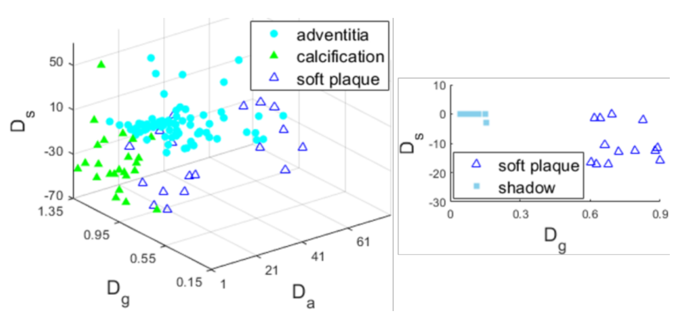
3.3.3. Tissue Recognition
3.3.4. LIB Extraction
3.4. Level Set Evolution
4. Experiments and Results
4.1. Materials and Evaluation Measures
4.2. Self-Evaluation
4.3. Comparative Evaluation
5. Discussion
6. Conclusions
Author Contributions
Funding
Conflicts of Interest
Nomenclature
| IVUS | Intravascular ultrasound |
| LIB | The lumen-intima border |
| MAB | The media-adventitia border |
| Probability density function | |
| SFCME | Superpixel-wise fuzzy clustering modified by edges |
| ROI | Region of interest |
| LSE | Level set evolution |
| FCM | Fuzzy c-means |
| SLIC | Simple linear iterative clustering |
| DRLSE | Distance regularized level set evolution |
| JM | Jaccard measure |
| PAD | Percentage of area difference |
| HD | Hausdorff distance |
| SNR | Signal-to-noise ratio |
References
- Frostegard, J. SLE, atherosclerosis and cardiovascular disease. J. Intern. Med. 2005, 257, 485–495. [Google Scholar] [CrossRef] [PubMed]
- Katouzian, A.; Angelini, E.D.; Carlier, S.G.; Suri, J.S.; Navab, N.; Laine, A.F. A state-of-the-art review on segmentation algorithms in intravascular ultrasound (IVUS) images. IEEE Trans. Inf. Technol. Biomed. 2012, 16, 823–834. [Google Scholar] [CrossRef] [PubMed]
- Wang, Y.; Peng, W.; Qiu, C.; Jiang, J.; Xia, S. Fractional-order Darwinian PSO-based feature selection for media-adventitia border detection in intravascular ultrasound images. Ultrasonics 2019, 92, 1–7. [Google Scholar] [CrossRef] [PubMed]
- Zakeri, F.S.; Setarehdan, S.K.; Norouzi, S. Automatic media-adventitia IVUS image segmentation based on sparse representation framework and dynamic directional active contour model. Comput. Biol. Med. 2017, 89, 561–572. [Google Scholar] [CrossRef] [PubMed]
- Chen, F.; Ma, R.; Liu, J.; Zhu, M.; Liao, H. Lumen and media-adventitia border detection in IVUS images using texture enhanced deformable model. Comput. Med. Imaging Graph. 2018, 66, 1–13. [Google Scholar] [CrossRef]
- Lo Vercio, L.; del Fresno, M.; Larrabide, I. Lumen-intima and media-adventitia segmentation in IVUS images using supervised classifications of arterial layers and morphological structures. Comput. Methods Programs Biomed. 2019, 177, 113–121. [Google Scholar] [CrossRef] [PubMed]
- Lo Vercio, L.; Orlando, J.I.; del Fresno, M.; Larrabide, I. Assessment of image features for vessel wall segmentation in intravascular ultrasound images. Int. J. Comput. Assist. Radiol. Surg. 2016, 11, 1397–1407. [Google Scholar] [CrossRef] [PubMed]
- Su, S.; Hu, Z.; Lin, Q.; Hau, W.K.; Gao, Z.; Zhang, H. An artificial neural network method for lumen and media-adventitia border detection in IVUS. Comput. Med. Imaging Graph. 2017, 57, 29–39. [Google Scholar] [CrossRef]
- Unal, G.; Bucher, S.; Carlier, S.; Slabaugh, G.; Fang, T.; Tanaka, K. Shape-driven segmentation of the arterial wall in intravascular ultrasound images. IEEE Trans. Inf. Technol. Biomed. 2008, 12, 335–347. [Google Scholar] [CrossRef]
- Plissiti, M.E.; Fotiadis, D.I.; Michalis, L.K.; Bozios, G.E. An automated method for lumen and media-adventitia border detection in a sequence of IVUS frames. IEEE Trans. Inf. Technol. Biomed. 2004, 8, 131–141. [Google Scholar] [CrossRef]
- Hammouche, A.; Cloutier, G.; Tardif, J.C.; Meunier, J. Space curve approach for IVUS image segmentation. In Proceedings of the IEEE Life Sciences Conference, Montreal, QC, Canada, 28–30 October 2018; pp. 37–40. [Google Scholar]
- Hammouche, A.; Cloutier, G.; Tardif, J.C.; Hammouche, K.; Meunier, J. Automatic IVUS lumen segmentation using a 3D adaptive helix model. Comput. Biol. Med. 2019, 107, 58–72. [Google Scholar] [CrossRef] [PubMed]
- Cardinal, M.H.R.; Meunier, J.; Soulez, G.; Maurice, R.L.; Therasse, É.; Cloutier, G.; Soulez, G.; Cloutier, G. Intravascular ultrasound image segmentation: A three-dimensional fast-marching method based on gray level distributions. IEEE Trans. Med. Imaging 2006, 25, 590–601. [Google Scholar] [CrossRef] [PubMed]
- Cardinal, M.H.R.; Soulez, G.; Tardif, J.C.; Meunier, J.; Cloutier, G. Fast-marching segmentation of three-dimensional intravascular ultrasound images: A pre- and post-intervention study. Med. Phys. 2010, 37, 3633–3647. [Google Scholar] [CrossRef] [PubMed]
- Destrempes, F.; Cardinal, M.H.R.; Allard, L.; Tardif, J.C.; Cloutier, G. Segmentation method of intravascular ultrasound images of human coronary arteries. Comput. Med. Imaging Graph. 2014, 38, 91–103. [Google Scholar] [CrossRef]
- Yan, J.; Lv, D.; Cui, Y. A novel segmentation approach for intravascular ultrasound images. J. Med. Biol. Eng. 2017, 37, 386–394. [Google Scholar] [CrossRef]
- Sun, S.; Sonka, M.; Beichel, R.R. Graph-based IVUS segmentation with efficient computer-aided refinement. IEEE Trans. Med. Imaging 2013, 32, 1536–1549. [Google Scholar]
- Essa, E.; Xie, X. Automatic segmentation of cross-sectional coronary arterial images. Comput. Vis. Image Underst. 2017, 165, 97–110. [Google Scholar] [CrossRef]
- Balocco, S.; Gatta, C.; Ciompi, F.; Pujol, O.; Carrillo, X.; Mauri, J.; Radeva, P. Combining growcut and temporal correlation for IVUS lumen segmentation. Lect. Notes Comput. Sci. 2011, 556–563. [Google Scholar] [CrossRef]
- Kermani, A.; Ayatollahi, A. A new nonparametric statistical approach to detect lumen and media-adventitia borders in intravascular ultrasound frames. Comput. Biol. Med. 2019, 104, 10–28. [Google Scholar] [CrossRef] [PubMed]
- Yang, J.; Tong, L.; Faraji, M.; Basu, A. IVUS-Net: An intravascular ultrasound segmentation network. Int. Conf. Smart Multimed. 2018, 367–377. [Google Scholar] [CrossRef]
- Yang, J.; Faraji, M.; Basu, A. Robust segmentation of arterial walls in intravascular ultrasound images using dual path U-Net. Ultrasonics 2019, 96, 24–33. [Google Scholar] [CrossRef] [PubMed]
- Bezdek, J.C. Pattern Recognition with Fuzzy Objective Function Algorithms; Plenum: New York, NY, USA, 1981. [Google Scholar]
- Ahmed, M.N.; Yamany, S.M.; Mohamed, N.; Farag, A.A.; Moriarty, T. A modified fuzzy c-means algorithm for MRI bias field estimation and adaptive segmentation. IEEE Trans. Med. Imaging 2002, 21, 193–199. [Google Scholar] [CrossRef] [PubMed]
- Chen, S.; Zhang, D. Robust image segmentation using FCM with spatial constraints based on new kernel-induced distance measure. IEEE Trans. Syst. Man Cybern. 2004, 34, 1907–1916. [Google Scholar] [CrossRef]
- Chuang, K.S.; Tzeng, H.L.; Chen, S.; Wu, J.; Chen, T.J. Fuzzy c-means clustering with spatial information for image segmentation. Comput. Med. Imaging Graph. 2006, 30, 9–15. [Google Scholar] [CrossRef] [PubMed]
- Krinidis, S.; Chatzis, V. A robust fuzzy local information c-means clustering algorithm. IEEE Trans. Image Process. 2010, 19, 1328–1337. [Google Scholar] [CrossRef]
- Liu, G.; Zhang, Y.; Wang, A. Incorporating adaptive local information into fuzzy clustering for image segmentation. IEEE Trans. Image Process. 2015, 24, 3990–4000. [Google Scholar]
- Lei, T.; Jia, X.; Zhang, Y.; Liu, S.; Meng, H.; Nandi, A.K. Superpixel-based fast fuzzy c-means clustering for color image segmentation. IEEE Trans. Fuzzy Syst. 2019, 27, 1753–1766. [Google Scholar] [CrossRef]
- Achanta, R.; Shaji, A.; Smith, K.; Lucchi, A.; Fua, P.; Süsstrunk, S. SLIC superpixels compared to state-of-the-art superpixel methods. IEEE Trans. Pattern Anal. Mach. Intell. 2012, 34, 2274–2281. [Google Scholar] [CrossRef]
- Caselles, V.; Kimmel, R.; Sapiro, G. Geodesic active contours. Int. J. Comput. Vis. 1997, 22, 61–79. [Google Scholar] [CrossRef]
- Li, C.; Xu, C.; Gui, C.; Fox, M.D. Distance regularized level set evolution and its application to image segmentation. IEEE Trans. Image Process. 2010, 19, 3243–3254. [Google Scholar]
- Chan, T.F.; Vese, L.A. Active contours without edges. IEEE Trans. Image Process. 2001, 10, 266–277. [Google Scholar] [CrossRef] [PubMed]
- Li, C.; Kao, C.Y.; Gore, J.C.; Ding, Z. Minimization of region-scalable fitting energy for image segmentation. IEEE Trans. Image Process. 2008, 17, 1940–1949. [Google Scholar] [PubMed]
- Ali, H.; Rada, L.; Badshah, N. Image segmentation for intensity inhomogeneity in presence of high noise. IEEE Trans. Image Process. 2018, 27, 3729–3738. [Google Scholar] [CrossRef] [PubMed]
- Zuiderveld, K. Contrast limited adaptive histograph equalization. In Graphic Gems IV; Academic Press Professional: San Diego, CA, USA, 1994; pp. 474–485. [Google Scholar]
- Kumar, K.; Li, J.P.; Khan, S.A.; Mustafa, N.; Shaikh, R.A.; Khan, A. Image edge detection scheme using wavelet transform. In Proceedings of the International Computer Conference on Wavelet Actiev Media Technology and Information Processing, Chengdu, China, 19–21 December 2014; pp. 261–265. [Google Scholar]
- Zhang, Q.; Wang, Y.; Wang, W.; Ma, J.; Qian, J.; Ge, J. Automatic segmentation of calcifications in intravascular ultrasound images using snakes and the contourlet transform. Ultrasound Med. Biol. 2010, 36, 111–129. [Google Scholar] [CrossRef] [PubMed]
- Balocco, S.; Gatta, C.; Ciompi, F.; Wahle, A.; Radeva, P.; Carlier, S.; Unal, G.; Sanidas, E.; Mauri, J.; Carillo, X.; et al. Standardized evaluation methodology and reference database for evaluating IVUS image segmentation. Comput. Med. Imaging Graph. 2014, 38, 70–90. [Google Scholar] [CrossRef] [Green Version]


| Step (1) | MAB = rectangular bounding border of ROIs in ; Vessel center = the image center (Figure 5a); = the largest ROI in ; ; Calcification recognition belongs to the adventitia; MAB update (Figure 5b); If , go to Step (3); else, go to Step (2). |
| Step (2) | = the largest ROI in ; ; Processing for guidewire artifact; Execute calcification, adventitia, soft plaque, and lumen recognition sequentially; MAB update (Figure 5c). |
| Step (3) | A circle is fitted to the MAB; Vessel center = the center of the fitted circle (Figure 5c); MAB update (Figure 5d); If , go to Step (5); else, go to Step (4). |
| Step (4) | = the largest ROI in ; ; ; Processing for guidewire artifact; If , go to Step (7); Else, execute adventitia, soft plaque, and lumen recognition sequentially; MAB update (Figure 5e–g); If , go to Step (5); else, go to Step (4). |
| Step (5) | ROIs outside MAB are deleted in . |
| Step (6) | = the largest ROI in ; ; ; Processing for guidewire artifact; If , go to Step (7); Else, execute soft plaque and lumen recognition sequentially; Go to Step (6). |
| Step (7) | ROIs outside MAB and the ROI containing are deleted in , the result is denoted by (Figure 5h). |
| Step (8) | = the largest ROI in ; ; ; If , go to Step (9); Else, execute shadow and soft plaque recognition sequentially (Figure 5i); Go to Step (8). |
| Step (9) | LIB extraction (Figure 5j). |
| Symbol | Definition | Reference | Value | |
|---|---|---|---|---|
| Dataset I | Dataset II | |||
| The radius of the catheter zone | Equation (4) | 43 pixels | ||
| The thickness of the shadow left by a calcification | Figure 4 | 23 pixels | ||
| The distance threshold for guidewire artifact processing | Section 3.3.1 | 3 pixels | ||
| The angle threshold | Section 3.3.2 | 281° | ||
| The area threshold | Section 3.3.2 | |||
| Thresholds for the anatomical indicator | Section 3.3.3 | 12.4 | 18.8 | |
| 9.4 | 12.4 | |||
| 11.5 | 17.8 | |||
| 38 | ||||
| The threshold for the spatial indicator | −20 | |||
| Thresholds for the grayscale indicator | 0.73 | 0.81 | ||
| 0.61 | 0.78 | |||
| 0.55 | ||||
| 0.16 | ||||
| Parameters controlling level set evolution | Equation (17) | 0.2 | ||
| 1 | ||||
| −0.03 (MAB)/0.03 (LIB) | ||||
| Experiment 1 | Experiment 2 | Experiment 3 | SFCME-LSE on Images with Different SNR | ||||||||
|---|---|---|---|---|---|---|---|---|---|---|---|
| 70 dB | 50 dB | 30 dB | 27 dB | 25 dB | 23 dB | 20 dB | |||||
| MAB | JM | 0.87 (0.06) | 0.89 (0.07) | 0.91 (0.07) | 0.87 (0.13) | 0.87 (0.12) | 0.87 (0.12) | 0.87 (0.12) | 0.84 (0.16) | 0.79 (0.25) | 0.66 (0.33) |
| HD | 0.90 (0.49) | 0.65 (0.44) | 0.65 (0.43) | 0.89 (0.80) | 0.88 (0.79) | 0.87 (0.81) | 0.89 (0.81) | 1.02 (1.05) | 1.17 (1.17) | 0.62 (0.37) | |
| PAD | 0.10 (0.09) | 0.09 (0.10) | 0.07 (0.09) | 0.11 (0.17) | 0.10 (0.14) | 0.12 (0.21) | 0.14 (0.26) | 1.19 (1.41) | 1.20 (1.29) | 0.33 (0.35) | |
| LIB | JM | 0.73 (0.10) | 0.80 (0.09) | 0.81 (0.09) | 0.80 (0.10) | 0.80 (0.09) | 0.80 (0.10) | 0.78 (0.12) | 0.77 (0.13) | 0.75 (0.15) | 0.67 (0.21) |
| HD | 1.41 (0.59) | 0.88 (0.57) | 0.86 (0.57) | 0.95 (0.61) | 0.97 (0.60) | 0.96 (0.58) | 1.06 (0.64) | 1.07 (0.68) | 1.11 (1.65) | 1.33 (0.76) | |
| PAD | 0.32 (0.23) | 0.15 (0.15) | 0.14 (0.13) | 0.15 (0.13) | 0.16 (0.14) | 0.18 (0.19) | 0.25 (0.47) | 1.27 (1.52) | 1.25 (1.23) | 0.33 (0.26) | |
| MAB | LIB | Time(s)/Frame | Hardware Used | |||||
|---|---|---|---|---|---|---|---|---|
| JM | HD | PAD | JM | HD | PAD | |||
| SFCME-LSE | 0.83 (0.10) | 1.20 (0.66) | 0.12 (0.11) | 0.78 (0.10) | 1.18 (0.70) | 0.16 (0.15) | 11.09 | Xeon 3.5 GHz |
| Kermani et al. [20] | 0.75 (0.13) | 1.32 (0.99) | 0.12 (0.12) | 0.77 (0.13) | 1.46 (1.23) | 0.16 (0.15) | --- | --- |
| Wang et al. [3] | 0.83 (0.09) | 1.27 (0.67) | 0.12 (0.13) | --- | --- | --- | 272.92 | Core 4, 2.67 GHz |
| Essa et al. [18] | 0.84 (0.10) | 1.22 (0.72) | 0.13 (0.15) | --- | --- | --- | --- | --- |
| Balocco et al. [19] | --- | --- | --- | 0.72 (0.12) | 1.70 (1.09) | 0.22 (0.14) | 13 | Core 2 Duo, 2.13 GHz |
| Intra-observer | 0.91 (0.07) | 0.85 (0.60) | 0.06 (0.07) | 0.92 (0.06) | 0.67 (0.52) | 0.05 (0.06) | ||
| Inter-observer | 0.87 (0.11) | 1.14 (1.00) | 0.11 (0.14) | 0.86 (0.10) | 1.04 (0.95) | 0.10 (0.10) | ||
© 2019 by the authors. Licensee MDPI, Basel, Switzerland. This article is an open access article distributed under the terms and conditions of the Creative Commons Attribution (CC BY) license (http://creativecommons.org/licenses/by/4.0/).
Share and Cite
Xia, M.; Yan, W.; Huang, Y.; Guo, Y.; Zhou, G.; Wang, Y. IVUS Image Segmentation Using Superpixel-Wise Fuzzy Clustering and Level Set Evolution. Appl. Sci. 2019, 9, 4967. https://doi.org/10.3390/app9224967
Xia M, Yan W, Huang Y, Guo Y, Zhou G, Wang Y. IVUS Image Segmentation Using Superpixel-Wise Fuzzy Clustering and Level Set Evolution. Applied Sciences. 2019; 9(22):4967. https://doi.org/10.3390/app9224967
Chicago/Turabian StyleXia, Menghua, Wenjun Yan, Yi Huang, Yi Guo, Guohui Zhou, and Yuanyuan Wang. 2019. "IVUS Image Segmentation Using Superpixel-Wise Fuzzy Clustering and Level Set Evolution" Applied Sciences 9, no. 22: 4967. https://doi.org/10.3390/app9224967
APA StyleXia, M., Yan, W., Huang, Y., Guo, Y., Zhou, G., & Wang, Y. (2019). IVUS Image Segmentation Using Superpixel-Wise Fuzzy Clustering and Level Set Evolution. Applied Sciences, 9(22), 4967. https://doi.org/10.3390/app9224967