
Brain Image Segmentation in Recent Years: A Narrative Review


Abstract
:1. Introduction
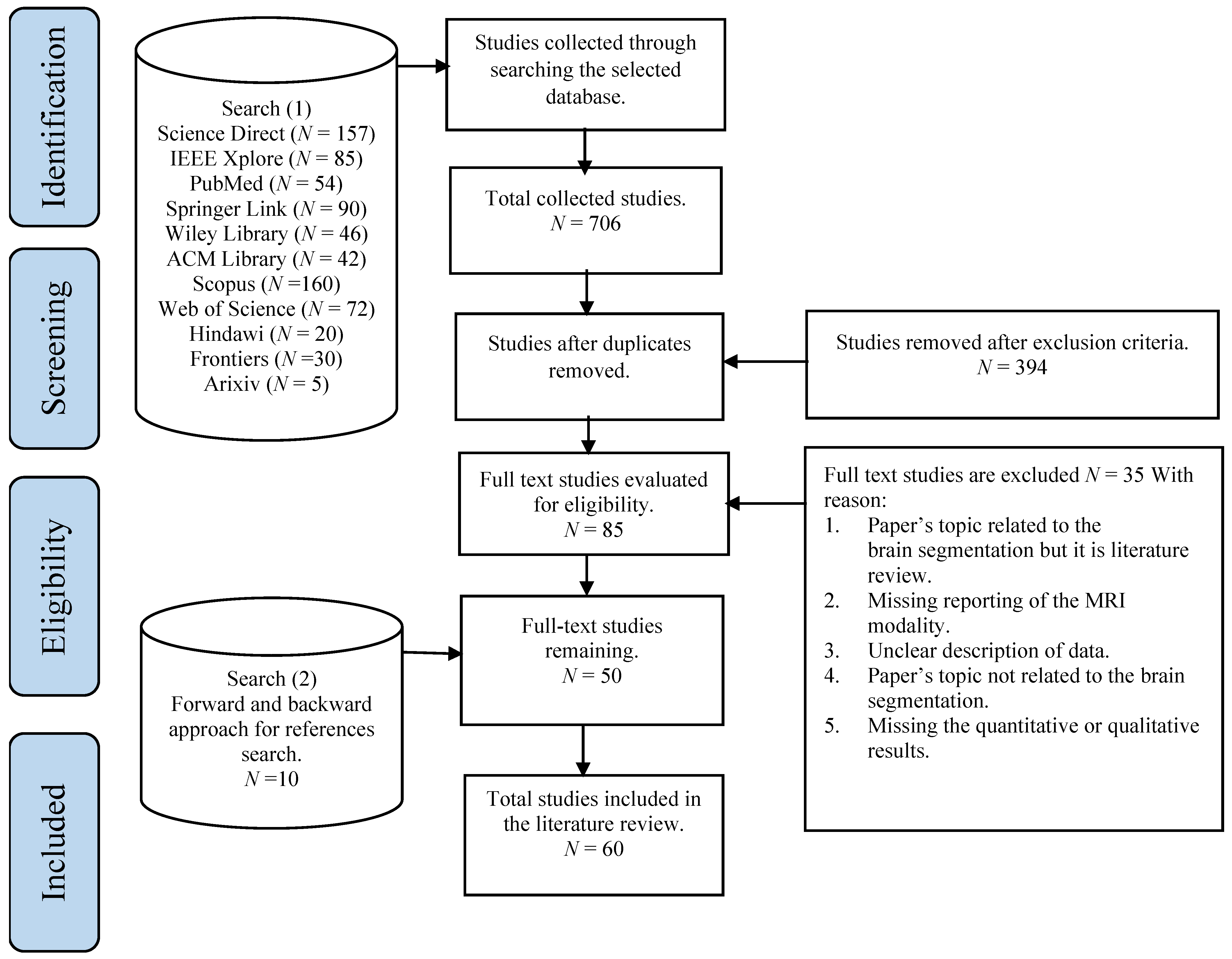
2. Search Strategy and Selection Criteria


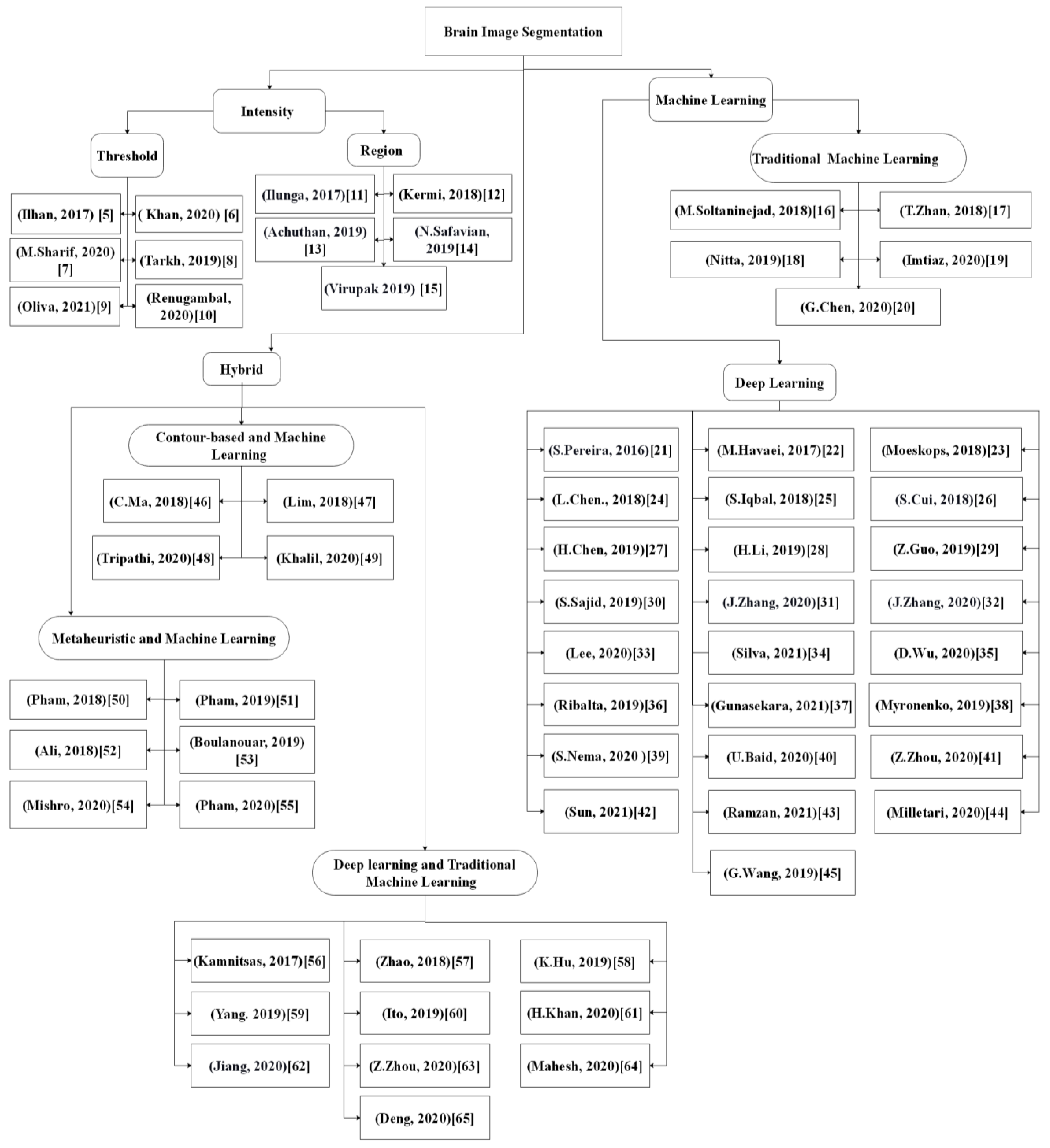
3. Brain Segmentation Approaches
3.1. Intensity-Based Approaches
3.1.1. Thresholding
3.1.2. Region-Based
3.2. Machine Learning
3.2.1. Traditional Machine Learning
3.2.2. Deep Learning
Deep Learning-Based Methods Using 2D Images
Deep Learning-Based Methods Using 3D Images
Deep Learning-Based Methods Using 2.5D Images
3.3. Hybrid Segmentation Approaches
3.3.1. Contour-Based and Machine Learning
3.3.2. Metaheuristic and Machine Learning
3.3.3. Deep Learning and Traditional Machine Learning
4. Discussion
4.1. Main Challenges in Segmenting Brain Structures
- Variation in brain tumor shapes: Brain tumors may occur anywhere in the brain tissue and could assume any shape and intensity. This poses difficulty in applying a model based on a shape without prior knowledge or a statistical model to estimate the tumor with a small variance. Besides, the tumor mass affects the arrangement of the surrounding normal tissues, which increases the intensity due to overlap between tumor regions and edema with healthy tissue.
- Intensity inhomogeneity: This is due to the intensity of non-homogeneity of homogeneous tissues during contrast injection and the variations of spatial intensity over each dimension.
- Bias field: The bias field is another challenge faced during the process of brain segmentation in MR images, which is caused by the defects in the acquisition sequences or radiofrequency coil imperfections. The various biases associated with MR images include shading, noise, artifacts, and partial volume effects.
- Non-standardized intensity: The intensity of MR modalities depends on the magnetic fields and radio wave parameters, which are, in turn, influenced by the MR system hardware requirements.
- Data scarcity: Data scarcity is the main weakness of supervised segmentation methods of medical images that leads to overfitting. This implies the model has a good result on the training data but fails to perform well on new data. Mostly, training labels are not available for brain medical image analysis as it requires specialists in this field to label MR images manually, which is a time-consuming process, subjective, and often vulnerable to error.
- Data imbalance: Imbalanced MR image training datasets are one of the main challenges in supervised-based segmentation, especially in the field of brain tumor segmentation or in lesions of the white matter. This is due to the fact that the healthy brain region is greater in region size than the abnormal region. In this case, the training model with imbalanced training datasets often results in an unreliable segmentation biased towards the dominant class with a larger region. For instance, in multimodal MR images, the region of normal brain tissues is larger in size than the abnormal regions that include brain intra-tumor regions. Generally, background and normal brain tissue regions occupy 98.46% of the whole image pixels, while approximately 1.54% of image pixels only belong to the tumor sub-regions. As a solution to tackle this data imbalance issue, several researchers have investigated the data resampling technique. Recently, GAN has been used to synthesize surrogates for the training dataset. This approach provides oversampling of the training dataset with synthetic samples [66,67]. In this work, GAN incorporates structural information of the original dataset to mitigate training data imbalance and scarcity issues. However, these approaches may add redundant data or remove some important details from the original sample. Besides that, a patch-wise sampling approach by Kamnitsas et al. [56] has been adapted to alleviate the data imbalance issue by randomly selecting patches of the normal and abnormal regions from the training datasets. However, this approach suffers the difficulty of determining the right patch size to generate the relevant training data samplings.
4.2. Trends in the Segmentation Methods
4.3. Types of Brain Structure Segmentation
4.4. Computation Time of Brain Structure Segmentation
5. Conclusions
Author Contributions
Funding
Institutional Review Board Statement
Acknowledgments
Conflicts of Interest
References
- Siegel, R.L.; Miller, K.D.; Jemal, A. Cancer statistics 2020. CA Cancer J. Clin. 2020, 70, 7–30. [Google Scholar] [CrossRef] [PubMed]
- Bauer, S.; Wiest, R.; Nolte, L.-P.; Reyes, M. A survey of MRI-based medical image analysis for brain tumor studies. Phys. Med. Biol. 2013, 58, R97–R129. [Google Scholar] [CrossRef] [PubMed] [Green Version]
- Webster, J.; Watson, R.T. Analyzing the past to prepare for the future: Writing a literature review. MIS Q. 2002, 26, 13–23. [Google Scholar]
- Moher, D.; Liberati, A.; Tetzlaff, J.; Altman, D.G.; PRISMA Group. Preferred reporting items for systematic reviews and meta-analyses: The PRISMA statement. Int. J. Surg. 2010, 8, 336–341. [Google Scholar] [CrossRef] [Green Version]
- Ilhan, A. Brain tumor segmentation based on a new threshold approach. Procedia Comput. Sci. 2017, 120, 580–587. [Google Scholar] [CrossRef]
- Khan, S.R.; Sikandar, M.; Almogren, A.; Din, I.U.; Guerrieri, A.; Fortino, G. IoMT-based computational approach for detecting brain tumor. Future Gener. Comput. Syst. 2020, 109, 360–367. [Google Scholar] [CrossRef]
- Sharif, M.; Amin, J.; Raza, M.; Yasmin, M.; Satapathy, S.C. An integrated design of particle swarm optimization (PSO) with fusion of features for detection of brain tumor. Pattern Recognit. Lett. 2020, 129, 150–157. [Google Scholar] [CrossRef]
- Tarkhaneh, O.; Shen, H. An adaptive differential evolution algorithm to optimal multi-level thresholding for MRI brain image segmentation. Expert Syst. Appl. 2019, 138, 112820. [Google Scholar] [CrossRef]
- Aranguren, I.; Valdivia, A.; Morales-Castañeda, B.; Oliva, D.; Elaziz, M.A.; Perez-Cisneros, M. Improving the segmentation of magnetic resonance brain images using the LSHADE optimization algorithm. Biomed. Signal Process. Control 2021, 64, 102259. [Google Scholar] [CrossRef]
- Renugambal, A.; Selva Bhuvaneswari, K. Image segmentation of brain MR images using Otsu’s based hybrid WCMFO algorithm. Comput. Mater. Contin. 2020, 64, 681–700. [Google Scholar] [CrossRef]
- Mbuyamba, E.I.; Avina-Cervantes, J.G.; Garcia–Perez, A.; Romero–Troncoso, R.D.J.; Aguirre-Ramos, H.; Cruz–Aceves, I.; Chalopin, C. Localized active contour model with background intensity compensation applied on automatic MR brain tumor segmentation. Neurocomputing 2017, 220, 84–97. [Google Scholar] [CrossRef]
- Kermi, A.; Andjouh, K.; Zidane, F. Fully automated brain tumour segmentation system in 3D-MRI using symmetry analysis of brain and level sets. IET Image Process. 2018, 12, 1964–1971. [Google Scholar] [CrossRef]
- Achuthan, A.; Rajeswari, M. Segmentation of hippocampus guided by assembled and weighted coherent point drift registration. J. King Saud Univ. Comput. Inf. Sci. 2019. [Google Scholar] [CrossRef]
- Safavian, N.; Batouli, S.A.H.; Oghabian, M.A. An automatic level set method for hippocampus segmentation in MR images. Comput. Methods Biomech. Biomed. Eng. Imaging Vis. 2019, 8, 400–410. [Google Scholar] [CrossRef]
- Amarapur, B. Cognition-based MRI brain tumor segmentation technique using modified level set method. Cogn. Technol. Work 2018, 21, 357–369. [Google Scholar] [CrossRef]
- Soltaninejad, M.; Yang, G.; Lambrou, T.; Allinson, N.; Jones, T.L.; Barrick, T.R.; Howe, F.A.; Ye, X. Supervised learning based multimodal MRI brain tumour segmentation using texture features from supervoxels. Comput. Methods Programs Biomed. 2018, 157, 69–84. [Google Scholar] [CrossRef] [PubMed]
- Zhan, T.; Shen, F.; Hong, X.; Wang, X.; Chen, Y.; Lu, Z.; Yang, G. A glioma segmentation method using cotraining and superpixel-based spatial and clinical constraints. IEEE Access 2018, 6, 57113–57122. [Google Scholar] [CrossRef]
- Nitta, G.R.; Sravani, T.; Nitta, S.; Muthu, B. Dominant gray level-based K-means algorithm for MRI images. Health Technol. 2019, 10, 281–287. [Google Scholar] [CrossRef]
- Imtiaz, T.; Rifat, S.; Fattah, S.A.; Wahid, K.A. Automated brain tumor segmentation based on multi-planar superpixel level features extracted from 3D MR images. IEEE Access 2019, 8, 25335–25349. [Google Scholar] [CrossRef]
- Chen, G.; Li, Q.; Shi, F.; Rekik, I.; Pan, Z. RFDCR: Automated brain lesion segmentation using cascaded random forests with dense conditional random fields. NeuroImage 2020, 211, 116620. [Google Scholar] [CrossRef] [PubMed]
- Pereira, S.; Pinto, J.A.; Alves, V.; Silva, C. Brain tumor segmentation using convolutional neural networks in MRI images. IEEE Trans. Med. Imaging 2016, 35, 1240–1251. [Google Scholar] [CrossRef] [PubMed]
- Havaei, M.; Davy, A.; Warde-Farley, D.; Biard, A.; Courville, A.; Bengio, Y.; Pal, C.; Jodoin, P.-M.; Larochelle, H. Brain tumor segmentation with deep neural networks. Med. Image Anal. 2017, 35, 18–31. [Google Scholar] [CrossRef] [Green Version]
- Moeskops, P.; de Bresser, J.; Kuijf, H.; Mendrik, A.M.; Biessels, G.J.; Pluim, J.P.; Išgum, I. Evaluation of a deep learning approach for the segmentation of brain tissues and white matter hyperintensities of presumed vascular origin in MRI. NeuroImage Clin. 2018, 17, 251–262. [Google Scholar] [CrossRef]
- Chen, L.; Bentley, P.; Mori, K.; Misawa, K.; Fujiwara, M.; Rueckert, D. DRINet for medical image segmentation. IEEE Trans. Med. Imaging 2018, 37, 2453–2462. [Google Scholar] [CrossRef]
- Iqbal, S.; Ghani, M.U.; Saba, T.; Rehman, A. Brain tumor segmentation in multi-spectral MRI using convolutional neural networks (CNN). Microsc. Res. Tech. 2018, 81, 419–427. [Google Scholar] [CrossRef]
- Cui, S.; Mao, L.; Jiang, J.; Liu, C.; Xiong, S. Automatic semantic segmentation of brain gliomas from MRI Images using a deep cascaded neural network. J. Health Eng. 2018, 2018, 1–14. [Google Scholar] [CrossRef]
- Chen, H.; Qin, Z.; Ding, Y.; Tian, L.; Qin, Z. Brain tumor segmentation with deep convolutional symmetric neural network. Neurocomputing 2020, 392, 305–313. [Google Scholar] [CrossRef]
- Li, H.; Li, A.; Wang, M. A novel end-to-end brain tumor segmentation method using improved fully convolutional networks. Comput. Biol. Med. 2019, 108, 150–160. [Google Scholar] [CrossRef] [PubMed]
- Guo, Z.; Li, X.; Huang, H.; Guo, N.; Li, Q. Deep learning-based image segmentation on multimodal medical imaging. IEEE Trans. Radiat. Plasma Med. Sci. 2019, 3, 162–169. [Google Scholar] [CrossRef]
- Sajid, S.; Hussain, S.; Sarwar, A. Brain tumor detection and segmentation in MR images using deep learning. Arab. J. Sci. Eng. 2019, 44, 9249–9261. [Google Scholar] [CrossRef]
- Zhang, J.; Jiang, Z.; Dong, J.; Hou, Y.; Liu, B. Attention gate ResU-Net for automatic MRI brain tumor segmentation. IEEE Access 2020, 8, 58533–58545. [Google Scholar] [CrossRef]
- Zhang, J.; Lv, X.; Zhang, H.; Liu, B. AResU-Net: Attention residual U-Net for brain tumor segmentation. Symmetry 2020, 12, 721. [Google Scholar] [CrossRef]
- Lee, B.; Yamanakkanavar, N.; Choi, J.Y. Automatic segmentation of brain MRI using a novel patch-wise U-net deep architecture. PLoS ONE 2020, 15, e0236493. [Google Scholar] [CrossRef]
- Silva, C.A.; Pinto, A.; Pereira, S.; Lopes, A. Multi-stage deep layer aggregation for brain tumor segmentation. Lect. Notes Comput. Sci. 2021, 179–188. [Google Scholar] [CrossRef]
- Wu, D.; Ding, Y.; Zhang, M.; Yang, Q.; Qin, Z. Multi-features refinement and aggregation for medical brain segmentation. IEEE Access 2020, 8, 57483–57496. [Google Scholar] [CrossRef]
- Lorenzo, P.R.; Nalepa, J.; Bobek-Billewicz, B.; Wawrzyniak, P.; Mrukwa, G.; Kawulok, M.; Ulrych, P.; Hayball, M.P. Segmenting brain tumors from FLAIR MRI using fully convolutional neural networks. Comput. Methods Programs Biomed. 2019, 176, 135–148. [Google Scholar] [CrossRef] [PubMed]
- Gunasekara, S.R.; Kaldera, H.N.T.K.; Dissanayake, M.B. A systematic approach for MRI brain tumor localization and segmentation using deep learning and active contouring. J. Health Eng. 2021, 2021, 1–13. [Google Scholar] [CrossRef]
- Myronenko, A. 3D MRI brain tumor segmentation using autoencoder regularization. In International MICCAI Brainlesion Workshop; Springer: Cham, Switzerland, 2019; pp. 311–320. [Google Scholar] [CrossRef] [Green Version]
- Nema, S.; Dudhane, A.; Murala, S.; Naidu, S. RescueNet: An unpaired GAN for brain tumor segmentation. Biomed. Signal Process. Control 2020, 55, 101641. [Google Scholar] [CrossRef]
- Baid, U.; Talbar, S.; Rane, S.; Gupta, S.; Thakur, M.H.; Moiyadi, A.; Sable, N.; Akolkar, M.; Mahajan, A. A novel approach for fully automatic intra-tumor segmentation with 3D U-Net architecture for gliomas. Front. Comput. Neurosci. 2020, 14, 10. [Google Scholar] [CrossRef] [PubMed] [Green Version]
- Zhou, Z.; He, Z.; Shi, M.; Du, J.; Chen, D. 3D dense connectivity network with atrous convolutional feature pyramid for brain tumor segmentation in magnetic resonance imaging of human heads. Comput. Biol. Med. 2020, 121, 103766. [Google Scholar] [CrossRef] [PubMed]
- Sun, J.; Peng, Y.; Guo, Y.; Li, D.; Sun, J.; Peng, Y.; Guo, Y.; Li, D. Segmentation of the multimodal brain tumor image used the multi-pathway architecture method based on 3D FCN. Neurocomputing 2021, 423, 34–45. [Google Scholar] [CrossRef]
- Ramzan, F.; Khan, M.U.G.; Iqbal, S.; Saba, T.; Rehman, A. Volumetric segmentation of brain regions from MRI scans using 3D convolutional neural networks. IEEE Access 2020, 8, 103697–103709. [Google Scholar] [CrossRef]
- Milletari, F.; Ahmadi, S.-A.; Kroll, C.; Plate, A.; Rozanski, V.; Maiostre, J.; Levin, J.; Dietrich, O.; Ertl-Wagner, B.; Bötzel, K.; et al. Hough-CNN: Deep learning for segmentation of deep brain regions in MRI and ultrasound. Comput. Vis. Image Underst. 2017, 164, 92–102. [Google Scholar] [CrossRef] [Green Version]
- Wang, G.; Li, W.; Ourselin, S.; Vercauteren, T. Automatic brain tumor segmentation based on cascaded convolutional neural networks with uncertainty estimation. Front. Comput. Neurosci. 2019, 13, 56. [Google Scholar] [CrossRef] [Green Version]
- Ma, C.; Luo, G.; Wang, K. Concatenated and connected random forests with multiscale patch driven active contour model for automated brain tumor segmentation of MR images. IEEE Trans. Med. Imaging 2018, 37, 1943–1954. [Google Scholar] [CrossRef]
- Lim, K.Y.; Mandava, R. A multi-phase semi-automatic approach for multisequence brain tumor image segmentation. Expert Syst. Appl. 2018, 112, 288–300. [Google Scholar] [CrossRef]
- Tripathi, P.; Singh, V.K.; Trivedi, M.C. Brain tumor segmentation in magnetic resonance imaging using OKM approach. Mater. Today Proc. 2021, 37, 1334–1340. [Google Scholar] [CrossRef]
- Khalil, H.A.; Darwish, S.; Ibrahim, Y.M.; Hassan, O.F. 3D-MRI brain tumor detection model using modified version of level set segmentation based on dragonfly algorithm. Symmetry 2020, 12, 1256. [Google Scholar] [CrossRef]
- Pham, T.X.; Siarry, P.; Oulhadj, H. Integrating fuzzy entropy clustering with an improved PSO for MRI brain image segmentation. Appl. Soft Comput. 2018, 65, 230–242. [Google Scholar] [CrossRef]
- Pham, T.X.; Siarry, P.; Oulhadj, H. A multi-objective optimization approach for brain MRI segmentation using fuzzy entropy clustering and region-based active contour methods. Magn. Reson. Imaging 2019, 61, 41–65. [Google Scholar] [CrossRef]
- Ali, H.A.M.; Ahmed, M.A.A.; Hussein, E.M. MRI brain tumour segmentation based on multimodal clustering and level-set method. In Proceedings of the 2018 International Conference on Computer, Control, Electrical, and Electronics Engineering (ICCCEEE), Khartoum, Sudan, 12–14 August 2018. [Google Scholar] [CrossRef]
- Boulanouar, S.; Lamiche, C. A new hybrid image segmentation method based on fuzzy c-mean and modified bat algorithm. Int. J. Comput. Digit. Syst. 2020, 9, 677–687. [Google Scholar] [CrossRef]
- Mishro, P.K.; Agrawal, S.; Panda, R.; Abraham, A. A novel type-2 fuzzy c-means clustering for brain MR image segmentation. IEEE Trans. Cybern. 2020, 1–12. [Google Scholar] [CrossRef]
- Pham, T.X.; Siarry, P.; Oulhadj, H. Segmentation of MR brain images through hidden Markov random field and hybrid metaheuristic algorithm. IEEE Trans. Image Process. 2020, 29, 6507–6522. [Google Scholar] [CrossRef]
- Kamnitsas, K.; Ledig, C.; Newcombe, V.; Simpson, J.P.; Kane, A.D.; Menon, D.K.; Rueckert, D.; Glocker, B. Efficient multi-scale 3D CNN with fully connected CRF for accurate brain lesion segmentation. Med. Image Anal. 2017, 36, 61–78. [Google Scholar] [CrossRef] [PubMed]
- Zhao, X.; Wu, Y.; Song, G.; Li, Z.; Zhang, Y.; Fan, Y. A deep learning model integrating FCNNs and CRFs for brain tumor segmentation. Med. Image Anal. 2018, 43, 98–111. [Google Scholar] [CrossRef] [PubMed]
- Hu, K.; Gan, Q.; Zhang, Y.; Deng, S.; Xiao, F.; Huang, W.; Cao, C.; Gao, X. Brain tumor segmentation using multi-cascaded convolutional neural networks and conditional random field. IEEE Access 2019, 7, 92615–92629. [Google Scholar] [CrossRef]
- Yang, T.; Song, J.; Li, L. A deep learning model integrating SK-TPCNN and random forests for brain tumor segmentation in MRI. Biocybern. Biomed. Eng. 2019, 39, 613–623. [Google Scholar] [CrossRef]
- Ito, R.; Nakae, K.; Hata, J.; Okano, H.; Ishii, S. Semi-supervised deep learning of brain tissue segmentation. Neural Netw. 2019, 116, 25–34. [Google Scholar] [CrossRef]
- Khan, H.; Shah, P.M.; Shah, M.A.; Islam, S.U.; Rodrigues, J. Cascading handcrafted features and convolutional neural network for IoT-enabled brain tumor segmentation. Comput. Commun. 2020, 153, 196–207. [Google Scholar] [CrossRef]
- Jiang, H.; Guo, Y. Multi-class multimodal semantic segmentation with an improved 3D fully convolutional networks. Neurocomputing 2020, 391, 220–226. [Google Scholar] [CrossRef]
- Zhou, Z.; He, Z.; Jia, Y. AFPNet: A 3D fully convolutional neural network with atrous-convolution feature pyramid for brain tumor segmentation via MRI images. Neurocomputing 2020, 402, 235–244. [Google Scholar] [CrossRef]
- Mahesh, K.M.; Renjit, J.A. Multiclassifier for severity-level categorization of glioma tumors using multimodal magnetic resonance imaging brain images. Int. J. Imaging Syst. Technol. 2019, 30, 234–251. [Google Scholar] [CrossRef]
- Deng, W.; Shi, Q.; Wang, M.; Zheng, B.; Ning, N. Deep learning-based HCNN and CRF-RRNN model for brain tumor segmentation. IEEE Access 2020, 8, 26665–26675. [Google Scholar] [CrossRef]
- Douzas, G.; Bacao, F. Effective data generation for imbalanced learning using conditional generative adversarial networks. Expert Syst. Appl. 2018, 91, 464–471. [Google Scholar] [CrossRef]
- Salazar, A.; Vergara, L.; Safont, G. Generative adversarial networks and Markov random fields for oversampling very small training sets. Expert Syst. Appl. 2021, 163, 113819. [Google Scholar] [CrossRef]


| Categories | Ref | Strengths | Limitations |
|---|---|---|---|
| Thresholding | [5,6,7,8,9,10] |
|
|
| Region based | [11,12,13,14,15] |
|
|
| Traditional machine learning | [16,17,18,19,20] |
|
|
| Deep learning | [21,22,23,24,25,26,27,28,29,30,31,32,33,34,35,36,37,38,39,40,41,42,43,44,45] |
|
|
| Categories | Approaches | Strengths | Limitations |
|---|---|---|---|
| Contour-based and machine learning | [46,47,48,49] |
|
|
| Metaheuristic and machine learning | [50,51,52,53,54,55] |
|
|
| Deep learning and clustering or classification | [56,57,58,59,60,61,62,63,64,65] |
|
|
| Approaches Employed | Objectives | Image Modality | Dataset Information | Performance Measure (Accuracy) | Computation Time | Ref |
|---|---|---|---|---|---|---|
| Thresholding | Complete tumor segmentation | MR | Cancer Imaging Archive, 2017 | 95% (Accuracy) | NA | [5] |
| Thresholding-based segmentation PART for grade-wise identification | Complete tumor segmentation Grade-wise classification | MR | Self-collected MRI images | 95% (Precision) | 0.02 s | [6] |
| PSO and thresholding LBP and deep features extraction GA ANN | Grade-wise classification | NA | RIDER BRATS 2018 | 99% (Accuracy) | 24.90334 s | [7] |
| ALDE | Healthy brain tissue segmentation | T2 | Autism Brain Imaging Data Exchange | NA | 1.2202 s | [8] |
| LSHADE with multilevel thresholding | Complete tumor segmentation | FLAIR | BRATS 2015 | 0.9172 (Accuracy) | NA | [9] |
| WCMFO Otsu | Healthy brain tissue segmentation | T2 | Harvard medical images | 55.834 (PSNR) | 2.3496 s | [10] |
| LACM-BIC k-means Hierarchical centroid shape descriptor | Complete tumor segmentation | T2 T1c | BRATS 2012 | 91% (DSC) | 15.8150 s | [11] |
| FBB Geodesic level set methods | Complete tumor segmentation | T2 FLAIR | BRATS 2017 | 89.01% FLAIR 81.59% T2 (True positive rate) | 5 min | [12] |
| Point set registration Level set function | Hippocampus segmentation | T1 | OASIS-MICCAI | 80.50% (DSC) | NA | [13] |
| Prior knowledge-based registration Level set function | Hippocampus segmentation | 3TMR | ADNI-HarP | 84.75% left Hippocampus 73.55% right Hippocampus | NA | [14] |
| Modified level set | Complete tumor segmentation | T1 | BRATS 2015 | 99% (Average accuracy of 5 images) | NA | [15] |
| SLIC algorithm RF Multimodal supervoxel | Tumor and intra-tumor segmentation | T1 FLAIR T1c T2 | BRATS 2012 BRATS 2013 | 0.89 whole tumor 0.80 tumor core (Dice Score) | 2200 ms | [16] |
| Multiple classifiers-based collaborative training Feature extraction along with clinical constraints | Tumor and intra-tumor segmentation | T1 FLAIR T1c T2 | BRATS 2012 BRATS 2013 | 0.88 whole tumor 0.81 tumor core 0.74 enhancing tumor (DSC) | NA | [17] |
| Dominant grey level-based k-means | Healthy brain tissue segmentation | MR | Clinical dataset | Qualitative results | NA | [18] |
| Multimodal supervoxel Feature selection (ERT) classification Planner voting | Complete tumor segmentation | FLAIR T1c T2 | BRATS 2013 | 0.88 (DSC) | 3.5 min prediction time per subject | [19] |
| Cascade RFs Dense CRF | Tumor and intra-tumor segmentation | T1 FLAIR T1c T2 | BRATS 2015 BRATS 2018 ISLES 2015 | 0.86 whole tumor 0.79 tumor core 0.75 enhancing tumor (DSC) | 8 h training 3–4 min inference | [20] |
| Parallel 2D deep CNN architecture | Tumor and intra-tumor segmentation | T1 FLAIR T1c T2 | BRATS 2013 BRATS 2015 | 88% whole 83% core 77% enhancing tumor (DSC) | 8 min testing | [21] |
| Two-CNN pathway architecture Cascade CNN architecture | Tumor and intra-tumor segmentation | T1 FLAIR T1c T2 | BRATS 2013 | 88% whole tumor 79% tumor core 73% enhancing tumor (DSC) | 3 min per epoch training 25 s per slice inference | [22] |
| Multi-scale CNN | WMH | T1 FLAIR T1IR T2 | MRBrainS13 | 0.87 WM, 0.85 cGM, 0.82 BGT, 0.93 CB, 0.92 BS, 0.93 lvCSF, 0.76 pCSF (DSC) | 3–4 min inference | [23] |
| Dense-Res-Inception Net (DRINet) | Intra-tumor segmentation CSF segmentation in CT images Multi-organ segmentation of abdominal CT images | T1 FLAIR T1c T2 CT | BRATS 2017 Two clinical datasets | 83.47% whole tumor 73.21% tumor core 64.98% enhancing tumor 83.42% pancreas 95.96% kidneys 96.57% liver 95.64% spleen (DSC) | 21.37 h training 44.46 s inference | [24] |
| Three modified versions of SEGNET | Tumor and intra-tumor segmentation | T1 FLAIR T1c T2 | BRATS 2015 | 0.87 whole tumor 0.86 tumor core 0.79 enhancing tumor (DSC) | 75 ms inference | [25] |
| Deep cascade neural network | Tumor and intra-tumor segmentation | T1 FLAIR T1c T2 | BRATS 2015 | 89% whole tumor 77% tumor core 77.2% enhancing tumor (DSC) | 1.54 s inference | [26] |
| Combined the symmetric masks in several layers of DCNN | Tumor and intra-tumor segmentation | T1 FLAIR T1c T2 | BRATS 2015 | 85.2% whole tumor 68.1% tumor core 58.1% enhancing tumor (DSC) | 9.7 s inference | [27] |
| Modified cascade 2D U-Net structure | Tumor and intra-tumor segmentation | T1 FLAIR T1c T2 | BRATS 2015 BRATS 2017 | 0.876 whole tumor 0.763 tumor core 0.642 enhancing tumor (DSC) | 10 h training 2 min inference | [28] |
| Deep CNN Cross-modality fusion | Complete tumor segmentation | PET CT T1 T2 | Soft-tissue sarcoma (STS) | 85% fusing at the feature level 85% fusing at the classifier level 84% fusing at the decision-making level | 740 s per epoch training | [29] |
| Combined two- and three-path CNNs Morphological operation | Tumor and intra-tumor segmentation | T1 FLAIR T1c T2 | BRATS 2013 | 0.86 whole tumor 0.86 tumor core 0.88 enhancing tumor (DSC) | 5–7 min inference | [30] |
| Attention gate residual U-Net model | Tumor and intra-tumor segmentation | T1 FLAIR T1c T2 | BRATS 2017 BRATS 2018 BRATS 2019 | 0.872 whole tumor 0. 808 tumor core 0.80 enhancing tumor (DSC) | NA | [31] |
| Attention residual U-Net | Tumor and intra-tumor segmentation | T1 FLAIR T1c T2 | BRATS 2017 BRATS 2018 | 87.6% WT, 81.0 TC, 77.3% ET BRATS 2018 | NA | [32] |
| Patch-wise U-net | Healthy brain tissue segmentation | T1 | OASIS IBSR | 93% in average for CSF, GM, WM OASIS (DSC) | 4 h training and inference | [33] |
| Three-stage cascade FCN | Tumor and intra-tumor segmentation | T1 FLAIR T1c T2 | 0.8858 whole tumor 0.8297 tumor core 0.7900 enhancing tumor (DSC) | NA | [34] | |
| MRANet | Tumor and intra-tumor segmentation | T1 FLAIR T1c T2 | BRATS2015 | 0.78 whole tumor 0. 68 tumor core 0.60 enhancing tumor (DSC) | 2 s inference | [35] |
| FCNN A battery augmentation | Complete tumor segmentation | FLAIR | MAGNETOM Prisma 3T Siemens | 85% average (DSC) | 27 ms inference | [36] |
| R-CNN Chan-Vese level set | Tumor classification and segmentation | T1 | Clinical dataset | 0.92 (DSC) | NA | [37] |
| Encoder-decoder based CNN architecture | Tumor and intra-tumor segmentation | T1 FLAIR T1c T2 | BRATS 2018 | 0.8839 whole tumor 0.8154 tumor core 0.7664 enhancing tumor (DSC) | 2 days training | [38] |
| RescueNet Unpaired GAN-based feature learning Scale-invariant | Tumor and intra-tumor segmentation | T1 FLAIR T1c T2 | BRATS 2015 BRATS 2017 | 94.63% whole tumor 85.6% tumor core 93.54% enhancing tumor (DSC) | 48 h training 60 s Inference | [39] |
| 3D U-Net architecture | Tumor and intra-tumor segmentation | T1 FLAIR T1c T2 | BRATS 2018 Local hospital dataset | 0.85 whole tumor 0.77 tumor core 0.67 enhancing tumor (DSC) | NA | [40] |
| Dense connectivity DCNNs Atrous convolutional feature pyramid | Tumor and intra-tumor segmentation | T1 FLAIR T1c T2 | BRATS 2015 BRATS 2017 BRATS 2018 | 0.8642 whole tumor 0.7738 tumor core 0.7525 enhancing tumor (DSC) | 10.616 s inference | [41] |
| Multi-pathway 3D FCN | Tumor and intra-tumor segmentation | T1 FLAIR T1c T2 | BRATS 2018 BRATS 2019 | 89% whole tumor 78% tumor core 76% enhancing tumor (DSC) | 979 s epoch training 86.1s inference | [42] |
| 3D CNN, Dilated convolution | Healthy brain tissue segmentation | T1 FLAIR T1c T2 | ADNI MRBrain18 MICCAI 2012 | 87.2%, WM, 87.2% GM, 89.6% CSF (DSC) | NA | [43] |
| CNNs test-time augmentation | Tumor and intra-tumor segmentation | T1 FLAIR T1c T2 | BRATS 2017 BRATS 2018 | 87.4% whole tumor 77.5% tumor core 8.3% enhancing tumor (DSC) | NA | [45] |
| RFs (ccRFs) mpAC | Tumor and intra-tumor segmentation | T1 FLAIR T1c T2 | TCGA-GBM TCGA-LGG | 90% whole tumor 80% tumor core 73% enhancing tumor (DSC) | 7 h training 5 m inference | [46] |
| Random walks algorithm Weighted averaging algorithm ITRS | Complete tumor segmentation | T1c T2 | BRATS 2015 | 70% HG 70% LG | 0.72 s inference | [47] |
| Otsu k-means | Tumor and intra-tumor segmentation | T2 FLAIR | BRATS 2013 | 0.8450 enhancing tumor 0.8450 necrotic 0.7834 edema | NA | [48] |
| Dragonfly algorithm k-Means Level set | Whole tumor | T1 FLAIR T1c T2 | BRATS 2017 | 85.67 (Accuracy) | 40–45 s | [49] |
| PSO-KFECSB | Healthy brain tissue segmentation | T1 | IBSR | 90.19% (Jaccard Index) | 300 s inference | [50] |
| MOPSO-KFECSB | Healthy brain tissue segmentation | T1 | Brain web IBSR | 98% CSF 94% GM 96% WM (DSC) 0.9560 (Sensitivity) | 4.76 ± 0.15 s per iteration | [51] |
| Hybrid FCM Particle swarm optimization Level set | Complete tumor segmentation | T1 FLAIR T1c T2 | BRATS 2013 | 93.1% low-grade glioma 89.6% high-grade glioma (DSC) | NA | [52] |
| MFBAFCM | Healthy brain tissue segmentation | T1 | Brainweb | 0.9580 CSF 0.9886 GM 0.9833 WM (DSC) | NA | [53] |
| Type-2 FCM | Healthy brain tissue segmentation | T1 T2 | IBSR | 0.8381 CSF 0.8381 GM 0.8381 WM (DSC) | 9.36 s inference | [54] |
| HMRF Hybridized of CS and PSO | Healthy brain tissue segmentation | T1 | MRBrainS18 | 0.9374 CSF 0.8744 GM 0.9200 WM (DSC) | 900 s inference | [55] |
| CNN CRF | Tumor and intra-tumor segmentation | T1 FLAIR T1 T2 | BRATS 2015 ISLES 2015 | 0.90 whole tumor 0.75 tumor core 0.73 enhancing Tumor (DSC) | NA | [56] |
| FCNNs CRF-RNN | Tumor and intra-tumor segmentation | FLAIR T1c T2 | BRATS 2013 BRATS 2015 BRATS 2016 | 0.84 whole tumor 0.73tumor core 0.62 enhancing tumor (DSC) BRATS 2016 | ~12 d training 2–4 m inference | [57] |
| Multi-cascaded convolution neural network Fully connected CRFs | Tumor and intra-tumor segmentation | FLAIR T1c T2 | BRATS 2013 BRATS 2015 BRATS 2018 | 88.24% whole tumor 74.81% tumor core 0.7178 enhancing tumor (DSC) BRATS 2018 | 3 d training 1.5–3 m per image inference | [58] |
| SK-TPCNN RF classifier Morphological operation | Tumor and intra-tumor segmentation | T1 FLAIR T1c T2 | BRATS 2015 | 0.89 whole tumor 0.80 tumor core 0.87 enhancing Tumor (DSC) | NA | [59] |
| DNN model using expectation maximization (EM) algorithm | Complete tumor segmentation | T1 | IBSR Marmoset | 94% (Mean Dice Coefficient) | ~65 h training 30 s inference | [60] |
| SVM Three path CNN | Intra-tumor segmentation | T1 FLAIR T1c T2 | BRATS 2015 | 81% whole tumor, 76% tumor core, 73% enhancing tumor (DSC) BRATS 2013 | NA | [61] |
| 3D fully CNN based on U-net CRF | Intra-tumor and hippocampus segmentation | T1 FLAIR T1c T2 | BRATS 2017 MNI-HISUB25 | 88.9% whole tumor 81.3% tumor core 73.49% enhancing tumor (DSC) BRATS2015 90.82 CA1-3 86.65 CA4-DG 95.88% whole MNI-HISUB25 | NA | [62] |
| DCNN 3D atrous 3D CRF | Complete tumor segmentation | T1 FLAIR T1c T2 | BRATS 2013 BRATS 2015 BRATS 2018 | 86% whole tumor 73% tumor core 68% enhancing tumor (DSC) BRATS 2013 | 7.21 s inference | [63] |
| PSO FJODCNN | Severity levels of glioma | T1 FLAIR T1c T2 | BRATS 2012 BRATS 2018 | 0.95 (Accuracy) | 5.5 s inference | [64] |
| HCNN CRF-RRNN | Intra-tumor segmentation | T1 FLAIR T1c T2 | BRATS 2013 BRATS 2015 | 96.5 (Precision) | NA | [65] |
Publisher’s Note: MDPI stays neutral with regard to jurisdictional claims in published maps and institutional affiliations. |
© 2021 by the authors. Licensee MDPI, Basel, Switzerland. This article is an open access article distributed under the terms and conditions of the Creative Commons Attribution (CC BY) license (https://creativecommons.org/licenses/by/4.0/).
Share and Cite
Fawzi, A.; Achuthan, A.; Belaton, B. Brain Image Segmentation in Recent Years: A Narrative Review. Brain Sci. 2021, 11, 1055. https://doi.org/10.3390/brainsci11081055
Fawzi A, Achuthan A, Belaton B. Brain Image Segmentation in Recent Years: A Narrative Review. Brain Sciences. 2021; 11(8):1055. https://doi.org/10.3390/brainsci11081055
Chicago/Turabian StyleFawzi, Ali, Anusha Achuthan, and Bahari Belaton. 2021. "Brain Image Segmentation in Recent Years: A Narrative Review" Brain Sciences 11, no. 8: 1055. https://doi.org/10.3390/brainsci11081055
APA StyleFawzi, A., Achuthan, A., & Belaton, B. (2021). Brain Image Segmentation in Recent Years: A Narrative Review. Brain Sciences, 11(8), 1055. https://doi.org/10.3390/brainsci11081055