

Multiple Machine Learning Approaches for Morphometric Parameters in Prediction of Hydrocephalus

Abstract
:1. Introduction
2. Materials and Methods
2.1. Patient Population
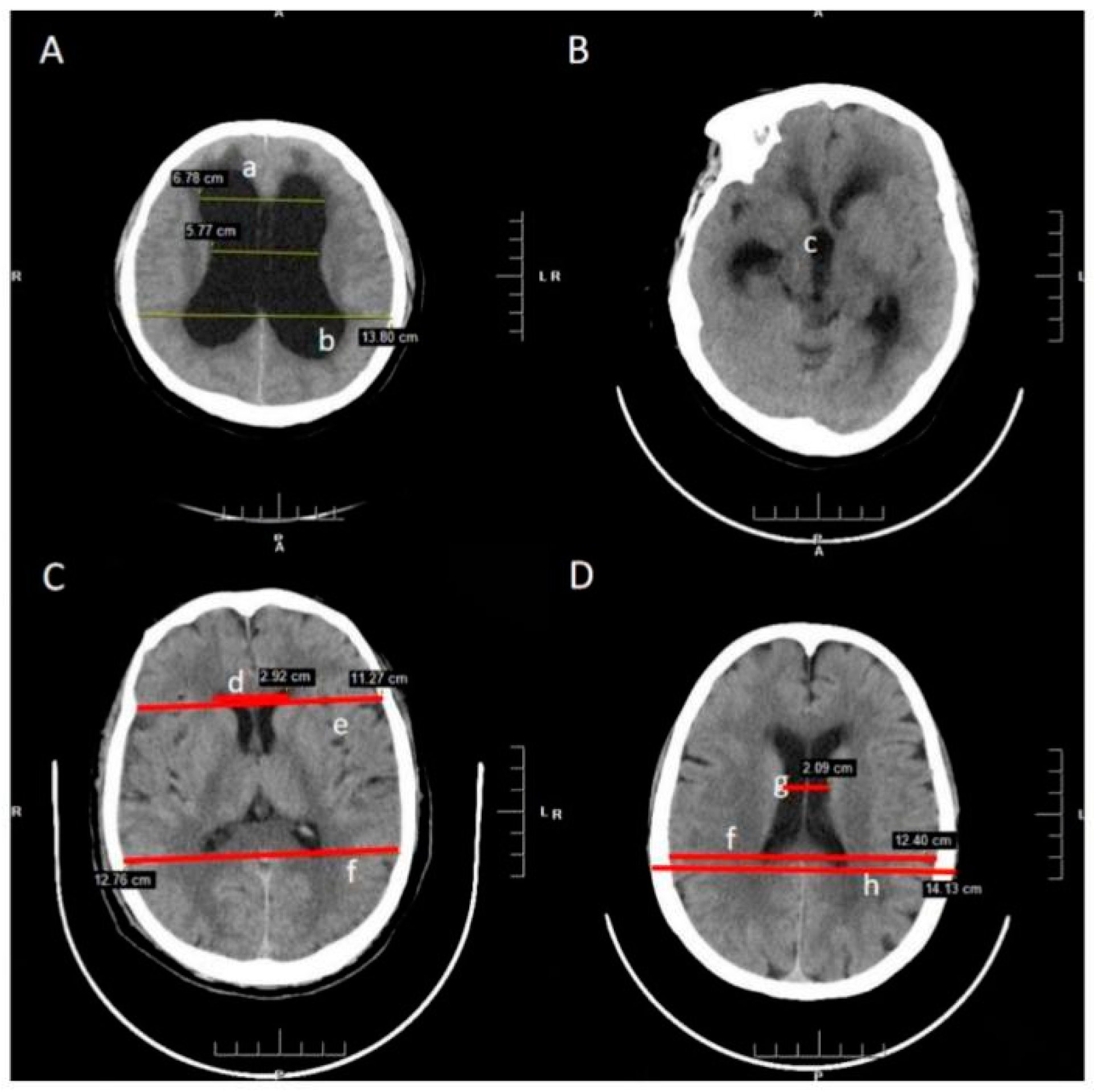
2.2. Measurements of Linear Parameters
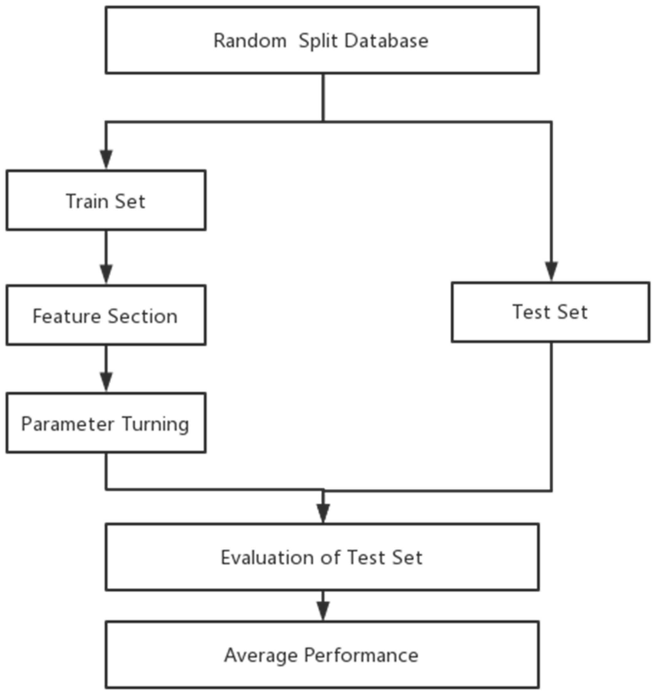
2.3. Model Construction
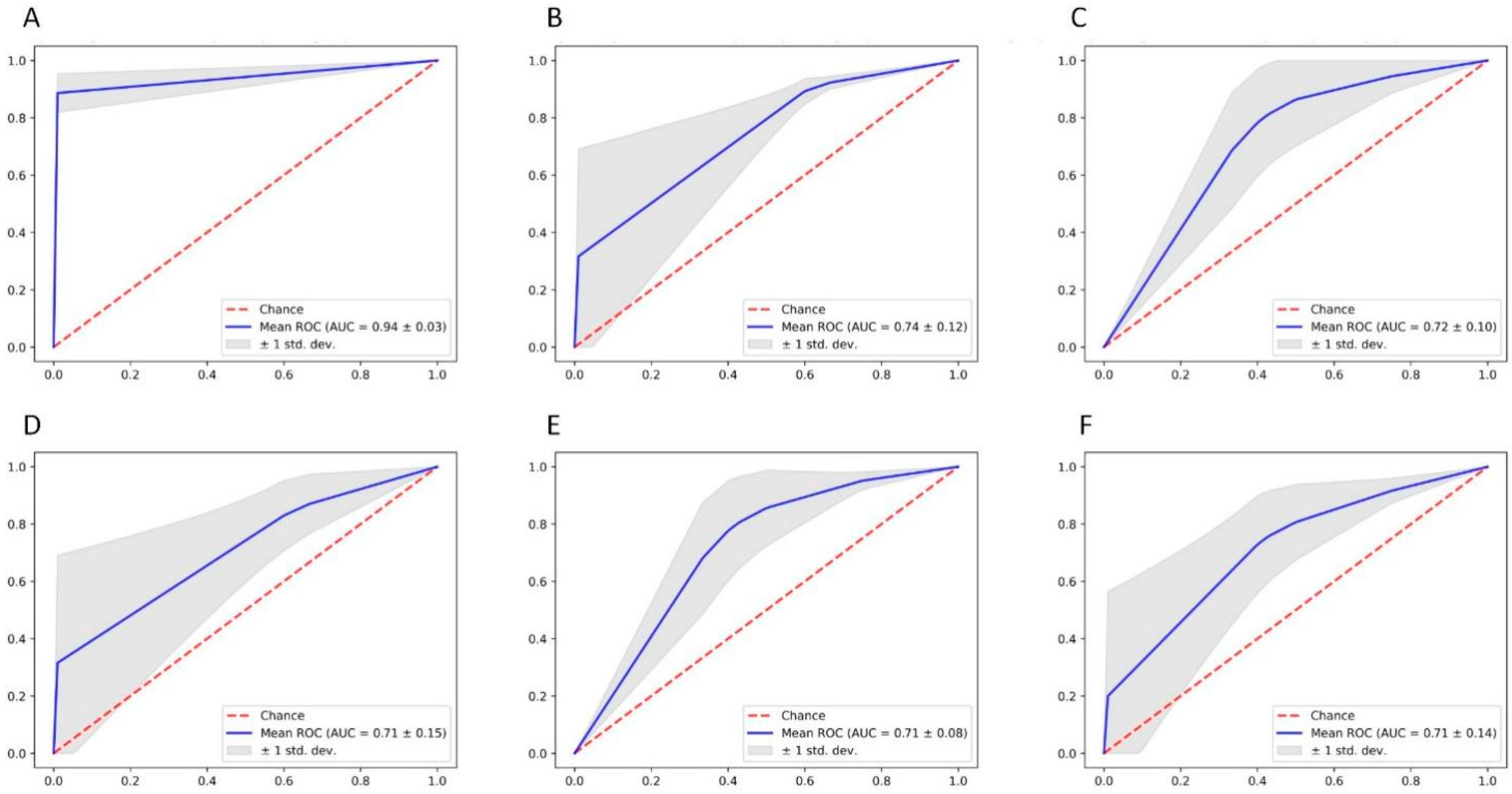
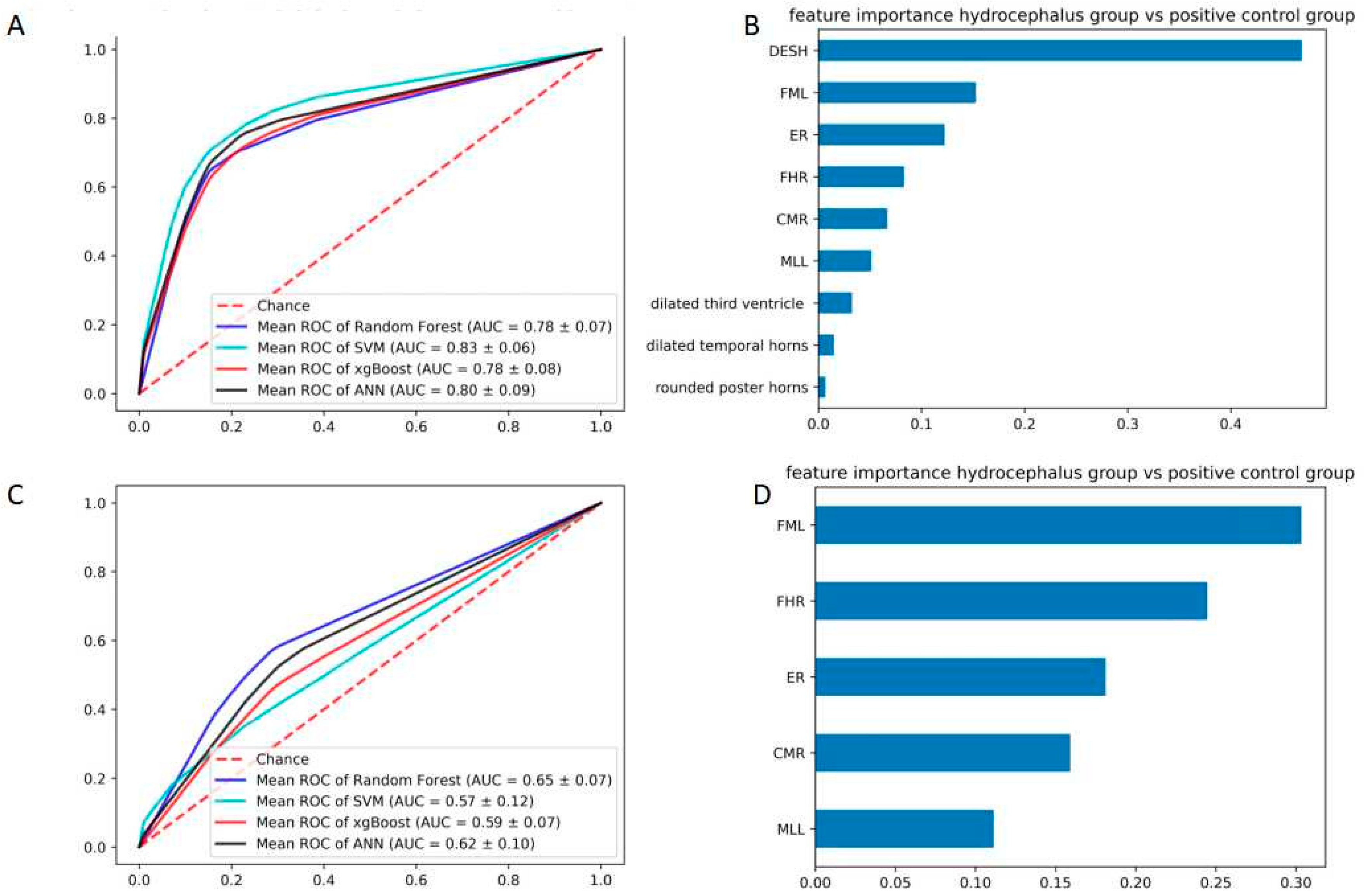
3. Results
4. Discussion
5. Conclusions and Future Work
Author Contributions
Funding
Institutional Review Board Statement
Data Availability Statement
Conflicts of Interest
References
- Kahle, K.T.; Kulkarni, A.V.; Limbrick, D.D., Jr.; Warf, B.C. Hydrocephalus in children. Lancet 2016, 387, 788–799. [Google Scholar] [CrossRef]
- Symss, N.P.; Oi, S. Theories of cerebrospinal fluid dynamics and hydrocephalus: Historical trend. J. Neurosurg. Pediatr. 2013, 11, 170–177. [Google Scholar] [CrossRef] [PubMed] [Green Version]
- Pisapia, J.M.; Rozycki, M.; Akbari, H.; Bakas, S.; Thawani, J.P.; Moldenhauer, J.S.; Storm, P.B.; Zarnow, D.M.; Davatzikos, C.; Heuer, G.G. Correlations of atrial diameter and frontooccipital horn ratio with ventricle size in fetal ventriculomegaly. J. Neurosurg. Pediatr. 2017, 19, 300–306. [Google Scholar] [CrossRef] [PubMed] [Green Version]
- Zhuravlova, I.; Kornieieva, M.J. Anatomic Variability of the Morphometric Parameters of the Third Ventricle of the Brain and Its Relations to the Shape of the Skull. Neurol. Surg. B Skull Base 2021, 82 (Suppl. S3), e315–e320. [Google Scholar] [CrossRef] [PubMed]
- Friedman, D.I.; Jacobson, D.M. Diagnostic criteria for idiopathic intracranial hypertension. Neurology 2002, 59, 1492–1495. [Google Scholar] [CrossRef] [PubMed]
- Dinçer, A.; Özek, M.M. Radiologic evaluation of pediatric hydrocephalus. Child’s Nerv. Syst. 2011, 27, 1543–1562. [Google Scholar] [CrossRef]
- Greitz, D. Radiological assessment of hydrocephalus: New theories and implications for therapy. Neurosurg. Rev. 2004, 27, 145–165. [Google Scholar] [CrossRef]
- Halperin, J.J.; Kurlan, R.; Schwalb, J.M.; Cusimano, M.D.; Gronseth, G.; Gloss, D. Practice guideline: Idiopathic normal pressure hydrocephalus: Response to shunting and predictors of response. Neurology 2015, 85, 2063–2071. [Google Scholar] [CrossRef]
- Wikkelso, C.; Hellstrom, P.; Klinge, P.M.; Tans, J.T.J.; European iNPH Multicentre Study Group. The European iNPH Multicentre Study on the predictive values of resistance to CSF outflow and the CSF Tap Test in patients with idiopathic normal pressure hydrocephalus. J. Neurol. Neurosurg. Psychiatry 2012, 84, 562–568. [Google Scholar] [CrossRef]
- Wikkelsø, C.; Andersson, H.; Blomstrand, C.; Lindqvist, G. The clinical effect of lumbar puncture in normal pressure hydrocephalus. J. Neurol. Neurosurg. Psychiatry 1982, 45, 64–69. [Google Scholar] [CrossRef]
- Wikkelsö, C.; Andersson, H.; Blomstrand, C.; Lindqvist, G.; Svendsen, P. Predictive value of the cerebrospinal fluid tap-test. Acta Neurol. Scand. 1986, 73, 566–573. [Google Scholar] [CrossRef] [PubMed]
- Swati, G.; Sanjay, G.; Pankaj, Y.; Saumya, M. CT evaluation of various linear indices in children with clinically suspected hydrocephalus. J. Evol. Med. Dent. Sci. 2017, 6, 3078–3082. [Google Scholar] [CrossRef]
- Yigin, B.O.; Algin, O.; Saygili, G. Comparison of morphometric parameters in prediction of hydrocephalus using random forests. Comput. Biol. Med. 2020, 116, 103547. [Google Scholar] [CrossRef]
- Kartal, M.G.; Algin, O. Evaluation of hydrocephalus and other cerebrospinal fluid disorders with MRI: An update. Insights Imaging 2014, 5, 531–541. [Google Scholar] [CrossRef] [PubMed] [Green Version]
- Lemay, M.; Hochberg, F.H. Ventricular differences between hydrostatic hydrocephalus and hydrocephalus ex vacuo by computed tomography. Neuroradiology 1979, 17, 191–195. [Google Scholar] [CrossRef]
- Relkin, N.; Marmarou, A.; Klinge, P.; Bergsneider, M.; Black, P.M. Diagnosing Idiopathic Normal-pressure Hydrocephalus. Neurosurgery 2005, 57 (Suppl. S3), S4–S16. [Google Scholar] [CrossRef] [PubMed] [Green Version]
- Demšar, J. Statistical comparisons of classifiers over multiple data sets. J. Mach. Learn. Res. 2006, 7, 1–30. [Google Scholar]
- Velez, D.R.; White, B.C.; Motsinger-Reif, A.; Bush, W.; Ritchie, M.D.; Williams, S.; Moore, J.H. A balanced accuracy function for epistasis modeling in imbalanced datasets using multifactor dimensionality reduction. Genet. Epidemiol. 2007, 31, 306–315. [Google Scholar] [CrossRef]
- Lundberg, S.M.; Lee, S.I. A unified approach to interpreting model predictions. Adv. Neural Inf. 2017, 30, 4765–4774. [Google Scholar]
- Li, Y.; Li, M.; Li, C.; Liu, Z. Forest aboveground biomass estimation using Landsat 8 and Sentinel-1A data with machine learning algorithms. Sci. Rep. 2020, 10, 9952. [Google Scholar] [CrossRef]
- Pedregosa, F.; Varoquaux, G.; Gramfort, A. Scikit-learn: Machine learning in Python. J. Mach. Learn. Res. 2012, 12, 2825–2830. [Google Scholar]
- Filis, A.K.; Aghayev, K.; Vrionis, F.D. Cerebrospinal Fluid and Hydrocephalus: Physiology, Diagnosis, and Treatment. Cancer Control. 2017, 24, 6–8. [Google Scholar] [CrossRef] [PubMed]
- Chen, Q.; Feng, Z.; Tan, Q.; Guo, J.; Tang, J.; Tan, L.; Feng, H.; Chen, Z. Post-hemorrhagic hydrocephalus: Recent advances and new therapeutic insights. J. Neurol. Sci. 2017, 375, 220–230. [Google Scholar] [CrossRef] [PubMed]
- Xu, H. New concept of the pathogenesis and therapeutic orientation of acquired communicating hydrocephalus. Neurol. Sci. 2016, 37, 1387–1391. [Google Scholar] [CrossRef]
- Kartal, M.G.; Ocakoglu, G.; Algin, O. Feasibility of 3-Dimensional Sampling Perfection with Application Optimized Contrast Sequence in the Evaluation of Patients with Hydrocephalus. J. Comput. Assist. Tomogr. 2015, 39, 321–328. [Google Scholar] [CrossRef] [Green Version]
- Jaeger, M.; Khoo, A.K.; Conforti, D.A.; Cuganesan, R. Relationship between intracranial pressure and phase contrast cine MRI derived measures of intracranial pulsations in idiopathic normal pressure hydrocephalus. J. Clin. Neurosci. 2016, 33, 169–172. [Google Scholar] [CrossRef]
- Bradley, W.G., Jr. Magnetic Resonance Imaging of Normal Pressure Hydrocephalus. In Seminars in Ultrasound, CT, and MR; WB Saunders: Philadelphia, PA, USA, 2016; Volume 37, pp. 120–128. [Google Scholar]
- Qvarlander, S.; Ambarki, K.; Wåhlin, A.; Jacobsson, J.; Birgander, R.; Malm, J.; Eklund, A. Cerebrospinal fluid and blood flow patterns in idiopathic normal pressure hydrocephalus. Acta Neurol. Scand. 2017, 135, 576–584. [Google Scholar] [CrossRef]
- Eide, P.K. Intracranial pressure parameters in idiopathic normal pressure hydrocephalus patients treated with ventriculo-peritoneal shunts. Acta Neurochir. 2006, 148, 21–29. [Google Scholar] [CrossRef]
- Evans, W.A. An encephalographic ratio for estimating ventricular enlargement and cerebral atrophy. Arch. Neurol. Psychiatry 1942, 47, 931–937. [Google Scholar] [CrossRef]
- Toma, A.K.; Holl, E.; Kitchen, N.D.; Watkins, L.D. Evans’ Index Revisited: The Need for an Alternative in Normal Pressure Hydrocephalus. Neurosurgery 2011, 68, 939–944. [Google Scholar] [CrossRef]
- Polat, S.; Oksuzler, F.; Oksuzler, M.; Kabakçi, A.; Yücel, A. Morphometric MRI study of the brain ventricles in healthy Turkish subjects. Drug Alcohol. Depend. 2013, 131, 222–229. [Google Scholar] [CrossRef] [Green Version]
- Hahn, F.; Rim, K. Frontal ventricular dimensions on normal computed tomography. Am. J. Roentgenol. 1976, 126, 593–596. [Google Scholar] [CrossRef] [PubMed] [Green Version]
- Gholipour, A.; Akhondi-Asl, A.; Estroff, J.A.; Warfield, S.K. Multi-atlas multi-shape segmentation of fetal brain MRI for volumetric and morphometric analysis of ventriculomegaly. NeuroImage 2012, 60, 1819–1831. [Google Scholar] [CrossRef] [PubMed]
- Lee, W.; Lee, A.; Li, H.; Ong, N.Y.X.; Keong, N.; Chen, R.; Chan, L.L. Callosal angle in idiopathic normal pressure hydrocephalus: Small angular mal-rotations of the coronal plane affect measurement reliability. Neuroradiology 2021, 63, 1659–1667. [Google Scholar] [CrossRef] [PubMed]
- Silva, M.A.; Patel, J.; Kavouridis, V.; Gallerani, T.; Beers, A.; Chang, K.; Hoebel, K.V.; Brown, J.; See, A.P.; Gormley, W.B.; et al. Machine Learning Models Can Detect Aneurysm Rupture and Identify Clinical Features Associated with Rupture. World Neurosurg. 2019, 131, e46–e51. [Google Scholar] [CrossRef]
- Liu, Q.; Jiang, P.; Jiang, Y.; Ge, H.; Li, S.; Jin, H.; Li, Y. Prediction of aneurysm stability using a machine learning model based on PyRadiomics-derived morphological features. Stroke 2019, 50, 2314–2321. [Google Scholar] [CrossRef]
- Sirsat, M.S.; Fermé, E.; Câmara, J. Machine Learning for Brain Stroke: A Review. J. Stroke Cerebrovasc. Dis. 2020, 29, 105162. [Google Scholar] [CrossRef]


| Features | Description |
|---|---|
| DESH | Disproportionately enlarged subarachnoid space hydrocephalus |
| Dilated temporal horns | Whether there is temporal horns dilated |
| Rounded third ventricle | Whether there is third ventricle rounded |
| horns | Whether there is posterior horns dilated |
| Morphometric parameters | |
| MLL | The narrowest width between the lateral walls |
| MTD | Maximum transverse diameter of the skull |
| DSL | The internal diameter of the skull in the same line as MLL |
| BPD | Maximum width of internal diameter of the skull |
| DM | Inner diameter of the skull in the same line as FHL |
| FHL | Width of greatest span of frontal horns |
| ER = FHL/MTD (d/f) | The ratio of the transverse diameter of the anterior horns of the lateral ventricles to the internal diameter |
| CMR = MLL/BPD (g/h) | The ratio of the minimum distance between lateral walls of lateral ventricles in cella media region |
| FHR = FHL/DM (d/e) | The ratio of maximum width of the frontal horns of the lateral ventricles |
| Hydrocephalus Group (n = 62) | Symptomatic Group (n = 36) | Normal Control Group (n = 200) | F/χ²/Z | p Value | |
|---|---|---|---|---|---|
| Sex (male) | 39/62.90 | 19/52.80 | 11/54.00 | 3.010 ** | 0.222 |
| age | 49.87 ± 15.53 | 70.37 ± 11.42 | 52.80 ± 11.36 | 14.498 * | 0.000 |
| DESH | 49/79.03 | 2/5.60 | 0/0 | 65.945 ** | 0.000 |
| Dilated temporal horns | 15/24.19 | 13/36.1 | 0/0 | 8.474 ** | 0.014 |
| Dilated third ventricle | 31/50.00 | 3/8.30 | 0/0 | 27.860 ** | 0.000 |
| Rounded poster horns | 18/29.03 | 8/22.20 | 0/0 | 8.157 ** | 0.017 |
| MLL | 3.71 ± 0.738 | 3.27 ± 0.615 | 2.81 ± 0.638 | 14.498 * | 0.000 |
| MTD | 14.81 ± 1.168 | 14.65 ± 0.720 | 15.03 ± 1.039 | 0.855 * | 0.428 |
| DM | 12.65 ± 1.15 | 12.34 ± 0.992 | 12.54 ± 0.815 | 1.200 * | 0.305 |
| ER | 0.307 ± 0.069 | 0.256 ± 0.035 | 0.229 ± 0.023 | 35.274 *** | 0.000 |
| CMR | 0.271 ± 0.052 | 0.238 ± 0.046 | 0.204 ± 0.047 | 14.835 * | 0.000 |
| FHR | 0.359 ± 0.082 | 0.305 ± 0.041 | 0.275 ± 0.038 | 26.548 *** | 0.000 |
| All Features Model | SVM | ANN | Random Forest | xgBoost |
|---|---|---|---|---|
| Test Set Percision | 0.928571 | 1.000000 | 0.818182 | 1.000000 |
| Test Set Recall | 1.000000 | 1.000000 | 0.900000 | 0.923077 |
| Test Set f1 | 0.962963 | 1.000000 | 0.857143 | 0.960000 |
| Morphometric parameters Model | ||||
| Test Set Percision | 0.857143 | 1.000000 | 0.857143 | 0.600000 |
| Test Set Recall | 1.000000 | 0.866667 | 1.000000 | 0.818182 |
| Test Set f1 | 0.923077 | 0.928571 | 0.923077 | 0.692308 |
| All Features Model | Radiographic Features | ER | ER + FHR | ER + CMR | FHR + CMR | ER + CMR + FHR |
|---|---|---|---|---|---|---|
| Test Set Percision | 1.000000 | 0.800000 | 0.588235 | 0.923077 | 0.923077 | 0.857143 |
| Test Set Recall | 0.714286 | 0.666667 | 1.000000 | 0.857143 | 0.857143 | 1.000000 |
| Test Set f1 | 0.833333 | 0.727273 | 0.740741 | 0.888889 | 0.888889 | 0.923077 |
| All Features Model | SVM | ANN | Random Forest | xgBoost |
|---|---|---|---|---|
| Test Set Percision | 0.900000 | 0.857143 | 0.666667 | 1.000000 |
| Test Set Recall | 1.000000 | 0.750000 | 0.800000 | 0.571429 |
| Test Set f1 | 0.947368 | 0.800000 | 0.727273 | 0.727273 |
| Morphometric parameters Model | ||||
| Test Set Percision | 0.583333 | 0.333333 | 0.375000 | 0.400000 |
| Test Set Recall | 1.000000 | 0.222222 | 0.428571 | 0.500000 |
| Test Set f1 | 0.736842 | 0.266667 | 0.400000 | 0.444444 |
| Hydrocephalus Group (n = 18) | Symptomatic Group (n = 29) | t/χ² | p Value | |
|---|---|---|---|---|
| age | 67.33 ± 4.46 | 73.90 ± 8.85 | −3.365 * | 0.002 |
| DESH | 14/77.78 | 1/3.45 | 28.239 ** | 0.000 |
| Dilated temporal horns | 4/22.22 | 10/34.48 | 0.798 ** | 0.372 |
| Dilated third ventricle | 10/55.56 | 3/10.34 | 11.346 ** | 0.001 |
| Rounded poster horns | 5/27.78 | 6/20.69 | 0.311 ** | 0.577 |
| MLL | 3.71 ± 0.738 | 3.27 ± 0.615 | 14.498 * | 0.000 |
| ER | 0.29 ± 0.05 | 0.26 ± 0.03 | 2.748 * | 0.011 |
| EMR | 0.27 ± 0.06 | 0.24 ± 0.05 | 2.268 * | 0.028 |
| FHR | 0.35 ± 0.07 | 0.31 ± 0.04 | 2.691 * | 0.010 |
Publisher’s Note: MDPI stays neutral with regard to jurisdictional claims in published maps and institutional affiliations. |
© 2022 by the authors. Licensee MDPI, Basel, Switzerland. This article is an open access article distributed under the terms and conditions of the Creative Commons Attribution (CC BY) license (https://creativecommons.org/licenses/by/4.0/).
Share and Cite
Xu, H.; Fang, X.; Jing, X.; Bao, D.; Niu, C. Multiple Machine Learning Approaches for Morphometric Parameters in Prediction of Hydrocephalus. Brain Sci. 2022, 12, 1484. https://doi.org/10.3390/brainsci12111484
Xu H, Fang X, Jing X, Bao D, Niu C. Multiple Machine Learning Approaches for Morphometric Parameters in Prediction of Hydrocephalus. Brain Sciences. 2022; 12(11):1484. https://doi.org/10.3390/brainsci12111484
Chicago/Turabian StyleXu, Hao, Xiang Fang, Xiaolei Jing, Dejun Bao, and Chaoshi Niu. 2022. "Multiple Machine Learning Approaches for Morphometric Parameters in Prediction of Hydrocephalus" Brain Sciences 12, no. 11: 1484. https://doi.org/10.3390/brainsci12111484
APA StyleXu, H., Fang, X., Jing, X., Bao, D., & Niu, C. (2022). Multiple Machine Learning Approaches for Morphometric Parameters in Prediction of Hydrocephalus. Brain Sciences, 12(11), 1484. https://doi.org/10.3390/brainsci12111484