
Spatial Memory Activity Distributions Indicate the Hippocampus Operates in a Continuous Manner


Abstract
:1. Introduction
2. Materials and Methods
2.1. Participants
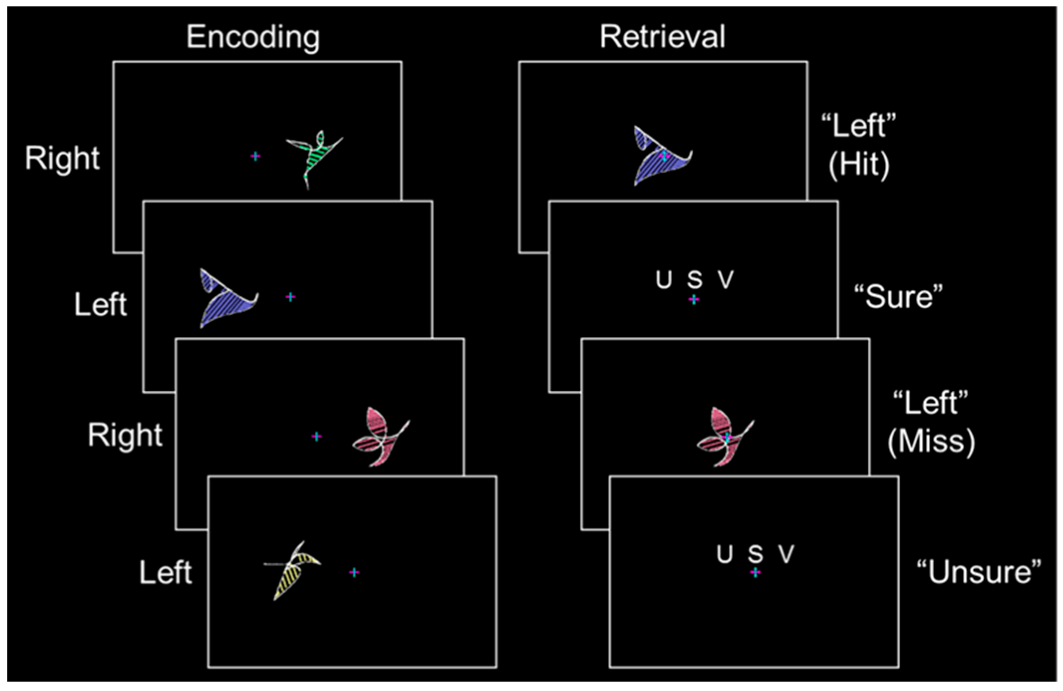
2.2. Stimulus Protocol
2.3. Data Acquisition and Pre-processing
2.4. General Linear Model Analysis
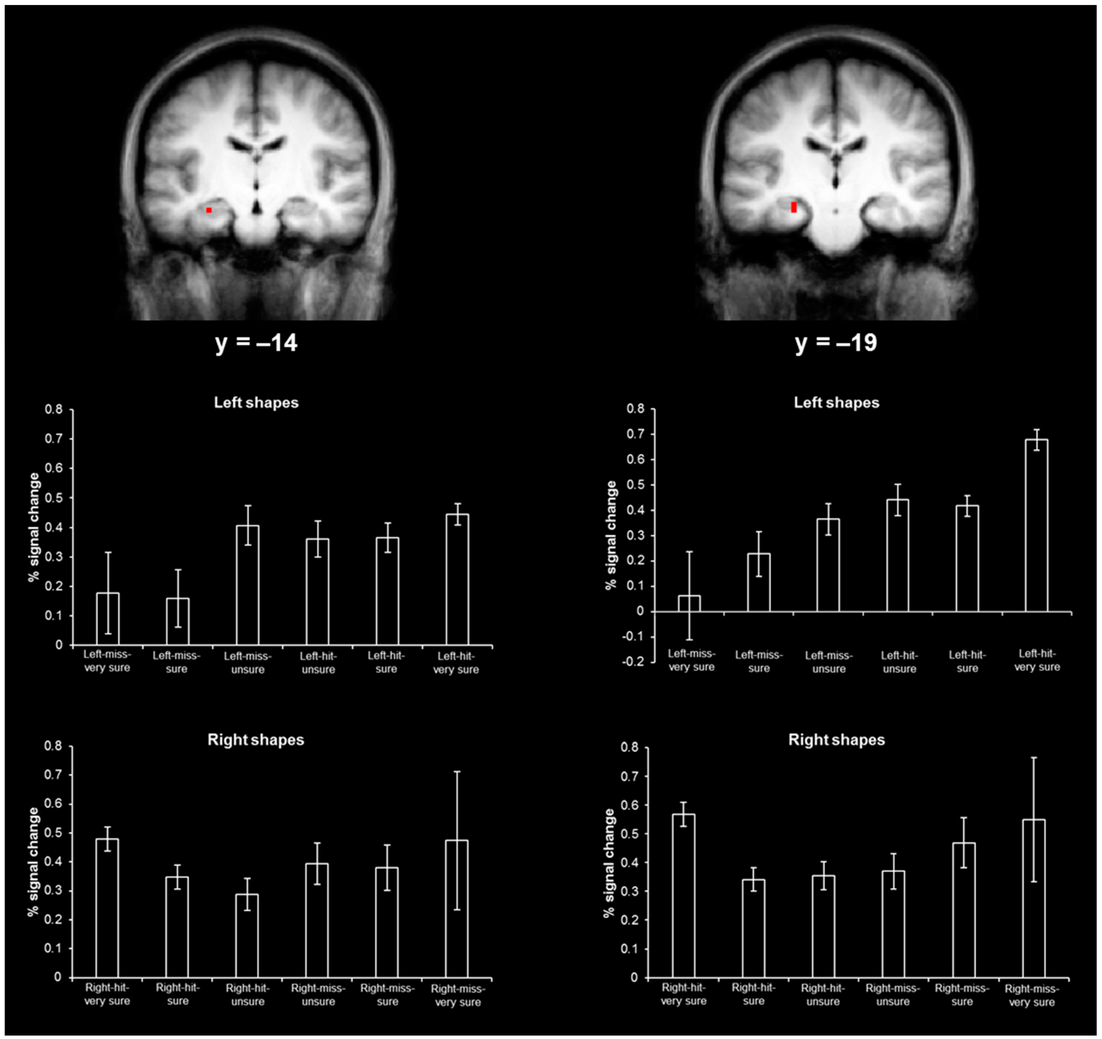
2.5. Hippocampal Activity Distribution Analysis
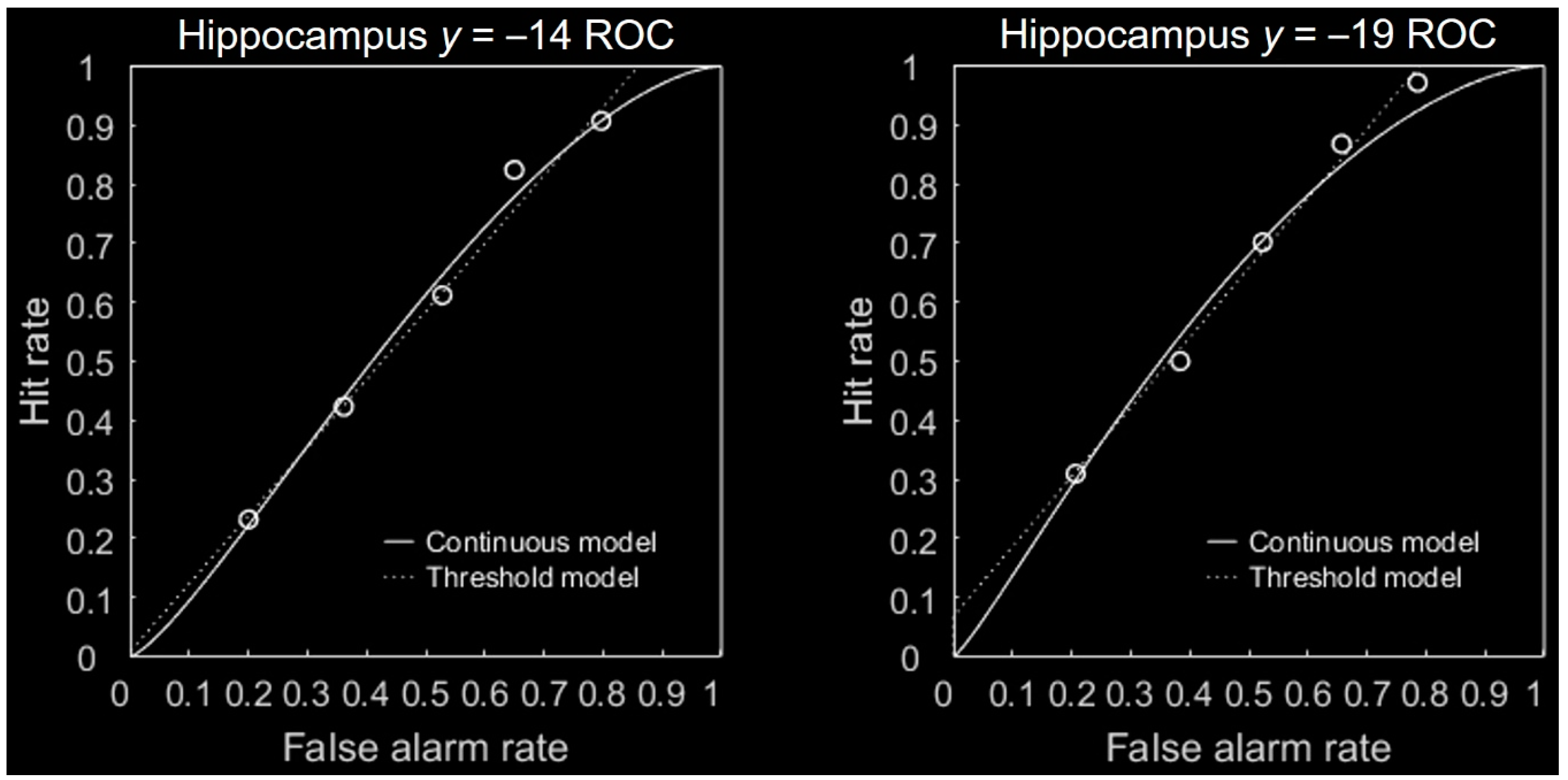
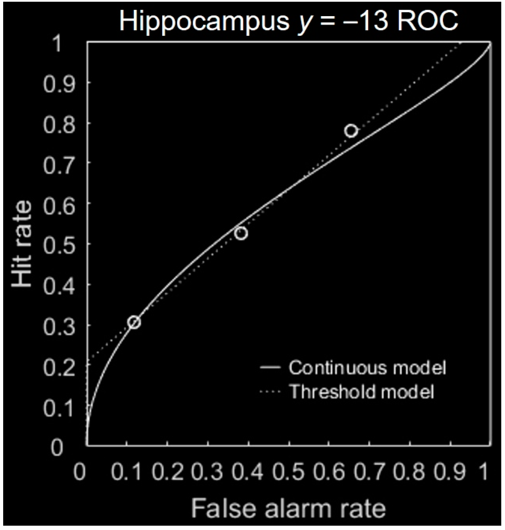
2.6. ROC Analysis
3. Results
3.1. Behavioral Results
3.2. Hippocampal Results
4. Discussion and Conclusions
Acknowledgments
Author Contributions
Conflicts of Interest
Appendix


References
- Mandler, G. Recognizing: The judgment of previous occurrence. Psychol. Rev. 1980, 87, 252–271. [Google Scholar] [CrossRef]
- Yonelinas, A.P. The nature of recollection and familiarity: A review of 30 years of research. J. Mem. Lang. 2002, 46, 441–517. [Google Scholar] [CrossRef]
- Yonelinas, A.P.; Parks, C.M. Receiver operating characteristics (ROCs) in recognition memory: A review. Psychol. Bull. 2007, 133, 800–832. [Google Scholar] [CrossRef] [PubMed]
- Wixted, J.T. Dual-process theory and signal-detection theory of recognition memory. Psychol. Rev. 2007, 114, 152–176. [Google Scholar] [CrossRef] [PubMed]
- Wixted, J.T.; Mickes, L. A continuous dual-process model of remember/know judgments. Psychol. Rev. 2010, 117, 1025–1054. [Google Scholar] [CrossRef] [PubMed]
- Slotnick, S.D. The nature of recollection in behavior and the brain. NeuroReport 2013, 24, 663–670. [Google Scholar] [CrossRef] [PubMed]
- Slotnick, S.D.; Dodson, C.S. Support for a continuous (single-process) model of recognition memory and source memory. Mem. Cogn. 2005, 33, 151–170. [Google Scholar] [CrossRef]
- Diana, R.A.; Yonelinas, A.P.; Ranganath, C. Imaging recollection and familiarity in the medial temporal lobe: A three-component model. Trends Cogn. Sci. 2007, 11, 379–386. [Google Scholar] [CrossRef] [PubMed]
- Slotnick, S.D.; Thakral, P.P. The hippocampus operates in a threshold manner during spatial source memory. NeuroReport 2013, 24, 265–269. [Google Scholar] [CrossRef] [PubMed]
- Slotnick, S.D.; Schacter, D.L. A sensory signature that distinguishes true from false memories. Nat. Neurosci. 2004, 7, 664–672. [Google Scholar] [CrossRef] [PubMed]
- Logan, B.R.; Rowe, D.B. An evaluation of thresholding techniques in fMRI analysis. NeuroImage 2004, 22, 95–108. [Google Scholar] [CrossRef] [PubMed]
- Insausti, R.; Juottonen, K.; Soininen, H.; Insausti, A.M.; Partanen, K.; Vainio, P.; Laakso, M.P.; Pitkänen, A. MR volumetric analysis of the human entorhinal, perirhinal, and temporopolar cortices. Am. J. Neuroradiol. 1998, 19, 659–671. [Google Scholar] [PubMed]
- Pruessner, J.C.; Li, L.M.; Serles, W.; Pruessner, M.; Collins, D.L.; Kabani, N.; Lupiem, S.; Evans, A.C. Volumetry of hippocampus and amygdala with high-resolution MRI and three-dimensional analysis software: Minimizing the discrepancies between laboratories. Cereb. Cortex 2000, 10, 433–442. [Google Scholar] [CrossRef] [PubMed]
- Bernasconi, N.; Bernasconi, A.; Caramanos, Z.; Antel, S.B.; Andermann, F.; Arnold, D.L. Mesial temporal damage in temporal lobe epilepsy: A volumetric MRI study of the hippocampus, amygdala and parahippocampal region. Brain 2003, 126, 462–469. [Google Scholar] [CrossRef] [PubMed]
- Malykhin, N.V.; Bouchard, T.P.; Ogilvie, C.J.; Coupland, N.J.; Seres, P.; Camicioli, R. Three-dimensional volumetric analysis and reconstruction of amygdala and hippocampal head, body and tail. Psychiatry Res. 2007, 155, 155–165. [Google Scholar] [CrossRef] [PubMed]
- Logothetis, N.K.; Pauls, J.; Augath, M.; Trinath, T.; Oeltermann, A. Neurophysiological investigation of the basis of the fMRI signal. Nature 2001, 412, 150–157. [Google Scholar] [CrossRef] [PubMed]
- Slotnick, S.D.; Klein, S.A.; Dodson, C.S.; Shimamura, A.P. An analysis of signal detection and threshold models of source memory. J. Exp. Psychol. Learn. Mem. Cogn. 2000, 26, 1499–1517. [Google Scholar] [CrossRef] [PubMed]
- Slotnick, S.D. “Remember” source memory ROCs indicate recollection is a continuous process. Memory 2010, 18, 27–39. [Google Scholar] [CrossRef] [PubMed]
- Jeye, B.M.; Karanian, J.M.; Slotnick, S.D. Distinct regions of the hippocampus are associated with memory for different spatial locations. Hippocampus 2016. under review. [Google Scholar]
- Elfman, K.W.; Aly, M.; Yonelinas, A.P. Neurocomputational account of memory and perception: Thresholded and graded signals in the hippocampus. Hippocampus 2014, 24, 1672–1686. [Google Scholar] [CrossRef] [PubMed]
- Schacter, D.L.; Addis, D.R.; Buckner, R.L. Remembering the past to imagine the future: The prospective brain. Nat. Rev. Neurosci. 2007, 8, 657–661. [Google Scholar] [CrossRef] [PubMed]
- Addis, D.R.; Schacter, D. The hippocampus and imagining the future: Where do we stand? Front. Hum. Neurosci. 2012, 5, 173. [Google Scholar] [CrossRef] [PubMed]


© 2016 by the authors; licensee MDPI, Basel, Switzerland. This article is an open access article distributed under the terms and conditions of the Creative Commons Attribution (CC-BY) license (http://creativecommons.org/licenses/by/4.0/).
Share and Cite
Jeye, B.M.; Karanian, J.M.; Slotnick, S.D. Spatial Memory Activity Distributions Indicate the Hippocampus Operates in a Continuous Manner. Brain Sci. 2016, 6, 37. https://doi.org/10.3390/brainsci6030037
Jeye BM, Karanian JM, Slotnick SD. Spatial Memory Activity Distributions Indicate the Hippocampus Operates in a Continuous Manner. Brain Sciences. 2016; 6(3):37. https://doi.org/10.3390/brainsci6030037
Chicago/Turabian StyleJeye, Brittany M., Jessica M. Karanian, and Scott D. Slotnick. 2016. "Spatial Memory Activity Distributions Indicate the Hippocampus Operates in a Continuous Manner" Brain Sciences 6, no. 3: 37. https://doi.org/10.3390/brainsci6030037
APA StyleJeye, B. M., Karanian, J. M., & Slotnick, S. D. (2016). Spatial Memory Activity Distributions Indicate the Hippocampus Operates in a Continuous Manner. Brain Sciences, 6(3), 37. https://doi.org/10.3390/brainsci6030037