Article

Solanum trilobatum L. Ameliorate Thioacetamide-Induced Oxidative Stress and Hepatic Damage in Albino Rats

Food Science and Technology Program, Beijing Normal University–Hong Kong Baptist University United International College, Zhuhai 519085, China
*
Author to whom correspondence should be addressed.
Antioxidants 2017, 6(3), 68; https://doi.org/10.3390/antiox6030068
Submission received: 26 July 2017 / Revised: 16 August 2017 / Accepted: 20 August 2017 / Published: 22 August 2017

Abstract

:
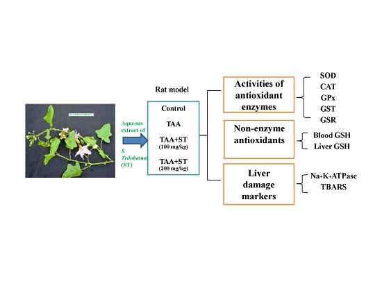
Solanum trilobatum L. (Solanaceae) has been well known as nightshade, commonly used by diverse populations to heal several disorders. Earlier studies in Solanum trilobatum were focused on different pharmacological activities and a few were concerned with antioxidant and hepatoprotective effects. Thus, the current study was focused to evaluate the antioxidant potential and hepatoprotective effects of S. trilobatum L. on thioacetamide (TAA) intoxication in Wistar albino rats. The rats were kept into four groups and six animals each. Group A was normal control. Group B was the TAA treated control. Groups C and D were pretreated with the aqueous extract from the leaves of S. trilobatum (100 mg, 200 mg/kg bw p.o.) once daily for 10 consecutive days administration followed by a single dose infusion of TAA (100 mg/kg s.c.). After 10 days, blood and livers were collected. The biochemical assay was carried out in the GSH (reduced glutathione), TBARS(thiobarbituric acid reactive substances), Na+-K+-ATPase, and antioxidant enzymes viz., SOD (superoxide dismutase), CAT (catalase), GPx (glutathione peroxidase), GST (glutathione-S-transferase), and GR (glutathione reductase) were analyzed in samples of blood and liver. Treatment with S. trilobatum reduced blood and liver TBARS, and Na+ K+ ATPase activity in TAA (thioacetamide)-induced hepatotoxicity rats. Furthermore, the above antioxidant enzymes were increased in the pretreatment of S. trilobatum in TAA intoxicated rats. Finally, we concluded that S. Trilobatum displayed potent antioxidant properties and alleviate oxidative stress induced hepatotoxic effects and possible engross mechanisms related to free radical scavenging properties.

Graphical Abstract

1. Introduction

Currently, oxidative stress associated free-radical biology have become niche research attention. Free radicals play a crucial function in the progress of tissue damage and pathological events in living beings [1]. In a cell, lipid peroxidation is restricted by several cellular defense mechanisms consisted of enzymatic and non-enzymatic scavenging systems [2]. Recent studies have also specified that there is a contrary connection between the intake of food rich in antioxidants and the occurrence of human illness [3]. Thus the investigations of new synthetic or natural antioxidants are urgently important.
The properties of medicinal plants have been investigated in the recent scientific progress due to its effective antioxidant potentials and ability in healing various ailments. Normally, antioxidants inhibit oxidative stress related tissue damage caused by free radicals; either obstruct oxidation mechanisms or scavenge the oxygen [4,5]. Potentially reactive oxygen derivatives are known to be recognized as reactive oxygen species (ROS), which is consistently produced by various biochemical mechanisms in the human cells. However, these generated ROS are detoxified by the antioxidants, either present in the body or in antioxidant rich food.
ROS generally caused by O2, HO• and H2O2 and led to the alteration of the enzymatic antioxidant defense systems in in vitro and in vivo models [6]. SOD, CAT, GSH, GPx, GR, and GST are the primary antioxidant enzymes serve as redox biomarkers as they are the first-line indicators of the antioxidant state through oxidation/reduction processes [7,8]. As the most abundant intracellular antioxidant, GSH is involved in the protection of cells against oxidative damage and in various detoxification mechanisms [9]. GSH primarily acts as a substrate and co-substrate in many essential enzymatic reactions involving GPx, GR, and GST, and a decrease in the GSH level usually impairs cells’ response to oxidants [10]. Conversely, excess ROS generation and/or insufficient antioxidant defense can simply influence oxidative injury to diverse biomolecules including proteins, lipids, and DNA [11]. These oxidative damages cause many chronic diseases in human including diabetes, cancer, arthritis, cardiovascular diseases, neurodegenerative diseases, and aging [12]. Numerous compounds have been used as hepatotoxic models, which facilitate investigation on a variety of pathological features. A single administration of thioacetamide (TAA) at a dose of 100 mg/kg causes hepatic centrilobular necrosis, cirrhosis, and fibrosis in the rodents [13,14,15]. Further, the study showed that prolonged administration of TAA along with diet causes liver cancer [16].
S. trilobatum Linn (Solanaceae) is a known shrub, familiar as ‘Tuduvelai’ and is distributed in various Asian countries, including India, Sri Lanka, Indonesia, Singapore, and Malaysia [17]. In Ayurveda and Siddha medicinal systems, the roots and leaves are prescribed to heal various respiratory tract problems, including acute and chronic bronchitis, asthma, sinusitis, tonsillitis, common cold, cough, and pulmonary infections [18,19,20,21,22]. The leaves are mainly used in treating dyspepsia, spermatorrhoea, tuberculosis, ear problems, and bacterial infections [23]. S. trilobatum has been extensively studied for various pharmacological activities including antibacterial, antifungal, anticancer [24,25,26,27,28,29,30,31,32,33], antioxidant [34], antidiabetic [35], hepatoprotective [27], antinociceptive [36], anti-ulcerogenic [37], anti-inflammatory [38,39,40,41,42,43], and mosquitocidal activity [41,42]. Studies showed that the leaves contain various metabolites, including sugar, fat, fiber, calcium, phosphorus, ferrous, and other minerals [38]. Phytochemical screening of the dried leaves found alkaloids, triterpenoids, phenolics, tannins, flavonoids, anthoquinones, phytosterols, saponins, cardiac glycosides, soladunalinidine, tomatidine, solanine, sobatum, solasodine, diosgenin, and β-solamarine [37,44]. Based on the literature, few studies were concerned with the investigation on antioxidant and hepatoprotective effects. Thus, the current study aimed to evaluate the antioxidant potential and hepatoprotective effects of S. trilobatum L. on TAA intoxication in Wistar albino rats.

2. Materials and Methods

2.1. Animals

Male Wister albino rats weighing about 180–200 g were used in the present study. Animals were procured from the animal house of Management and Science University, Malaysia and were kept in wire-floored cages under normal laboratory settings in 12 h light/dark cycles at 25–28 °C and 60–80% relative humidity. Animals were nurtured at robust health by the supply of a normal pellet diet and water ad libitum. Animal studies were carried out in accordance with the National Institute of Health Guide [45]. The study was initially approved by the ethics committee (The committee for the purpose of control and supervision of experiments on animals-CPCSEA) and ethical norms were strictly followed during all experimental procedures (Reg No. 12/2011/CPCSEA, proposal No. 77).

2.2. Plant Material

The fresh leaves of S. trilobatum L. were collected during January–February 2014 from Sri Muda, Shah Alam, Selangor, Malaysia. The plant was authenticated by Dr. Sujit, Taxonomist in the Department of Pharmacognosy and compared with reference specimens preserved in the Herbarium and Center for Molecular Systematics in Management and Science University, Malaysia. Voucher specimens are maintained in the institution.

2.3. Preparation of Plant Extract

Fresh powdered leaves (500 g) were subjected to 2 L of distilled water and extraction was kept in a cold room with constant stirring overnight. The extraction was filtered using cheesecloth, and Whatmann filter paper followed by centrifugation (1200× g for 10 min). The supernatant was evaporated under reduced pressure using a vacuum rotary evaporator and residues were kept under refrigeration until used (yield: 210 w/w).

2.4. Chemicals

Thioacetamide (TAA), thiobarbituric acid (TBA), 1,1-diphenyl-2-picrylhydrazyl (DPPH), 5,5-dithiobis-2-nitrobenzoic acid (DTNB), nicotinamide adenine dinucleotide hydrogen (NADH), phenazine methosulphate, trichloroacetic acid (TCA), 1-chloro-2, 4-dinitrobenzene (CDNB), nicotinamide adenine dinucleotide hydrogen phosphate (NADPH) were obtained from Sigma-Aldrich Co., St. Louis, MO, USA, and Merck (Darmstadt, Germany). All chemicals and reagents used in the experiments were of analytical grade (AR).

2.5. Experimental Design

Animals were kept into four groups and six rats each. Group A was normal control. Group B was TAA treated control. Group C and D were pretreated with the aqueous extract from the leaves of S. trilobatum (100 mg, 200 mg/kg bw p.o.) once daily for 10 consecutive days administration followed by a single dose infusion of TAA (100 mg/kg s.c.) as a 2% W/V solution in distilled water. The control group received the vehicle as distilled water. The method of acute hepatotoxicity induction was followed according to the method of Kumar et al. [46]. After 24 h of toxin administration, all rats were sacrificed by cervical dislocation; blood was collected and used for the measurement of antioxidant enzymes. Livers were dissected immediately and homogenized. The homogenates were then centrifuged at 3200× g for 20 min at 4 °C and supernatant obtained was used for the assay of various enzymes.

2.6. Assay of Antioxidant Enzymes

2.6.1. Reduced Glutathione (GSH) Assay

GSH was estimated using DTNB by the method of Sedlak and Lindsay [47]. 1 mL sample of 10% homogenate was precipitated with 1 mL of (4%) sulfosalicylic acid. The samples were maintained at 4 °C for 1 h and then centrifuged at 1200× g for 20 min at 4 °C. Added 0.1 mL filtered aliquot, 0.2 mL of 100 mM DTNB, 2.7 mL phosphate buffer (0.1 M, pH 7.4) in the cuvette. The yellow color of the mixture was developed, read immediately at 412 nm on the spectrophotometer and expressed as μM GSH/g tissue.

2.6.2. Superoxide Dismutase (SOD) Assay

SOD activity was estimated by the method of Kakkar et al. [48]. Reaction mixture of this method contained 0.1 mL of phenazine methosulphate (186 μM), 1.2 mL of sodium pyrophosphate buffer (0.05 mM, pH 7.0), 0.3 mL of supernatant after centrifugation (1500× g, 10 min followed by 10,000× g, 15 min) of 10% homogenate was added to the reaction mixture. The enzyme reaction was initiated by adding 0.2 mL of NADH (780 μM) and stopped after 1 min by adding 1 mL of glacial acetic acid. The changes in the absorbance were measured at 560 nm using the spectrophotometer. Results are expressed in units/mg protein.

2.6.3. Catalase Activity (CAT)

CAT activity was measured by following decomposition of H2O2 according to the method of Aebi and Vergmeyer [49]. The reaction solution contained 0.1 mL enzyme extract, 2.5 mL of 50 mM phosphate buffer (pH 5.0), and 0.4 mL of 6 mM H2O2. After one min, the absorbance of the reaction solution was read at 240 nm. One unit of CAT activity was defined as an absorbance change of as 0.01 units/min.

2.6.4. Glutathione Peroxidase (GPx) Assay

GPx activity was assayed by the method of Rotruck et al. [50] using H2O2 as substrate. The reaction mixture consisted of 1.49 mL phosphate buffer (0.1 M, pH 7.4), 0.1 mL sodium azide (1 mM), 0.05 mL glutathione reductase (1 IU/mL), 0.05 mL GSH (1 mM), 0.1 mL EDTA (1 mM), 0.1 mL NADPH (0.2 mM), 0.01 mL H2O2 (0.25 mM), and 0.1 mL 10% homogenate in a total volume of 2 mL. The discoloration of NADPH at 340 nm was recorded at 25 °C. Enzyme activity was calculated as nM NADPH oxidized/min/mg protein using a molar extinction coefficient of 6.22 × 10³/M cm.

2.6.5. Glutathione-S-Transferase (GST) Assay

GST activity was measured using CDNB as substrate according to Habig et al. [51]. The reaction mixture consisted of 1.475 mL phosphate buffer (0.1 M, pH 6.5), 0.025 mL CDNB (1 mM), 0.2 mL reduced GSH (1 mM), and 0.3 mL of 10% homogenate in a total volume of 2.0 mL. The absorbance was measured at 340 nm using a spectrophotometer and GST activity was determined as nM CDNB conjugate formed/min/mg protein using a molar extinction coefficient of 9.6 × 10³/M cm.

2.6.6. Glutathione Reductase (GSR) Assay

GSR activity was determined by the method of James et al. [52]. The reaction solution composed of 1.65 mL phosphate buffer: (0.1 M, pH 7.6), 0.1 mL EDTA (0.5 mM), 0.1 mL NADPH (0.1 mM), 0.05 mL oxidized glutathione (1 mM), and 0.1 mL 10% homogenate in a total volume of 2 mL. Enzyme activity was estimated at 25 °C by measuring the disappearance of NADPH at 340 nm and was calculated as nM NADPH oxidized/min/mg protein using a molar extinction coefficient of 6.22 × 10³/M cm.

2.6.7. Estimation of Lipid Peroxidation (TBARS) Assay

The TBARS assay for lipid peroxidation was carried out by following the method of Ohkawa et al. [53]. The reaction mixture in a total volume of 1 mL contained 0.58 mL phosphate buffer (0.1 M, pH 7.4), 0.2 mL homogeneous samples, 0.2 mL ascorbic acid (100 mM), and 0.02 mL ferric chloride (100 mM). The composite was kept at 37 °C for 1 h in a water bath. The reaction was stopped by the addition of 1.0 mL 10% TCA. After addition of 1.0 mL 0.67% TBA, all the tubes were boiled in a water bath for 20 min and then shifted to an ice-bath before centrifuging at 2500× g for 10 min. The quantity of TBARS was analyzed by determining the optical density of the supernatant at 535 nm using a spectrophotometer. The results were expressed as nM TBARS/min/mg tissue at 37 °C using a molar extinction coefficient of 1.56 × 10⁵/M cm. The concentration of TBARS was expressed as nmol of malondialdehyde per mg of protein using 1,1,3,3-tetraethoxypropane as the standard.

2.6.8. Measurement of Na+ K+ ATPase Activity

Na+ K+ ATPase activity was measured by Bonting method [54]. This method is based on the measurement of orthophosphate released from ATP during incubation of membranes with a medium containing: 1 mM ATP, 10 mM MgCl2, 100 mM Tris-HCl buffer (pH 7.4), and 0.1 mM ouabain, which is added to block the Na+ K+ ATPase. Samples were incubated for 30 min at 37 °C and of 0 °C. After the incubation, 0.6 M TCA was added. The concentration of the orthophosphate was determined in the supernatant by the Veldhoven and Mannaers’ method.

2.7. Statistical Analysis

All data obtained in the study were expressed as Mean ± S.D., using the Statistical Package for Social Sciences (SPSS) software made for Windows Version 20.0 and statistically assessed by one-way analysis of variance (ANOVA). The difference between test animals and controls were evaluated by Student’s t-test [55].

3. Results

The level of glutathione (GSH) in the blood and in liver homogenate, liver Na+ K+ ATPase and liver TBARS in TAA intoxicated rats were given in Table 1. Animals treated with TAA elevated blood and liver GSH, and Na+K+ATPase level. Pretreatment of rats with 100 mg, 200 mg/kg bw of aqueous leaf extracts of S. trilobatum showed a significant reclamation in the reduction of blood (75%) and liver GSH (70%), liver Na+ K+ ATPase level (75.7%). While TBARS of TAA treated animals were significantly higher than the control animals. Administration of aqueous extract of leaves of S. trilobatum markedly decreased the level of TBARS (85.6%). The protection was maximal with the treatment at a higher dose of the extract.
The effect of aqueous extract of leaves of S. trilobatum on GSR, GST, GPX, SOD, and CAT in TAA intoxicated rats were shown in Table 2. Levels of these antioxidant enzymes were decreased significantly (p < 0.001) in TAA-induced rats when compared with those of control animals. Treatment of rats with aqueous extract from leaves of S. trilobatum (100 mg, 200 mg/kg bw p.o.) showed an elevated level of those antioxidant enzymes such as GSR (69.6%), GST (67.7%), GPx (87.5%), SOD (75.7%), and CAT (88.7%) in TAA intoxicated rats. The hepatoprotection was higher with the treatment at a higher dose of the plant extracts.

4. Discussion

Generally, glutathione plays a noteworthy cellular antioxidant defense mechanism that scavenges singlet oxygen, superoxide, and hydroxyl radicals [1,2]. The degradation of the GSH-dependent antioxidant defense mechanism enhances the intracellular flux of oxygen radicals [3] which creates cellular oxidative stress and promoting apoptosis [56]. The protective functions of GSH on cellular lipid peroxidation have been well documented [5]. An increase hepatic lipid peroxidation was evident by lifting MDA in liver homogenate and its reduction to nearly normal levels by S. trilobatum administration.
Thioacetamide was the well-known hepatotoxin and causes hepatocellular carcinoma in rodents [46]. The studies further suggested that the acute or chronic exposure of TAA caused liver cirrhosis, encephalopathy, and fibrosis in rats [13,14,15,16]. In the present findings, TAA produced depletion of blood and liver GSH and elevation of hepatic TBARS. Pretreatment of rats with S. trilobatum (100, 200 mg/kg bw) significantly abridged the TBARS levels and increased the concentration of GSH. These findings demonstrated that a high concentration of GSH in blood and liver could provide a strong tissue defense system against oxidative stress, and thus contribute to attenuating the hepatotoxicity effect of TAA. In addition, detoxification of TAA in the liver could be triggered by GST-catalyzed conjugation with GSH [57], the elevated levels of GST in the liver persuaded by the administration of crude extracts of S. trilobatum, as a result, diminish the acute hepatotoxicity effect of TAA. The GSR is a member of cytosolic enzymes, mainly involved in the detoxification of xenobiotic compounds by their GSH conjugation [58]. It has been reported that TAA produces an increased blood GSR activity, which occurs at a maximum of 24 h after administration. Pre-treatment of S. trilabatum significantly reduced GSR in TAA administered rats.
The enzyme activities of Na+ K+ ATPase, SOD, and CAT were significantly decreased (p < 0.001), the experiment was positively correlated with previous studies associated with hepatotoxin-treated animals [57,59]. The administration of plant extract showed the reversal of these enzyme activities and could be helpful to reduce the hepatotoxin-induced liver damage. These findings suggested that pre-treatment of leaf extract of S. trilabatum elevated the activity of SOD and CAT, and thereby inhibited the generation of lipid peroxides and reduced hepatic damage. In addition, these elevated levels of enzymes in S. trilobatum treated animals has prominent free radical scavenging activity, which may exert a beneficial effect against pathological alterations, and protect the membrane damage in liver caused by ROS. Antioxidants may be molecules/substances that can neutralize free radicals by accepting or donating electron(s) to eliminate the unpaired condition of the radical [7,8,59]. S. trilobatum possesses enormous bioactive substances, acts as antioxidant molecules, which may directly react with the reactive radicals and destroy or neutralize them [60]. Hence, the present findings strongly suggest that the hepatoprotective action of the plant extract might be due to its antioxidant potential. However, the potential of antioxidant agents and active principles can only be realized after further comprehensive pharmacological and molecular mechanisms.

5. Conclusions

In conclusion, the current finding highlights that S. trilobatum is known to be possess potent antioxidants and ameliorate the effect of TAA-induced oxidative stress and liver toxicity in rats appears to be associated with the inhibition of lipid peroxides and elevation the antioxidant enzymes in the blood and liver.

Acknowledgments

The authors sincerely acknowledge to the faculty members and technical staffs in Faculty of Medicine, International Medical School, Management and Science University, Shah Alam, Malaysia who provided a great deal of support and cooperation in the study.

Author Contributions

Kumar Ganesan conceived, designed, and performed the experiments; Kumeshini Sukalingam analyzed the data as well as wrote the paper. Baojun Xu critically read and improved the manuscript.

Conflicts of Interest

The authors declare that there is no conflict of interest.

Abbreviation

ATPadenosine triphosphate
b.wbody weight
CATcatalase
CDNB1 chloro 2, 4 dinitrobenzene
cmcentimeter
DNAdeoxy ribonucleic acid
DPPH1,1-diphenyl-2-picrylhydrazyl
DTNB5,5-dithiobis-2-nitrobenzoic acid
EDTAethylene diamine tetra acetic acid
GPxglutathione peroxidase
GRglutathione reductase
GSHreduced glutathione
GSTglutathione-S-transferase
HO•hydroxyl radicals
H2O2hydrogen peroxide
Kgkilogram
MDAmelondialdehyde
mgmilligram
MgCl2magnesium chloride
mMmillimolar
Na+-K+-ATPasesodium-potassium adenosine triphosphatase
NADHnicotinamide adenine dinucleotide hydrogen
NADPHnicotinamide adenine dinucleotide hydrogen phosphate; nm-nanometer
O2oxygen radicals
°Cdegree Celsius
p.o.per oral
ROSreactive oxygen species
s.c.subcutaneous
S.D.standard deviation
SODsuperoxide dismutase
TAAthioacetamide
TBA-thiobarbituric acid
TBARSthiobarbituric acid reactive substances
TCAtrichloroacetic acid
μMmicro molar

References

  1. Gutteridge, J.M.; Halliwell, B. Antioxidants: Molecules, medicines, and myths. Biochem. Biophys. Res. Common. 2010, 393, 561–564. [Google Scholar] [CrossRef] [PubMed]
  2. Halliwell, B.; Gutteridge, J.M.C. Lipid peroxidation oxygen radicals, cell damage, and antioxidant therapy. Lancet 1994, 1, 1396–1397. [Google Scholar] [CrossRef]
  3. Scholz, R.W.; Reddy, P.V.; Wynn, M.K.; Graham, K.S.; Liken, A.D.; Gumpricht, E.; Reddy, C.C. Glutathione-dependent factors, and inhibition of rat liver microsomal lipid peroxidation. Free Radic. Biol. Med. 1997, 23, 815–828. [Google Scholar] [CrossRef]
  4. Papoutsis, K.; Vuong, Q.V.; Pristijono, P.; Golding, J.B.; Bowyer, M.C.; Scarlett, C.J.; Stathopoulos, C.E. Enhancing the total phenolic content and antioxidants of lemon pomace aqueous extracts by applying UV-C irradiation to the dried powder. Foods 2016, 5, 55. [Google Scholar] [CrossRef] [PubMed]
  5. Gonçalves, S.; Moreira, E.; Grosso, C.; Andrade, P.B.; Valentão, P.; Romano, A. Phenolic profile, antioxidant activity and enzyme inhibitory activities of extracts from aromatic plants used in the Mediterranean diet. J. Food Sci. Technol. 2017, 54, 219–227. [Google Scholar] [CrossRef] [PubMed]
  6. Yang, H.Y.; Lee, T.H. Antioxidant enzymes as redox-based biomarkers: A brief review. BMB Rep. 2015, 48, 200–208. [Google Scholar] [CrossRef] [PubMed]
  7. Wang, X.; Wu, Q.; Liu, A.; Anadón, A.; Rodríguez, J.L.; Martínez-Larrañaga, M.R.; Yuan, Z.; Martínez, M.A. Paracetamol: Overdose-induced oxidative stress toxicity, metabolism, and protective effects of various compounds in vivo and in vitro. Drug Metab. Rev. 2017, 2, 1–83. [Google Scholar] [CrossRef] [PubMed]
  8. Foster, S.R.; Dilworth, L.L.; Thompson, R.K.; Alexander-Lindo, R.L.; Omoruyi, F.O. Effects of combined inositol hexakisphosphate and inositol supplement on antioxidant activity and metabolic enzymes in the liver of streptozotocin-induced type 2 diabetic rats. Chem. Biol. Interact. 2017, 275, 108–115. [Google Scholar] [CrossRef] [PubMed]
  9. Shi, J.; Sun, B.; Shi, W.; Zuo, H.; Cui, D.; Ni, L.; Chen, J. Decreasing GSH and increasing ROS in chemosensitivity gliomas with IDH1 mutation. Tumour Biol. 2015, 36, 655–662. [Google Scholar] [CrossRef] [PubMed]
  10. Aydin, B. Effects of thiacloprid, deltamethrin and their combination on oxidative stress in lymphoid organs, polymorphonuclear leukocytes and plasma of rat. Pestic. Biochem. Physiol. 2011, 100, 165–171. [Google Scholar] [CrossRef]
  11. Rodrigues, B.P.; Campagnaro, B.P.; Balarini, C.M.; Pereira, T.M.; Meyrelles, S.S.; Vasquez, E.C. Sildenafil ameliorates biomarkers of genotoxicity in an experimental model of spontaneous atherosclerosis. Lipids Health Dis. 2013, 12, 128. [Google Scholar] [CrossRef] [PubMed]
  12. Patel, V.R.; Patel, P.R.; Kajal, S.S. Antioxidant activity of some selected medicinal plants in western region of India. Adv. Biol. Res. 2010, 4, 23–26. [Google Scholar]
  13. Li, X.; Benjamin, I.S.; Alexander, B. Reproducible production of thioacetamide-induced macronodular cirrhosis in the rat with no mortality. J. Hepatol. 2002, 36, 488–493. [Google Scholar] [CrossRef]
  14. Okuyama, H.; Makamura, H.; Shimahara, Y.; Uyama, N.; Kwon, Y.W.; Yamaoka, N.; Yodoi, J. Overexpression of thioredoxin prevents thioacetamide-induced hepatic fibrosis in mice. J. Hepatol. 2002, 42, 117–123. [Google Scholar] [CrossRef] [PubMed]
  15. Hajovsky, H.; Hu, G.; Koen, Y.; Sarma, D.; Cui, W.; Moore, D.S.; Staudinger, J.L.; Hanzlik, R.P. Metabolism and toxicity of thioacetamide and thioacetamide S-oxide in rat hepatocytes. Chem. Res. Toxicol. 2012, 25, 1955–1963. [Google Scholar] [CrossRef] [PubMed]
  16. Yeh, C.-N.; Maitra, A.; Lee, K.-F.; Jan, Y.-Y.; Chen, M.-F. Thioacetamide-induced intestinal-type cholangiocarcinoma in rat: An animal model recapitulating the multi-stage progression of human cholangiocarcinoma. Carcinogenesis 2004, 25, 631–636. [Google Scholar] [CrossRef] [PubMed]
  17. Nadkarni, K.M. The Indian Materia Medica, 3rd ed.; Popular Book Depot: Bombay, India, 1976; Volume 1, pp. 1153–1154. [Google Scholar]
  18. Kiritikar, K.R.; Basu, B.D. Indian Medicinal Plants, 2nd ed.; Bishen Singh Mahendra Pal Singh: Dehradun, India, 1999; Volume 3, p. 1762. [Google Scholar]
  19. Govindan, S.; Viswanathan, S.; Vijayasekaran, V.; Alagappan, R. A pilot study on the clinical efficacy of Solanum xanthocarpum and Solanum trilobatum in bronchial asthma. J. Ethnopharmacol. 1999, 66, 205–210. [Google Scholar] [CrossRef]
  20. Govindan, S.; Viswanathan, S.; Vijayasekaran, V.; Alagappan, R. Further studies on the clinical efficacy of Solanum trilobatum in bronchial asthma. Phytother. Res. 2004, 18, 805–809. [Google Scholar] [CrossRef] [PubMed]
  21. Sastri, B.N. The Wealth of India. A Dictionary of Indian Raw Materials and Industrial Products; CSIR: New Delhi, India, 1972; Volume 6, pp. 395–396. [Google Scholar]
  22. Pandurangan, A.; Lal Khosa, R.; Hemalatha, S. Evaluation of analgesic potential of Solanum trilobatum roots. Iran. J. Pharm. Res. 2009, 8, 269–273. [Google Scholar]
  23. Mohanan, P.V.; Rao, J.M.; Kutty, M.A.S.; Devi, K.S. Cytotoxicity and anti-carcinogenic activity of Sobatum. Biomedicine 1998, 18, 106–111. [Google Scholar]
  24. Mohan, P.V.; Devi, K.S. Cytotoxic potential of the preparations from Solanum trilobatum and the effect of Sobatum on tumor reduction in mice. Cancer Lett. 1996, 110, 71–76. [Google Scholar] [CrossRef]
  25. Mohanan, P.V.; Devi, K.S. Toxicological evaluation of sobatum. Cancer Lett. 1998, 127, 135–140. [Google Scholar] [CrossRef]
  26. Shahjahan, M.; Sabitha, K.E.; Mallika Devi, R.; Shyamala, C.S. Effect of medicinal plants on tumourigenesis. Indian J. Med. Res. 2004, 123, 23–27. [Google Scholar]
  27. Shahjahan, M.; Vani, G.; Shyamaladevi, C.S. Effect of Solanum trilobatum on the antioxidant status during diethyl nitrosamine-induced and phenobarbital promoted hepatocarcinogenesis in rat. Chem. Biol. Interact. 2005, 156, 113–119. [Google Scholar] [CrossRef] [PubMed]
  28. Latha, P.S.; Kannabiran, K. Antimicrobial activity, and phytochemicals of Solanum trilobatum Linn. Afr. J. Biotechnol. 2006, 5, 2402–2404. [Google Scholar]
  29. Jahan, M.S.; Vani, G.; Shyamaladevi, C.S. Effect of Solanum trilobatum on hepatic drug metabolizing enzymes during diethylnitrosamine-induced hepatocarcinogenesis promoted by phenobarbital in rat. Hepatol. Res. 2007, 37, 35–49. [Google Scholar] [CrossRef] [PubMed]
  30. Venkatesan, P.N.; Rajendran, P.; Ekambaram, G.; Sakthisekaran, D. Combination therapeutic effect of cisplatin along with Solanum trilobatum on benzo(a)pyrene induced experimental lung carcinogenesis. Nat. Prod. Res. 2008, 22, 1094–1106. [Google Scholar] [CrossRef] [PubMed]
  31. Nagarajan, S.M.; Kandasamy, S.; Chinnappa, R. Comparative antimicrobial activity of callus and natural plant extracts of Solanum trilobatum L. Anc. Sci. Life 2009, 28, 3–5. [Google Scholar] [PubMed]
  32. Govindarajan, P.; Chinnachamy, C. Phytochemical and therapeutic evaluation of leaf and in vitro derived callus and shoot of Solanum trilobatum. Pak. J. Pharm. Sci. 2014, 27, 2101–2107. [Google Scholar] [PubMed]
  33. Ramar, M.; Manikandan, B.; Marimuthu, P.N.; Raman, T.; Mahalingam, A.; Subramanian, P.; Karthick, S.; Munusamy, A. Synthesis of silver nanoparticles using Solanum trilobatum fruits extract and its antibacterial, cytotoxic activity against human breast cancer cell line MCF 7. Spectrochim. Acta A Mol. Biomol. Spectrosc. 2015, 140, 223–228. [Google Scholar] [CrossRef] [PubMed]
  34. Sini, H.; Devi, K.S. Antioxidant activities of the chloroform extract of Solanum trilobatum. Pharm. Biol. 2004, 42, 462–466. [Google Scholar] [CrossRef]
  35. Ahmed, K.Z.; Sidhra, S.Z.; Ponmurugan, P.; Kumar, B.S. Ameliorative potential of Solanum trilobatum leaf extract and fractions on lipid profile and oxidative stress in experimental diabetes. Pak. J. Pharm. Sci. 2016, 29, 1578. [Google Scholar] [PubMed]
  36. Pandurangan, A.; Khosa, R.L.; Hemalatha, S. Antinociceptive activity of steroid alkaloids isolated from Solanum trilobatum Linn. J. Asian Nat. Prod. Res. 2010, 12, 691–695. [Google Scholar] [CrossRef] [PubMed]
  37. Amir, M.; Kumar, S. Possible Industrial application of genus Solanum in twenty-first century—A review. J. Sci. Ind. Res. 2004, 63, 116–124. [Google Scholar]
  38. Emmanuel, S.; Ignacimuthu, S.; Perumalsamy, R.; Amalraj, T. Anti-inflammatory activity of Solanum trilobatum. Fitoterapia 2006, 77, 611–612. [Google Scholar] [CrossRef] [PubMed]
  39. Pandurangan, A.; Lal Khosa, R.; Hemalatha, S. Evaluation of anti-inflammatory and analgesic activity of root extract of Solanum Trilobatum Linn. Iran. J. Pharm. Res. 2008, 7, 217–221. [Google Scholar]
  40. Pandurangan, A.; Khosa, R.L.; Hemalatha, S. Anti-inflammatory activity of an alkaloid from Solanum trilobatum on acute and chronic inflammation models. Nat. Prod. Res. 2011, 25, 1132–1141. [Google Scholar] [CrossRef] [PubMed]
  41. Premalatha, S.; Elumalai, K.; Jeyasankar, A. Mosquitocidal properties of Solanum trilobatum L. (Solanaceae) leaf extracts against three important human vector mosquitoes (Diptera: Culicidae). Asian Pac. J. Trop. Med. 2013, 6, 854–858. [Google Scholar] [CrossRef]
  42. Rajakumar, G.; Rahuman, A.A.; Jayaseelan, C.; Santhoshkumar, T.; Marimuthu, S.; Kamaraj, C.; Bagavan, A.; Zahir, A.A.; Kirthi, A.V.; Elango, G.; et al. Solanum trilobatum extract-mediated synthesis of titanium dioxide nanoparticles to control Pediculus humanus capitis, Hyalomma anatolicum anatolicum and Anopheles subpictus. Parasitol. Res. 2014, 113, 469–479. [Google Scholar] [CrossRef] [PubMed]
  43. Jawhar, M.; Amalan Rabert, G.; Jeyaseelan, D. Rapid proliferation of multiple shoots in Solanum trilobatum L. Plant Tissue Cult. 2004, 14, 107–112. [Google Scholar]
  44. Vijaimohan, K.; Mallika, J.; Shyamala, D.C. Chemoprotective effect of Sobatum against lithium-induced oxidative damage in rats. J. Young Pharm. 2010, 2, 68–73. [Google Scholar] [CrossRef] [PubMed]
  45. National Institutes of Health. Guide for the Care and Use of Laboratory Animals, 8th ed.; National Academies Press: Washington, DC, USA, 2011.
  46. Kumar, G.; Sharmila Banu, G.; Vanitha Pappa, P.; Sundararajan, M.; Rajasekara Pandian, M. Hepatoprotective activity of Trianthema portulacastrum L against paracetamol and thioacetamide intoxication in albino rats. J. Ethnopharmacol. 2004, 92, 37–40. [Google Scholar] [CrossRef] [PubMed]
  47. Sedlak, J.; Lindsay, R.H. Estimation of blood protein-bound sulfhydryl groups in tissue with Ellman’s reagent. Anal. Biochem. 1968, 25, 192–197. [Google Scholar] [CrossRef]
  48. Kakkar, P.; Das, B.; Viswanathan, P.N. A modified spectrophotometric assay of SOD. Indian J. Biochem. Biophys. 1978, 21, 130–132. [Google Scholar]
  49. Aebi, H.; Vergmeyer, H.U. Catalase in Method Enzymatic Analysis; Academic Press: New York, NY, USA, 1974; p. 673. [Google Scholar]
  50. Rotruck, J.; Pope, A.; Ganther, H.; Swanson, A. Selenium: Biochemical roles as a component of glutathione peroxidase. Science 1973, 179, 588–590. [Google Scholar] [CrossRef] [PubMed]
  51. Habig, W.; Pabst, M.; Jackoby, W. Glutathione-S-transferases. The first enzymatic step in mercapturic acid formation. J. Biol. Chem. 1974, 249, 7130–7139. [Google Scholar] [PubMed]
  52. James, N.; Gay, R.; Hill, R. Influence of estrogen on glutathione levels and glutathione metabolizing enzymes in uteri and R3230 AC mammary tumors of rats. Biochem. Biophys. Acta 1980, 630, 485–496. [Google Scholar]
  53. Ohkawa, H.; Ohishi, N.; Yagi, K. Assay of lipid peroxidation in animal tissues by thiobarbituric acid reaction. Anal. Biochem. 1979, 95, 351–358. [Google Scholar] [CrossRef]
  54. Bonting, S.L. Sodium-potassium activated adenosine triphosphatase and cation transport. Memb. Ion Transp. 1970, 1, 257–363. [Google Scholar]
  55. Scheff’e, H.A. A method for judging all contrasts in the analysis of variance. Biometrika 1953, 40, 87–104. [Google Scholar]
  56. Ahmed, M.B.; Khater, M.R. Evaluation of the protective potential of Ambrosia maritima extract on acetaminophen-induced liver damage. J. Ethnopharmacol. 2001, 75, 169–174. [Google Scholar] [CrossRef]
  57. Kumar, G.; Sharmila Banu, G.; Pandian, M.R. Evaluation of the antioxidant activity of Trianthema portulacastrum L. Indian J. Pharmacol. 2005, 37, 331–335. [Google Scholar] [CrossRef]
  58. Eaton, D.L.; Bammler, T.K. Concise review of the glutathione S-transferases and their significance to toxicology. Toxicol. Sci. 1999, 49, 156–164. [Google Scholar] [CrossRef] [PubMed]
  59. Chattopadhyay, R.R. Possible mechanism of hepatoprotective activity of Azadirachta indica leaf extract: Part II. J. Ethnopharmacol. 2003, 89, 217–219. [Google Scholar] [CrossRef] [PubMed]
  60. Doss, A.; Anand, S.P. Free Radical Scavenging Activity of Solanum trilobatum Linn. on Alloxan-Induced Diabetic Rats. Biochem. Anal. Biochem. 2012, 1, 115. [Google Scholar]
Table 1. Effect of aqueous extract of Solanum trilobatum on GSH (blood & liver), liver Na+ K+ ATPase, and liver TBARS in rats subjected to TAA toxicity.
TreatmentBlood GSH (mg%)Liver GSH
(μmol/g of Liver)
Liver Na+K+ATPase
(U/g prOtein)
TBARS
(Nmol MDA/g of Wet Tissue/h)
Control2.82 ± 0.0611.2 ± 0.5613.6 ± 1.23359.42 ± 18.45
TAA0.98 ± 0.02 a7.89 ± 0.56 b8.56 ± 1.03 b498.5 ± 15.69 a
S. trilobatum
(100 mg/kg bw)
1.96 ± 0.04 c (53.3)8.95 ± 1.06 d (32)10.57 ± 1.13 c (40)409.6 ± 19.46 c (64)
S. trilobatum
(200 mg/kg bw)
2.36 ± 0.56 c (75)10.2 ± 1.23 c (70)12.65 ± 1.64 c (75.7)379.4 ± 19.49 c (85.6)
Values are mean ± S.D.; n = 6 animals per group. Values in the parental protection in individual biochemical parameters from their elevated values caused by the hepatoprotection. The percentage of protection is calculated as 100 × (values of TAA control values of sample)/(values of TAA control − values of control). a p < 0.001 when compared with Group A; b p < 0.01 when compared with Group A; c p < 0.001 when compared with Group B; d p < 0.01 when compared with Group B; MDA: melondialdehyde.
Table 2. Effect of aqueous extract of Solanum trilobatum. L on GSR, GPX, GST, SOD, and CAT in rats subjected to TAA toxicity.
TreatmentGSR (µmol NADPH/min/g/of Wet Liver)GPx (U/mg Protein)GST (U/g of Wet Weight)SOD (U/mg Protein)CAT (H2O2 Decomposed/min/mg Protein)
Control189.5 ± 10.813.46 ± 1.78156.4 ± 9.4310.56 ± 1.8990.25 ± 8.2
TAA122.6 ± 12.8 a8.97 ± 1.02 b88.9 ± 5.6 b5.57 ± 0.96 a55.96 ± 8.7 a
S. trilobatum
(100 mg/kg bw)
155.8 ± 15.6 d (50)10.9 ± 1.46 d (43)116.9 ± 9.87 d (41.4)8.9 ± 1.72 d (66.7)72.8 ± 9.8 c (49.1)
S. trilobatum
(200 mg/kg bw)
169.2 ± 10.3 c (69.6)12.9 ± 1.08 c (87.5)134.6 ± 10.6 d (67.7)9.35 ± 0.27 c (75.7)86.4 ± 9.89 c (88.7)
Values are mean ± S.D.; n = 6 animals per group. Values in the parental protection in individual biochemical parameters from their elevated values caused by the hepatoprotection. The percentage of protection is calculated as 100 × (values of TAA control values of sample)/(values of TAA control − values of control). a p < 0.001 when compared with Group A; b p < 0.01 when compared with Group A; c p < 0.001 when compared with Group B; d p < 0.01 when compared with Group B.