18F-FDG PET/MR versus MR Alone in Whole-Body Primary Staging and Restaging of Patients with Rectal Cancer: What Is the Benefit of PET?

 , , , , and
Abstract
:1. Introduction
2. Materials and Methods
2.1. Patient Selection
2.2. 18F-FDG PET/MR Imaging Protocol
2.3. Imaging Analysis and Interpretation
2.4. Reference Standard
2.5. Statistics
3. Results
3.1. Patient’s Characteristics
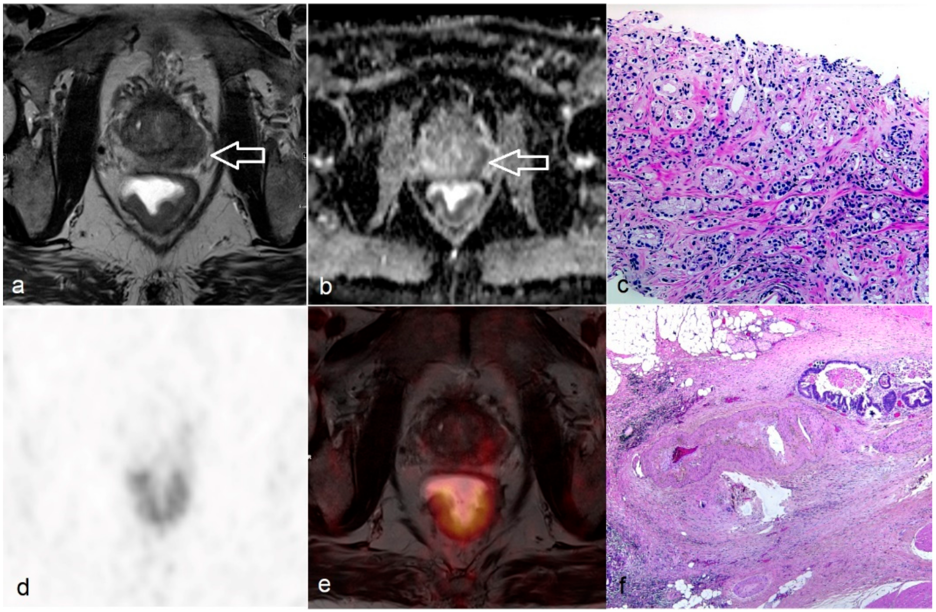
3.2. Diagnostic Performance of PET/MR and MR Alone in Locoregional T and N Staging
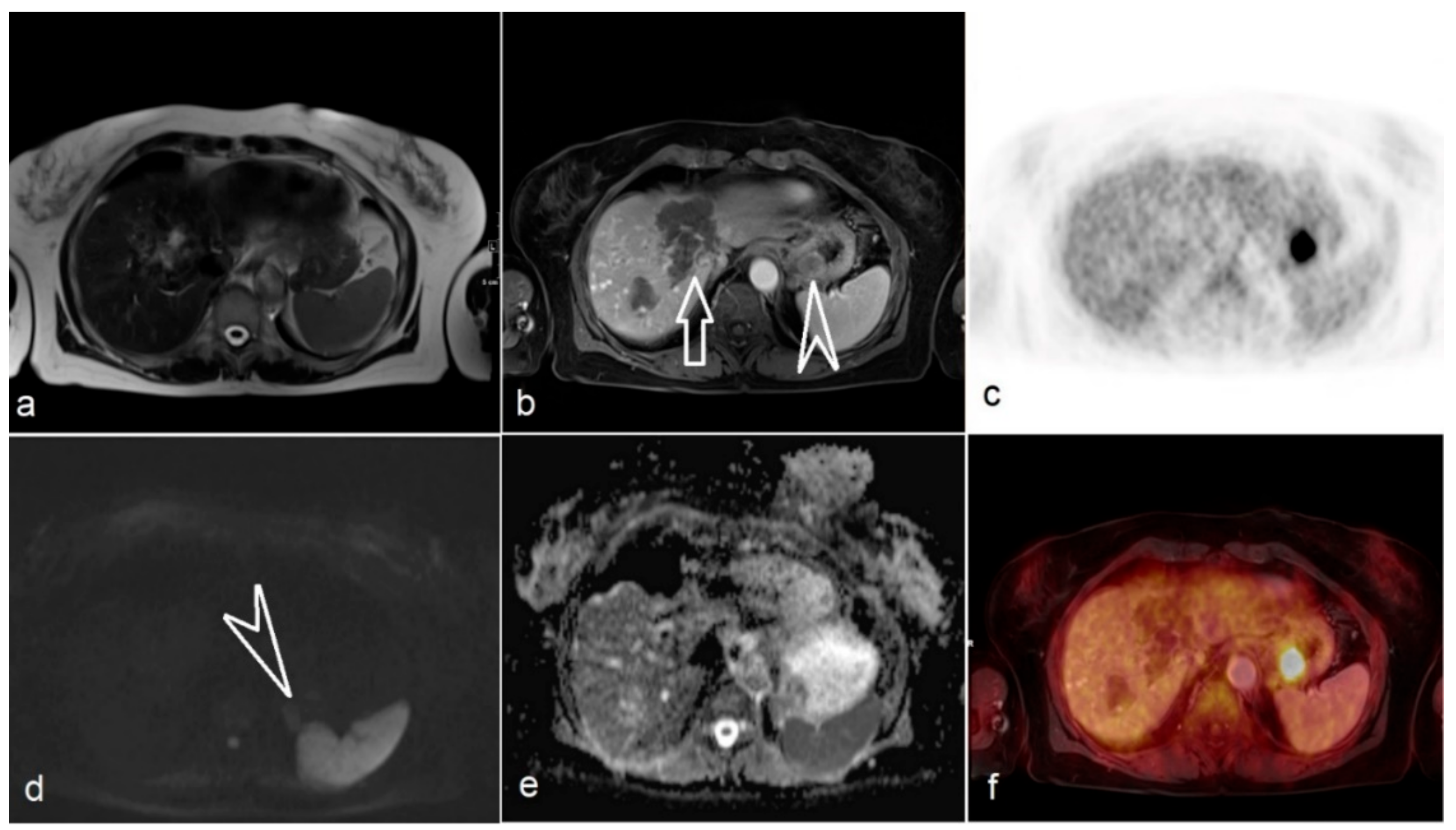
3.3. Diagnostic Performance of PET/MR and MR Alone in M Staging
3.4. Metabolic Information of Untreated and Post-CRT Rectal Tumor
3.5. Additional Findings and Their Clinical Management
4. Discussion
5. Conclusions
Supplementary Materials
Author Contributions
Funding
Conflicts of Interest
References
- Ferlay, J.; Colombet, M.; Soerjomataram, I.; Dyba, T.; Randi, G.; Bettio, M.; Gavin, A.; Visser, O.; Bray, F. Cancer incidence and mortality patterns in Europe: Estimates for 40 countries and 25 major cancers in 2018. Eur. J. Cancer (Oxf. Engl. 1990) 2018, 103, 356–387. [Google Scholar] [CrossRef] [PubMed]
- Beets-Tan, R.G.H.; Lambregts, D.M.J.; Maas, M.; Bipat, S.; Barbaro, B.; Curvo-Semedo, L.; Fenlon, H.M.; Gollub, M.J.; Gourtsoyianni, S.; Halligan, S.; et al. Magnetic resonance imaging for clinical management of rectal cancer: Updated recommendations from the 2016 European Society of Gastrointestinal and Abdominal Radiology (ESGAR) consensus meeting. Eur. Radiol. 2018, 28, 1465–1475. [Google Scholar] [CrossRef] [Green Version]
- Zech, C.J.; Korpraphong, P.; Huppertz, A.; Denecke, T.; Kim, M.J.; Tanomkiat, W.; Jonas, E.; Ba-Ssalamah, A. Randomized multicentre trial of gadoxetic acid-enhanced MRI versus conventional MRI or CT in the staging of colorectal cancer liver metastases. Br. J. Surg. 2014, 101, 613–621. [Google Scholar] [CrossRef] [PubMed] [Green Version]
- van de Velde, C.J.; Boelens, P.G.; Borras, J.M.; Coebergh, J.W.; Cervantes, A.; Blomqvist, L.; Beets-Tan, R.G.; van den Broek, C.B.; Brown, G.; Van Cutsem, E.; et al. EURECCA colorectal: Multidisciplinary management: European consensus conference colon & rectum. Eur. J. Cancer (Oxf. Engl. 1990) 2014, 50, e18–e34. [Google Scholar] [CrossRef]
- Quaia, E.; De Paoli, L.; Angileri, R.; Cabibbo, B.; Cova, M.A. Indeterminate solid hepatic lesions identified on non-diagnostic contrast-enhanced computed tomography: Assessment of the additional diagnostic value of contrast-enhanced ultrasound in the non-cirrhotic liver. Eur. J. Radiol. 2014, 83, 456–462. [Google Scholar] [CrossRef] [PubMed]
- Laghi, F.; Catalano, O.; Maresca, M.; Sandomenico, F.; Siani, A. Indeterminate, subcentimetric focal liver lesions in cancer patients: Additional role of contrast-enhanced ultrasound. Ultraschall in der Med. (Stuttg. Ger. 1980) 2010, 31, 283–288. [Google Scholar] [CrossRef] [PubMed]
- Lee, D.H.; Lee, J.M. Whole-body PET/MRI for colorectal cancer staging: Is it the way forward? J. Magn. Reson. Imaging JMRI 2017, 45, 21–35. [Google Scholar] [CrossRef]
- Kang, B.; Lee, J.M.; Song, Y.S.; Woo, S.; Hur, B.Y.; Jeon, J.H.; Paeng, J.C. Added Value of Integrated Whole-Body PET/MRI for Evaluation of Colorectal Cancer: Comparison with Contrast-Enhanced MDCT. Am. J. Roentgenol. 2015, 206, W10–W20. [Google Scholar] [CrossRef]
- Yoon, J.H.; Lee, J.M.; Chang, W.; Kang, H.-j.; Bandos, A.; Lim, H.-j.; Kang, S.Y.; Kang, K.W.; Ryoo, S.-B.; Jeong, S.-Y.; et al. Initial M Staging of Rectal Cancer: FDG PET/MRI with a Hepatocyte-specific Contrast Agent versus Contrast-enhanced CT. Radiology 2020, 294, 310–319. [Google Scholar] [CrossRef]
- Amorim, B.J.; Hong, T.S.; Blaszkowsky, L.S.; Ferrone, C.R.; Berger, D.L.; Bordeianou, L.G.; Ricciardi, R.; Clark, J.W.; Ryan, D.P.; Wo, J.Y.; et al. Clinical impact of PET/MR in treated colorectal cancer patients. Eur. J. Nucl. Med. Mol. Imaging 2019, 46, 2260–2269. [Google Scholar] [CrossRef]
- Catalano, O.A.; Coutinho, A.M.; Sahani, D.V.; Vangel, M.G.; Gee, M.S.; Hahn, P.F.; Witzel, T.; Soricelli, A.; Salvatore, M.; Catana, C.; et al. Colorectal cancer staging: Comparison of whole-body PET/CT and PET/MR. Abdom. Radiol. 2017, 42, 1141–1151. [Google Scholar] [CrossRef]
- Martin, O.; Schaarschmidt, B.M.; Kirchner, J.; Suntharalingam, S.; Grueneisen, J.; Demircioglu, A.; Heusch, P.; Quick, H.H.; Forsting, M.; Antoch, G.; et al. PET/MRI versus PET/CT in whole-body staging: Results from a unicenter observational study in 1003 subsequent examinations. J. Nucl. Med. Off. Publ. Soc. Nucl. Med. 2019. [Google Scholar] [CrossRef]
- Beets-Tan, R.G.; Lambregts, D.M.; Maas, M.; Bipat, S.; Barbaro, B.; Caseiro-Alves, F.; Curvo-Semedo, L.; Fenlon, H.M.; Gollub, M.J.; Gourtsoyianni, S.; et al. Magnetic resonance imaging for the clinical management of rectal cancer patients: Recommendations from the 2012 European Society of Gastrointestinal and Abdominal Radiology (ESGAR) consensus meeting. Eur. Radiol. 2013, 23, 2522–2531. [Google Scholar] [CrossRef]
- Kim, J.H.; Beets, G.L.; Kim, M.J.; Kessels, A.G.; Beets-Tan, R.G. High-resolution MR imaging for nodal staging in rectal cancer: Are there any criteria in addition to the size? Eur. J. Radiol. 2004, 52, 78–83. [Google Scholar] [CrossRef]
- Brown, G.; Richards, C.J.; Bourne, M.W.; Newcombe, R.G.; Radcliffe, A.G.; Dallimore, N.S.; Williams, G.T. Morphologic predictors of lymph node status in rectal cancer with use of high-spatial-resolution MR imaging with histopathologic comparison. Radiology 2003, 227, 371–377. [Google Scholar] [CrossRef]
- Nougaret, S.; Reinhold, C.; Mikhael, H.W.; Rouanet, P.; Bibeau, F.; Brown, G. The Use of MR Imaging in Treatment Planning for Patients with Rectal Carcinoma: Have You Checked the “DISTANCE”? Radiology 2013, 268, 330–344. [Google Scholar] [CrossRef]
- Beets-Tan, R.G.; Beets, G.L.; Vliegen, R.F.; Kessels, A.G.; Van Boven, H.; De Bruine, A.; von Meyenfeldt, M.F.; Baeten, C.G.; van Engelshoven, J.M. Accuracy of magnetic resonance imaging in prediction of tumour-free resection margin in rectal cancer surgery. Lancet (Lond. Engl.) 2001, 357, 497–504. [Google Scholar] [CrossRef]
- Jia, X.; Zhang, Y.; Wang, Y.; Feng, C.; Shen, D.; Ye, Y.; Hong, N. MRI for Restaging Locally Advanced Rectal Cancer: Detailed Analysis of Discrepancies with the Pathologic Reference Standard. Am. J. Roentgenol. 2019, 213, 1081–1090. [Google Scholar] [CrossRef]
- van der Paardt, M.P.; Zagers, M.B.; Beets-Tan, R.G.; Stoker, J.; Bipat, S. Patients who undergo preoperative chemoradiotherapy for locally advanced rectal cancer restaged by using diagnostic MR imaging: A systematic review and meta-analysis. Radiology 2013, 269, 101–112. [Google Scholar] [CrossRef] [PubMed]
- Park, M.J.; Kim, S.H.; Lee, S.J.; Jang, K.M.; Rhim, H. Locally advanced rectal cancer: Added value of diffusion-weighted MR imaging for predicting tumor clearance of the mesorectal fascia after neoadjuvant chemotherapy and radiation therapy. Radiology 2011, 260, 771–780. [Google Scholar] [CrossRef]
- Kim, S.H.; Song, B.-I.; Kim, B.W.; Kim, H.W.; Won, K.S.; Bae, S.U.; Jeong, W.K.; Baek, S.K. Predictive Value of [18F]FDG PET/CT for Lymph Node Metastasis in Rectal Cancer. Sci. Rep. 2019, 9, 4979. [Google Scholar] [CrossRef] [PubMed]
- Lu, Y.-Y.; Chen, J.-H.; Ding, H.-J.; Chien, C.-R.; Lin, W.-Y.; Kao, C.-H. A systematic review and meta-analysis of pretherapeutic lymph node staging of colorectal cancer by 18F-FDG PET or PET/CT. Nucl. Med. Commun. 2012, 33, 1127–1133. [Google Scholar] [CrossRef] [PubMed]
- Findlay, M.; Young, H.; Cunningham, D.; Iveson, A.; Cronin, B.; Hickish, T.; Pratt, B.; Husband, J.; Flower, M.; Ott, R. Noninvasive monitoring of tumor metabolism using fluorodeoxyglucose and positron emission tomography in colorectal cancer liver metastases: Correlation with tumor response to fluorouracil. J. Clin. Oncol. 1996, 14, 700–708. [Google Scholar] [CrossRef] [PubMed] [Green Version]
- de Geus-Oei, L.F.; Vriens, D.; van Laarhoven, H.W.; van der Graaf, W.T.; Oyen, W.J. Monitoring and predicting response to therapy with 18F-FDG PET in colorectal cancer: A systematic review. J. Nucl. Med. Off. Publ. Soc. Nucl. Med. 2009, 50 (Suppl. 1), 43s–54s. [Google Scholar] [CrossRef] [Green Version]
- Murcia Duréndez, M.J.; Frutos Esteban, L.; Luján, J.; Frutos, M.D.; Valero, G.; Navarro Fernández, J.L.; Mohamed Salem, L.; Ruiz Merino, G.; Claver Valderas, M.A. The value of 18F-FDG PET/CT for assessing the response to neoadjuvant therapy in locally advanced rectal cancer. Eur. J. Nucl. Med. Mol. Imaging 2013, 40, 91–97. [Google Scholar] [CrossRef]
- Avallone, A.; Aloj, L.; Pecori, B.; Caraco, C.; De Stefano, A.; Tatangelo, F.; Silvestro, L.; Granata, V.; Bianco, F.; Romano, C.; et al. (18)F-FDG PET/CT Is an Early Predictor of Pathologic Tumor Response and Survival After Preoperative Radiochemotherapy with Bevacizumab in High-Risk Locally Advanced Rectal Cancer. J. Nucl. Med. Off. Publ. Soc. Nucl. Med. 2019, 60, 1560–1568. [Google Scholar] [CrossRef] [Green Version]


| Characteristics | Value (Percentage) |
|---|---|
| Sex | |
| Male | n = 17 (50%) |
| Female | n = 17 (50%) |
| Age | |
| Mean | 58 |
| Range | 18–78 |
| Histopathology of rectal cancer | |
| Adenocarcinoma | n = 30 (88.2%) |
| Mucinous adenocarcinoma | n = 4 (11.8%) |
| Location of rectal cancer | |
| Upper (12–16 cm from anal verge) | n = 7 (20.6%) |
| Middle (6–12 cm) | n = 14 (41.2%) |
| Lower (≤6 cm) | n = 13 (28.2%) |
| Indications for PET/MR | |
| Primary staging | n = 9 (23.1%) |
| Preoperative restaging after CRT * | n = 14 (35.9%) |
| Restaging under surveillance with indices of local recurrence/distant metastasis | n = 16 (41.0%) |
| Untreated Patients (n = 9) | Post-CRT * Patients (n = 14) | All Patients (n = 23) | ||||||||
|---|---|---|---|---|---|---|---|---|---|---|
| T1/2 | T3/4 | Total Number | T1/2 | T3/4 | Total Number | T1/2 | T3/4 | Total Number | ||
| Reader 1 | T1/2 | 1 | 1 | 2 | 5 | 3 | 8 | 6 | 4 | 10 |
| T3/4 | 1 | 6 | 7 | 0 | 6 | 6 | 1 | 12 | 13 | |
| Total number | 2 | 7 | 9 | 5 | 9 | 14 | 7 | 16 | 23 | |
| Reader 2 | T1/2 | 1 | 1 | 2 | 2 | 5 | 7 | 3 | 6 | 9 |
| T3/4 | 1 | 6 | 7 | 3 | 4 | 7 | 4 | 10 | 14 | |
| Total number | 2 | 7 | 9 | 5 | 9 | 14 | 7 | 16 | 23 | |
| Inter-observer agreement for T stage | κ **= 1, p < 0.001 | κ = 0.29, p > 0.05 | κ = 0.553, p = 0.008 | |||||||
| Untreated Patients (n = 9) | Post-CRT * Patients (n = 14) | All Patients (n = 23) | |||||||||
|---|---|---|---|---|---|---|---|---|---|---|---|
| N0 | N+ | Total Number | N0 | N+ | Total Number | N0 | N+ | Total Number | |||
| Reader 1 | MR alone | N0 | 4 | 0 | 4 | 6 | 3 | 9 | 10 | 3 | 13 |
| N+ | 2 | 3 | 5 | 0 | 5 | 5 | 2 | 8 | 10 | ||
| Total Number | 6 | 3 | 9 | 6 | 8 | 14 | 12 | 11 | 23 | ||
| Combined PET/MR | N0 | 5 | 0 | 5 | 6 | 4 | 10 | 11 | 4 | 15 | |
| N+ | 1 | 3 | 4 | 0 | 4 | 4 | 1 | 7 | 8 | ||
| Total Number | 6 | 3 | 9 | 6 | 8 | 14 | 12 | 11 | 23 | ||
| Reader 2 | MR alone | N0 | 3 | 0 | 3 | 6 | 3 | 9 | 9 | 3 | 12 |
| N+ | 3 | 3 | 6 | 0 | 5 | 5 | 3 | 8 | 11 | ||
| Total Number | 6 | 3 | 9 | 6 | 8 | 14 | 12 | 11 | 23 | ||
| Combined PET/MR | N0 | 4 | 0 | 4 | 6 | 3 | 9 | 10 | 3 | 13 | |
| N+ | 2 | 3 | 5 | 0 | 5 | 5 | 2 | 8 | 1 | ||
| Total Number | 6 | 3 | 9 | 6 | 8 | 14 | 12 | 11 | 23 | ||
| Inter-observer agreement for N stage | κ ** = 0.768 for MR, p = 0.018 κ = 0.780 for PET/MR, p = 0.016 | κ = 0.689 for MR, p = 0.01 κ = 0.837 for PET/MR, p = 0.001 | κ = 0.738 for MR, p < 0.001 κ = 0.819 for PET/MR, p < 0.001 | ||||||||
| M Stage (n = 39) | |||||
|---|---|---|---|---|---|
| M0 | M1 | Total Number | |||
| Reader 1 | MR alone | M0 | 21 | 0 | 21 |
| M1 | 2 | 16 | 18 | ||
| Total number | 23 | 16 | 39 | ||
| Combined PET/MR | M0 | 20 | 0 | 20 | |
| M1 | 3 | 16 | 19 | ||
| Total number | 23 | 16 | 39 | ||
| Reader 2 | MR alone | M0 | 20 | 1 | 21 |
| M1 | 3 | 15 | 18 | ||
| Total number | 23 | 16 | 39 | ||
| Combined PET/MR | M0 | 19 | 1 | 20 | |
| M1 | 4 | 15 | 19 | ||
| Total number | 23 | 16 | 39 | ||
| Inter-observer agreement for M stage | κ * = 0.637 for MR and 0.64 for PET/MR, both p < 0.001 | ||||
| Untreated T0-2 Stage (n = 2) | Untreated T3-4 Stage (n = 7) | Post-CRT ** T0-2 Stage (n = 5) | Post-CRT T3-4 Stage (n = 9) | |
|---|---|---|---|---|
| SUVmax | 17.25 (± 10.11) (mean ±SD) | 15.9 (± 4.5) | 2.89 (± 3.76) | 7.98 (± 4.88) |
| p * = 0.125 | ||||
| SUVmax_Ratio | 6.01 (± 4.41) | 4.29 (± 1.8) | 0.85 (± 0.6) | 2.1 (± 4) |
| p * = 0.142 | ||||
© 2020 by the authors. Licensee MDPI, Basel, Switzerland. This article is an open access article distributed under the terms and conditions of the Creative Commons Attribution (CC BY) license (http://creativecommons.org/licenses/by/4.0/).
Share and Cite
Li, Y.; Mueller, L.I.; Neuhaus, J.P.; Bertram, S.; Schaarschmidt, B.M.; Demircioglu, A.; Ludwig, J.M.; Kirchner, J.; Rischpler, C.; Herrmann, K.; et al. 18F-FDG PET/MR versus MR Alone in Whole-Body Primary Staging and Restaging of Patients with Rectal Cancer: What Is the Benefit of PET? J. Clin. Med. 2020, 9, 3163. https://doi.org/10.3390/jcm9103163
Li Y, Mueller LI, Neuhaus JP, Bertram S, Schaarschmidt BM, Demircioglu A, Ludwig JM, Kirchner J, Rischpler C, Herrmann K, et al. 18F-FDG PET/MR versus MR Alone in Whole-Body Primary Staging and Restaging of Patients with Rectal Cancer: What Is the Benefit of PET? Journal of Clinical Medicine. 2020; 9(10):3163. https://doi.org/10.3390/jcm9103163
Chicago/Turabian StyleLi, Yan, Laura Isabel Mueller, Jan Peter Neuhaus, Stefanie Bertram, Benedikt Michael Schaarschmidt, Aydin Demircioglu, Johannes Maximilian Ludwig, Julian Kirchner, Christoph Rischpler, Ken Herrmann, and et al. 2020. "18F-FDG PET/MR versus MR Alone in Whole-Body Primary Staging and Restaging of Patients with Rectal Cancer: What Is the Benefit of PET?" Journal of Clinical Medicine 9, no. 10: 3163. https://doi.org/10.3390/jcm9103163
APA StyleLi, Y., Mueller, L. I., Neuhaus, J. P., Bertram, S., Schaarschmidt, B. M., Demircioglu, A., Ludwig, J. M., Kirchner, J., Rischpler, C., Herrmann, K., Catalano, O. A., & Umutlu, L. (2020). 18F-FDG PET/MR versus MR Alone in Whole-Body Primary Staging and Restaging of Patients with Rectal Cancer: What Is the Benefit of PET? Journal of Clinical Medicine, 9(10), 3163. https://doi.org/10.3390/jcm9103163