Article

Novel Rapid Protein Coating Technique for Silicon Photonic Biosensor to Improve Surface Morphology and Increase Bioreceptor Density

by
Suruk Udomsom
1,2,*,
Ukrit Mankong
1,3,*,
Pathinan Paengnakorn
1,4 and
Nipon Theera-Umpon
1,3
1
Biomedical Engineering Institute, Chiang Mai University, Chiang Mai 50200, Thailand
2
PhD Program in Biomedical Engineering, Biomedical Engineering Institute, Chiang Mai University, Chiang Mai 50200, Thailand
3
Department of Electrical Engineering, Faculty of Engineering, Chiang Mai University, Chiang Mai 50200, Thailand
4
Center of Excellence for Innovation in Analytical Science and Technology, Chiang Mai University, Chiang Mai 50200, Thailand
*
Authors to whom correspondence should be addressed.
Coatings 2021, 11(5), 595; https://doi.org/10.3390/coatings11050595
Submission received: 26 March 2021 / Revised: 10 May 2021 / Accepted: 11 May 2021 / Published: 18 May 2021
(This article belongs to the Special Issue Thin Films and Structures for Optical Sensing)

Abstract

:
Silicon photonic devices with either silicon or silicon nitride waveguides have increasingly been used in many applications besides communications, especially as sensors in label-free biosensing, where guided light signals are affected by biorecognition molecules immobilized on the surface. The coating of protein (i.e., bioreceptors) by biochemical process on the waveguide surface is a crucial step in creating a functionalized device that can be used for biosensing. As a conventional method that uses 3-aminopropryltriethoxysilane (APTES) and glutaraldehyde (GA), the APTES-GA method has the limitation of using a GA crosslink, of which the two functional groups can bind to nonspecific proteins, causing irregular binding. In this study, we proposed a new coating technique to avoid such problem by applying APTES silanization with 1-ethyl-3-(3-dimethyl aminopropyl)-carbodiimide (EDC)-N-hydroxysuccinimide (NHS) protein crosslink, denoted by the APTES-(EDC/NHS) method. The EDC/NHS reaction was shown to be able to immobilize protein in ordered orientation due to consistent arrangement between a carboxylic group of protein molecules and an amine group of covalent-linked APTES on surface. By applying APTES silanization, we circumvented the use of hazardous cleaning agent in the conventional EDC/NHS technique. Several surface characterization techniques were carried out to assess and compare the two biocoating techniques, including scanning electron microscopy (SEM), energy-dispersive X-ray spectroscopy (EDS), spectroscopic ellipsometry (SE), and atomic force microscopy (AFM). On silicon, the results of antihuman TNF-alpha antibody coating showed that the proposed APTES-(EDC/NHS) technique has better repeatability in terms of less roughness of the coated protein at 1.5 nm compared with 6.3 nm, due to the ordered arrangement of coated antibody molecules. On a silicon nitride waveguide device, the proposed APTES-(EDC/NHS) technique exhibits dense antibody immobilization on a waveguide in SEM images due to stable amide bond formation via EDC/NHS crosslink mechanism. The specificity of the immobilized antibodies was confirmed by enzyme-linked immunosorbent assays (ELISA), with an average optical density at 450 nm of 0.175 ± 0.01 compared with 0.064 ± 0.009 of negative control. The proposed technique also reduced the overall process time since proteins are crosslinked to the silanized waveguide surface in a single step.

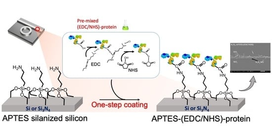
Graphical Abstract

1. Introduction

In recent years, advances in compact biosensor and chemical sensor technologies, artificial intelligence, and the Internet of Things have led to the growth of personalized health monitoring and diagnosis. For instance, high-performance “wearable sensors” have been developed and increasingly utilized in health monitoring such as blood monitoring, pH sensors, and sweat glucose analysis [1,2,3,4]. In addition, wearable silicon photonic based devices are now used in physical sensing, chemical/gas sensing, and glucose and real-time pH monitoring [5,6,7,8]. Silicon photonic resonator-based devices using either silicon (Si) or silicon nitride (Si3N4) waveguides can also be applied in point-of-care testing, enabling timely diagnosis and patient care [9,10].
Silicon photonic resonator-based devices rely on the resonant property of light signal transmission. The resonant wavelength shift of light through the resonator is responsive to the effective refractive index change that occurs through biomaterial binding on the waveguide surface, such as antibody–antigen [11,12,13] or enzyme–substrate [14,15]. Therefore, the coating of a bioreceptor—such as an antibody or enzyme—on silicon is the essential stage where a high density of protein molecules must be immobilized onto silicon surface uniformly. The quality and physical properties of such coating have a direct effect on the sensitivity and accuracy of the measurement [16,17,18]. Generally, several steps are required in protein coating by a biochemical process, including device cleaning, silanization, and bioreceptor crosslinking. Silanization is the process where an aminosilane is used to create a chemical functional group of Si that is able to react with the functional group of bioreceptors. The organosilane reagent 3-aminopropyltriethoxysilane (APTES) is widely used in silanization where it reacts with the −OH group on the silicon surface and leaves its amino group (–NH2) available to react with a crosslink [16,19,20,21,22,23,24,25,26,27]. However, the uniformity and repeatability of APTES layer has been difficult to control since APTES can attach the silicon surface in a different position [28]. Polyethylene glycol (PEG) is another popular alternative because it is able to react with -OH directly on the silicon surface due to the presence of alkylthiol groups such as (HS–C11–(EG)4–OH) and (HS–C11–(EG)6–COOH). PEG can also bind to the primary amine (–NH2) of APTES after silanization [29]. The advantages of PEG are hydrophobicity and specificity to the crosslink functional group to reduce the occurrence of irregular binding in the system. However, it is also difficult to control the coated thickness of PEG due to its large molecular size. APTES silanization on silicon requires an additional crosslink agent to connect with the bioreceptor protein molecules. The most common crosslinker is glutaraldehyde (GA) [13,30], the structure of which consists of the two arms of aldehydes (–CHO) groups, which can react well with the amino groups of proteins. Due to the nonspecific binding sites of the two arms, this can potentially cause an undesired mesh structure between the proteins.
Another silanization and crosslinking system is 1-Ethyl-3-(3-dimethyl aminopropyl)-carbodiimide (EDC) in combination with N-hydroxysuccinimide (NHS) [31,32]. EDC specifically binds to carboxyl (–COOH) groups of protein, and it can react accurately with NHS to crosslink proteins, which can reduce the occurrence of irregular binding in the system. When NHS reacts with EDC, its form becomes active to bind specific primary amine (–NH2) of functional proteins. Therefore, regular binding in C-terminal to N-terminal on the surface can be assured [33]. However, to bind EDC onto the silicon surface, additional steps are required, as well as the use of harmful substances such as hydrogen fluoride (HF). A comparison of different techniques are summarized in Table 1.
The aim of this study is to develop a suitable protein coating technique for silicon photonic biosensor devices, in which repeatability, uniformity, and sensitivity are especially needed. We propose a rapid bioreceptor protein coating technique on Si and Si3N4 by combining APTES-based silanization with EDC/NHS for the first time. Experiments are carried out to compare the proposed APTEST-(EDC/NHS) technique with the conventional APTES-GA technique. The results of surface morphology measurements and element composition analysis are obtained using scanning electron microscopes (SEM), energy-dispersive X-ray spectroscopy (EDS), spectroscopic ellipsometry (SE), and atomic force microscope (AFM). Using the proposed technique, we can avoid the disadvantage of using a GA crosslink and concurrently eliminate the HF cleaning process in a conventional EDC/NHS system. Therefore, both benefits of APTES silanization and the specificity of EDS/NHS may be obtained. In this proposed technique, the silanized surface is crosslinked with NHS, which is attached to functional proteins beforehand, thus enabling a simplified and rapid process. In addition, to confirm the presence of an immobilized bioreceptor protein on Si and Si3N4 surfaces, ELISA experiments are carried out using an antihuman TNF-alpha coating to represent the antibody–antigen system. Glucose oxidase (GOx) enzyme coating experiments are also carried out to represent the enzyme–substrate system.

2. Differences of APTES-GA, EDC-NHS and the Proposed APTES-(EDC/NHS) Coating Systems

2.1. Conventional APTES-GA and EDC-NHS Coating Techniques

Figure 1 describes the steps in the conventional APTES-GA method, which is based on the condensation between the siloxanes of the organosilane and hydroxyl groups that present on the surface. The device surface must firstly be cleaned with a Piranha solution treatment to produce hydroxyl groups (–OH) on Si (or Si3N4) by a hydroxylation process. Hydroxyl groups (–OH) are used by the silanization step by the hydrolysis of ethoxy (–C 2H 5) groups from APTES, leading to the formation of silanols (Si–OH). APTES silanols can condense with surface silanols and express amine groups, which can further crosslink with a bioreceptor protein (e.g., antibody) using a GA linker [11,12,13,14,15,16,17,18,19]. There is a potential issue with crosslinking by GA since both functional groups (aldehydes) of GA molecules are identical, and nonspecific to proteins. Therefore, irregular binding may occur. For example, both aldehydes of some GA molecules may bind with APTES, thereby preventing the desired protein crosslink.
Figure 2 shows the conventional (EDC/NHS) method, based on the coupling reaction between EDC and carboxyl (–COOH) groups on the modified Si (or Si3N4) device surface. Firstly, the surface must be free of oxide by cleaning it with a HF solution, leading to an atomically hydrogenated surface (SiH). Then, it is modified with acid (acrylic acid) to produce carboxyl (–COOH) groups or an acid-terminated silicon surface (Si–COOH), which can be coupled with the reactive O-acylisourea functional group of EDC. Subsequently, NHS is used to crosslink with a bioreceptor protein [41,42,43]. The EDC/NHS technique does not have the irregular binding issue as in the APTES-GA method since the reaction sites of EDC/NHS are unique. However, the step to produce a hydrogenated surface requires hazardous HF acid, which is not suitable for our laboratory.

2.2. The Proposed APTES-(EDC/NHS) Coating Technique

We propose an APTES-(EDC/NHS) coating technique to combine APTES silanization with the modified EDC/NHS crosslinking as shown in Figure 3. Firstly, the silicon surface was treated with Piranha solution using the chemical reaction between sulfuric acid (H2SO4) and ≈30% hydrogen peroxide (H2O2). This produced Caro’s acid (H2SO5), which can cause dissociation and generate a hydroxyl radical (OH*) to produce hydroxyl groups (–OH) by a hydroxylation process. Then, APTES silanization was performed to prepare primary amine on the surface. Contrary to the conventional EDC/NHS technique in Figure 2, we prepared a premixed protein with EDC and NHS in an activation buffer (0.1 M MES, 0.5 M NaCl, pH 6.0), thus the process was greatly simplified as in Figure 3. This step resulted in carboxyl (–COOH) groups on the bioreceptor protein that is active to EDC and formed an amine-reactive O-acylisourea intermediate that can conjugate with NHS for stabilization and release an iso-urea by-product. Finally, when applying the premixed protein to an APTES silanized surface, NHS changes itself to be a substance that is active to the primary amine (–NH2) of APTES on silicon, resulting in an amide bond.

3. Materials and Methods

Experiments were carried out to compare the conventional APTES-GA biocoating technique with the proposed combined APTES with the modified EDC/NHS coating technique (to be called APTES-(EDC/NHS)). Concentrations of chemical and reagents, the incubation time, and temperature were the same as in other references [30,31,46,47,48], except where the coating antibody concentration are varied (in Section 4.3).

3.1. Chemicals and Reagents

All chemical and reagents were of analytical grade and were used without further purification. Deionized (DI) water were used throughout. Chemicals in the cleaning and silanization were a phosphate buffer solution (PBS, Sigma-Aldrich, Queenstown, Singapore) and 3-aminopropryltriethoxysilane (APTES, Sigma-Aldrich, Queenstown, Singapore), while 30% hydrogen peroxide (H2O2 [49]) and ethanol (Merck Ltd., Queenstown, Singapore) were used to prepare a Piranha solution for the cleaning step. In addition, 4-amino antipyrine for GOx enzyme activity measurement, bovine serum albumin (BSA), and Tween 20 were obtained from Sigma-Aldrich (St. Louis, MO, USA). Glutaraldehyde (GA, Thermo Scientific Ltd., New York, NY, USA) was used only in the APTES-GA technique. In the APTES-(EDC/NHS) coating technique, additional chemicals were N-ethyl-N’-(3-dimethylaminopropyl) carbodiimide hydrochloride (EDC, Sigma-Aldrich, Queenstown, Singapore) and N-hydroxysuccinimide (NHS, Sigma-Aldrich, Queenstown, Singapore), while 2-(N-morpholino) ethanesulfonic acid (MES, Sigma-Aldrich, Singapore) solution was used as an activation buffer. The bioreceptor proteins and detected analytes were the antihuman TNF-alpha, TNF-alpha (Human TNF alpha Uncoated ELISA, Invitrogen, Woodlands, Singapore), and glucose oxidase enzyme (GOx, Sigma-Aldrich, Queenstown, Singapore). Coating experiments were performed on two types of samples: (i) the device-grade (100) silicon wafer (cut into chips of roughly 5 × 5 mm2) and (ii) chips with silicon and silicon nitride waveguide devices. The silicon wafer had the same surface orientation as those used to fabricate silicon photonic biosensors.

3.2. Procedure for Protein Coating

3.2.1. Cleaning and Silanization

The same cleaning and silanization steps were applied in both the APTES-GA and the APTES-(EDC/NHS) techniques, as summarized in this section. The silicon surface was cleaned with acetone for 10 min at room temperature. Then, it was cleaned with methanol for 5 min at room temperature and thoroughly rinsed with DI water and dried with nitrogen gas. To remove the organic compound on Si or Si3N4 surfaces, the chips were cleaned with Piranha solution (H2SO4:H2O2 = 3:1) by immersing them in the solution for 30 min, then thoroughly rinsed with DI water and dried with nitrogen gas. In the silanization stage, the hydroxylated silicon surface was immersed in APTES solution (2% APTES in 95% ethanol) for 1 h at 68 °C, then rinsed thoroughly with DI water and dried with nitrogen gas.

3.2.2. APTES-GA Coating Procedure

This is the conventional procedure that is compared with the proposed technique. Firstly, the silanized silicon devices were coupled with 2.5% GA for 45 min at room temperature and rinsed with DI water and dried with nitrogen gas. Secondly, the functionalized APTES-GA was incubated with bioreceptor proteins. Two functional proteins, i.e., antibody and enzyme, were demonstrated as model systems. Either antihuman TNF-alpha antibody solution in dilution 1:250 (also other dilutions in Section 4.3) in PBS (denoted by Ab-TNF-alpha) or 100 U/mL glucose oxidase enzyme (denoted by GOx) was added and incubated for 2 h at room temperature, then thoroughly rinsed with DI water and dried with nitrogen gas. The idle time between consecutive steps was under 150 min, while the samples were kept in PBS.

3.2.3. The Proposed APTES-(EDC/NHS) Coating Procedure

In this technique, a solution of bioreceptor protein conjugated with EDC and NHS was prepared before being applied onto the silanized silicon in a single step. Each protein was conjugated with 0.7 mg/mL of EDC and 0.6 mg/mL of NHS in the activation buffer (0.1 M MES, 0.5 M NaCl, pH 6.0) for 15 min at room temperature (premixing). Then, the silanized silicon was immersed in the prepared EDC-NHS-protein solution for 2 h at room temperature and thoroughly rinsed with DI water and dried with nitrogen gas. The bioreceptor protein was either an antihuman TNF-alpha antibody solution in dilution 1:250 (also other dilutions in Section 4.3) in PBS or 100 U/mL glucose oxidase enzyme as in the previous technique. The APTES-(EDC/NHS) coating process is summarized in Figure 4.

3.3. Measurements of Coating Effectiveness

Several measurements were performed to assess and compare the two biocoating techniques, which are categorized into 3 groups: surface morphology measurements, element composition analysis, and bioreaction measurement.

3.3.1. Surface Morphology Measurements

Images of the protein-coated silicon surface were taken using scanning electron microscopes (SEM). All samples were dried and coated with gold before scanning with JCM-7000 field-emission scanning electron microscope (JEOL Ltd., Tokyo, Japan).
The thickness of coating layers was measured by spectroscopic ellipsometry (Sopra GES 5, Semilab USA LLC., Billerica, MA, USA) after each step of surface modification and biofunctionalization. Ellipsometry is an optical technique for determining dielectric properties, complex refractive index or dielectric function, of thin films. Ellipsometry measures the polarization changes from reflections or transmissions and compares them with models [50]. All measurements were performed at an incidence angle of 74° and with varying wavelengths between 210 and 920 nm. The data were processed using Winelli software. The ellipsometric parameters were estimated using dispersion law fitting (Cauchy Law) [50,51]. The best fit for the experimental data and for the theoretical model was obtained with a refractive index of 1.42. The extinction coefficient was set to 0 and 0.01 for silane (APTES silanized) and glutaraldehyde (GA), respectively [52]. For the antibody layer and EDC/NHS, the best fit was obtained for a refractive index of 1.45 [53,54,55] and an extinction coefficient of 0. Five measurements were taken for each sample at different locations to obtain the mean and the standard deviation values.
Surface morphology was measured to study the biocoated surface roughness using an atomic force microscope (AFM), XE 70 model (Park system, Suwon, Korea) in contact mode with NCS36 cantilevers, with a tip apex radius of curvature under 10 nm, a scan rate of 1 Hz, and scan area 2 × 2 μm2 [49].

3.3.2. Surface Element Composition

Energy-dispersive X-ray spectroscopy (EDS) measurements were performed coupled with SEM imaging to analyze the element composition on the surface of the coated silicon based on their emitted elemental characteristic X-ray.

3.3.3. Bioreaction Measurements

In addition to surface morphology and spectroscopy measurements, the specificity of coated bioreceptor on the silicon surface was obtained by the measurements of reaction between the coated receptor protein and its target analyte. Two experiments were performed: ELISA for the coated TNF-alpha antibody and an enzyme activity measurement for the coated GOx enzyme. Changes in absorption due to the reaction were measured using a visible spectrophotometer.
The procedure for ELISA detection of TNF-alpha was modified from previous reports [56,57,58]. The antihuman TNF-alpha antibody coated samples and negative controls were placed into a 96-well plate. The procedure consisted of the following steps, and before each step, the samples were washed with a 200 μL/well wash buffer (0.05% Tween 20 in PBS) with a microplate shaker for 5 times to remove excess Ab or other molecules. Firstly, a blocking step was required to block nonspecific binding and reduce the background. An aliquot of 200 μL assay diluent was added per well and left for incubation at room temperature (RT) for 1 h while shaking. Secondly, an aliquot of 100 µL of the TNF-alpha recognition molecule was added per well and incubated for 90 min at RT while shaking. Then, an aliquot of 100 µL of the biotin-conjugated antihuman TNF-alpha antibody was added per well and incubated for 1 h at RT while shaking. After this, an aliquot of 100 μL of avidin-HRP solution was added to each well, incubated for 30 min at RT while shaking. Finally, an aliquot of 100 μL of TMB substrate solution was added and incubated for 15–30 min in the dark or until the desired color developed. The reaction was stopped by adding 100 μL of stop solution (2N H2SO4). The absorbance was measured at 450 nm and subtracted with absorbance at 570 nm within 30 min.
The procedure for GOx enzyme activity measurement was modified from previous reports [59,60]. It consists of the following steps. The GOx coated silicon samples and uncoated silicon samples (negative control) were incubated by being immersed in 500 µL of 10 mg/mL glucose solution for 1 h and 24 h. An aliquot of 2 µL of incubated solution was mixed with 200 µL of H2O2 measurement reagent (peroxidase enzyme in 1.6 mM 4-aminoantipyrine with 22 mM phenol solution) and incubated for 10 min at RT. The activity was measured by the absorbance at 490 nm at incubation time 1 h and 24 h.

4. Results and Discussions

4.1. Biocoating of Silicon Wafer (Nondevice)

Surface Morphology Measurement
The SEM images show that the APTES and APTES-GA layers were distributed on the silicon surface (Figure 5b,c). The final Ab-TNF-alpha protein films were observed on the surface using both conventional (APTES-GA) and the proposed (APTES-(EDC/NHS)) coating techniques, as shown in Figure 6a,b, respectively. It was found that the quantity of aggregated particles in the APTES-GA technique was greater than those in the APTES-(EDC/NHS) technique. This result indicates that the APTES-GA method has a strong crosslink between the proteins from the aggressive aldehyde functional group of GA. Similar SEM images of GOx coating using APTES-GA and APTES-(EDC/NHS) techniques are shown in Figure 7a,b, respectively. The surface of the APTES-(EDC/NHS) technique appeared to be smoother compared with that of the APTES-GA technique.
The spectroscopic ellipsometer was used to measure the thickness to compare the increased average thickness after each stage of coating (see Supplementary Materials, Figures S1–S3). In the conventional APTES-GA technique, we measured the increased thickness at four stages: (i) initial SiO2 layer due to Si surface oxidization of the sample, (ii) APTES layer, (iii) APTES-GA layers, and (iv) APTES-GA-protein layers. On the other hand, in the proposed APTES-(EDC/NHS) technique, we measured the increased thickness at three stages: (i) initial SiO2 layer due to Si surface oxidization of the sample, (ii) APTES layer, and (iii) APTES-(EDC/NHS)-protein layers, which was the final stage after the premixed EDC/NHS-protein solution had been applied. The protein was either anti-TNF-alpha Ab or GOx enzyme.
The thickness results are presented in Figure 8 and Figure 9. The results show that for anti-TNF-alpha coating using the APTES-GA and APTES-(EDC/NHS) techniques, the overall thickness values were 10.8 nm and 8.6 nm, respectively. For the GOx coating using the APTES-GA and APTES-(EDC/NHS) techniques, the overall thickness values were 10.12 nm and 9.2 nm, respectively.
Thicker coated layers were observed in the APTES-GA technique than in the APTES-(EDC/NHS) technique, and this was likely caused by the self-polymerization of GA to an agglomeration of the protein layer, causing irregular protein reaction. This agrees with previous studies that explained the relationship between silanization reaction and GA [49].
The atomic force microscope (AFM) was utilized to determine the surface topology, which was presented in terms of root mean square (RMS) roughness (see Supplementary Materials, Figures S4–S6). The observed agglomeration of the molecule on the surface was a result of the silanization process mechanism, as described in a previous study [6]. Table 2 summarizes the RMS roughness values of the APTES-GA method compared with the APTES-(EDC/NHS) method. The values of roughness of uncoated and APTES silanized silicon surfaces were similar in both techniques. However, it was found that the APTES-(EDC/NHS) technique yielded less roughness of the coated anti-TNF-alpha and GOx than the APTES-GA technique at 1.5 nm and 3.9 nm compared with 6.3 nm and 8.7 nm, respectively.

4.2. Surface Element Composition

The relative weight percentages of Si, N, O, and C atoms on the surface were measured after each coating step by energy-dispersive X-ray spectroscopy (EDS). The results are shown in Figure 10 and Figure 11. All results show that, as the protein and crosslink layers are increased, the percentages of N, O, and C, which are indicative of organic compounds, increase on the surface while the percentage of Si drops. EDS can penetrate at ca. 1–10 nm in depth; therefore, these changes indicate the presence of bioreceptor proteins on top of the silicon surface.

4.3. Bioreaction Measurement

Bioactivity experiments were used to demonstrate the quantity of the coated bioreceptor. We applied ELISA to the antigen–antibody system. The anti-TNF-alpha antibody concentration that was used to coat on Si was varied (1:128, 1:250, 1:500, 1:1000). The antigen concentration is constant at 500 μg/mL. The results in Figure 12 show that the average values of optical density (OD) at 450 nm using the antibody-coated Si by both the proposed APTES-(EDC/NHS) (n = 5) and the conventional APTES-GA techniques (n = 5) are similar at all conditions. The reactivity of the immobilized protein by both techniques are approximately 16 and 20% of the positive control at all concentrations. Thus, the results suggest that the proposed coating technique is able to bind the antibody onto the silicon and generate an antibody–antigen reaction in the same order as the conventional method.
Furthermore, the higher anti-TNF-alpha antibody concentration generally corresponds to a lower standard deviation in reactivity, i.e., 0.167 ± 0.03 (1:128), 0.165 ± 0.04 (1:250), 0.191 ± 0.05 (1:500), and 0.179 ± 0.02 (1:1000) in the APTES-(EDC/NHS) technique, and 0.198 ± 0.02 (1:128), 0.156 ± 0.05 (1:250), 0.163 ± 0.05 (1:500), and 0.181 ± 0.07 (1:1000) using the APTES-GA technique. The deviations could be a result of protein loss during the coating process and nonspecific binding on Si. By increasing the antibody concentration, a better repeatability of the quantity of immobilized antibody on the Si surface should be achieved.
The reaction results of glucose oxidase coated silicon to glucose solution are shown in Figure 13. An increase in absorbance at 490 nm is directly proportional to GOx activity for the oxidation of glucose to gluconic acid and H2O2. This indicates the presence of enzyme being successfully coated on the surface by both methods. The higher enzyme activity in the APTES-(EDC/NHS) technique was observed (both after 1 h and 24 h incubation). This may be the effect of a consistent arrangement of GOx by the APTES-(EDC/NHS) technique, which does not affect the active site of the enzyme. The incubation time was long in these experiments due to the thin enzyme film on the surface, unlike standard enzyme experiments.

4.4. Results of Silicon and Silicon Nitride Waveguide Devices

The coating of an antibody on a silicon-based device with waveguide(s) was performed in order to investigate the structure of the antibody layer on waveguides as part of sensor devices. The SEM images of Ab-TNF-alpha coating on Si and Si3N4 waveguides using both APTES-GA and APTES-(EDC/NHS) techniques are shown in Figure 14 and Figure 15. It is shown that surface modification with APTES for antibody immobilization can be applied to both Si and Si3N4 waveguides, consistent with a previous report [61]. On the Si waveguide devices, it was observed that the antibody coating using the APTES-(EDC/NHS) technique exhibited distinct protein clusters on the waveguide (Si), while the antibody coating using the APTES-GA technique showed more scattered proteins on both the waveguide (Si) and the substrate (SiO2), as in Figure 14. On the Si3N4 waveguide devices, similarly, it was found that the proposed APTES-(EDC/NHS) method shows a greater amount of immobilized antibody on the waveguide (Si3N4) compared with the substrate (SiO2), as in Figure 15. The reason should be that EDC/NHS facilitates the formation of a stable amide bond between a carboxylic group of protein molecules and an amine group of covalent-linked APTES on Si3N4 surface. While in case of the APTES-GA technique, a glutaraldehyde crosslinker can cause a self-crosslink between protein molecules which competes with protein immobilization on the Si3N4 waveguide [62]. Therefore, our proposed method is attractive for the sensor devices using Si3N4 waveguides, as the higher density of the antibody on the waveguide corresponds to increased sensor sensitivity.

5. Conclusions

We proposed and experimentally verified a new technique to biochemically coat a bioreceptor protein on silicon chips and a silicon photonic device using either silicon or silicon nitride waveguides. The technique combines APTES silanization with a modified EDC/NHS crosslink. The protein can be bound to the silanized silicon surface in a single step since the EDC/NHS-protein solution is premixed, thus simplifying the process. On plain silicon chips, the proposed APTEST-(EDS/NHS) technique showed smoother and more consistent layers compared with the APTES-GA technique due to the specificity of protein binding in the EDC/NHS system. Similar average immobilized bioreceptor densities were observed by both techniques, as evidenced by ELISA results. However, increased protein concentration is associated with improved repeatability. On the waveguide devices, the proposed APTEST-(EDS/NHS) technique showed a higher immobilized bioreceptor density on the Si3N4 waveguides. Therefore, a higher sensitivity of biosensing can be expected in Si3N4 waveguide sensors using our method. Further study is required to apply this protein coating technique in actual bioanalyte sensing and to determine the necessary sensor characteristics, including sensitivity and detection limit, in real world applications such as point-of-care testing.

Supplementary Materials

The following are available online at https://www.mdpi.com/article/10.3390/coatings11050595/s1, Figure S1: Figure S1. SE data of (a) uncoated silicon surface (b) APTES layer (c) APTES-GA layer, Figure S2: SE data of anti-TNF-alpha coating using (a) APTES-GA technique; (b) APTES-(EDC/NHS) tech-nique, Figure S3: SE data of GOx enzyme coating using (a) APTES-GA technique (b) APTES-(EDC/NHS) technique, Figure S4: AFM results of (a) uncoated silicon surface (b) APTES layer (c) APTES-GA layer, Figure S5: AFM results of anti-TNF-alpha coating using (a) APTES-GA technique (b) APTES-(EDC/NHS) technique, Figure S6: AFM results of GOx enzyme coating using (a) APTES-GA technique (b) APTES-(EDC/NHS) technique.

Author Contributions

Conceptualization, U.M. and N.T.-U.; methodology, S.U.; validation, S.U., P.P. and U.M.; formal analysis, S.U., P.P. and U.M.; investigation, S.U.; resources, U.M. and N.T.-U.; data curation, S.U.; writing original draft preparation, S.U., P.P. and U.M.; writing review and editing, U.M. and N.T.-U.; visualization, S.U.; supervision, U.M.; project administration, U.M. and N.T.-U.; funding acquisition, U.M. and N.T.-U. All authors have read and agreed to the published version of the manuscript.

Funding

This research was funded by the Capacity Building Program for New Researcher 2018 from National Research Council of Thailand (NRCT), grant number 24834.

Institutional Review Board Statement

Not applicable.

Informed Consent Statement

Not applicable.

Acknowledgments

We are grateful for the collaborative efforts in silicon photonics technology development between Chiang Mai University, Chulalongkorn University, Thai Microelectronics Center (TMEC) and National Institute of Information and Communications Technology, Japan. we would like to thank Chatchai Tayapiwattana from the Center of Biomolecular Therapy and Diagnostic at the Faculty of Associated Medical Sciences, Chiang Mai University for his suggestion in the ELISA technique. We would like to thank TMEC and NICT for providing the devices used in the experiments.

Conflicts of Interest

The authors declare no conflict of interest. The funder had no role in the design of the study; in the collection, analyses, or interpretation of data; in the writing of the manuscript, or in the decision to publish the results.

References

  1. Nasiri, S.; Khosravani, M.R. Progress and Challenges in Fabrication of Wearable Sensors for Health Monitoring. Sens. Actuat. A Phys. 2020, 312, 112105. [Google Scholar] [CrossRef]
  2. Hashem, M.; Al Kheraif, A.A.; Fouad, H. Design and Development of Wireless Wearable Bio-tooth Sensor for Monitoring of Tooth Fracture and Its Bio metabolic Components. Comput. Commun. 2020, 150, 278–285. [Google Scholar] [CrossRef]
  3. Lee, H.; Choi, T.K.; Lee, Y.B.; Cho, H.R.; Ghaffari, R.; Wang, L.; Choi, H.J.; Chung, T.D.; Lu, N.; Hyeon, T.; et al. A Graphene-based Electrochemical Device with Thermoresponsive Microneedles for Diabetes Monitoring and Therapy. Nat. Nanotechnol. 2016, 11, 566–572. [Google Scholar] [CrossRef] [PubMed]
  4. Nakata, S.; Shiomi, M.; Fujita, Y.; Arie, T.; Akita, S.; Takei, K. A Wearable pH Sensor with High Sensitivity Based on a Flexible Charge-coupled Device. Nat. Electron. 2018, 1, 596–603. [Google Scholar] [CrossRef]
  5. Shi, Q.; Dong, B.; He, T.; Sun, Z.; Zhu, J.; Zhang, Z.; Lee, C. Progress in Wearable Electronics/photonics—Moving toward the Era of Artificial Intelligence and Internet of Things. InfoMat 2020, 2, 1131–1162. [Google Scholar] [CrossRef]
  6. Farandos, N.M.; Yetisen, A.K.; Monteiro, M.J.; Lowe, C.R.; Yun, S.H. Contact Lens Sensors in Ocular Diagnostics. Adv. Healthc. Mater. 2015, 4, 792–810. [Google Scholar] [CrossRef]
  7. Lee, H.; Hong, Y.J.; Baik, S.; Hyeon, T.; Kim, D.-H. Enzyme-Based Glucose Sensor: From Invasive to Wearable Device. Adv. Healthc. Mater. 2018, 7, 1701150. [Google Scholar] [CrossRef] [Green Version]
  8. Zhu, H.; Goi, K.; Ishikura, N.; Omichi, K. Silicon Photonics Based System-On-Chip Gas Sensor. In Proceedings of the 26th International Conference on Optical Fiber Sensors, Lausanne, Switzerland, 24–28 September 2018; p. ThE46. [Google Scholar]
  9. Mi Kyoung, P.; Qing, L.; Kyung Woo, K.; Yong, S.; Jack Sheng, K.; Junfeng, S.; Guo-Qiang, L.; Dim-Lee, K. Integrated Silicon Microring Resonator Devices for Point-of-care Diagnostic Applications. In Proceedings of the SPIE Photonics West, San Francisco, CA, USA, 1–6 February 2014. [Google Scholar]
  10. Luan, E.; Shoman, H.; Ratner, D.M.; Cheung, K.C.; Chrostowski, L. Silicon Photonic Biosensors Using Label-Free Detection. Sensors 2018, 18, 3519. [Google Scholar] [CrossRef] [Green Version]
  11. Taniguchi, T.; Hirowatari, A.; Ikeda, T.; Fukuyama, M.; Amemiya, Y.; Kuroda, A.; Yokoyama, S. Detection of Antibody-antigen Reaction by Silicon Nitride Slot-ring Biosensors Using Protein G. Opt. Commun. 2016, 365, 16–23. [Google Scholar] [CrossRef]
  12. Gandolfi, D.; Guider, R.; Chalyan, T.; Pavesi, L.; Pasquardini, L.; Pederzolli, C.; Samusenko, A.; Pucker, G. Sensitivity and Limit of Detection of Biosensors Based on Ring Resonators. In Proceedings of the 2015 XVIII AISEM Annual Conference, Trento, Italy, 3–5 February 2015; pp. 1–4. [Google Scholar]
  13. Chen, Y.Q.; Yu, F.; Yang, C.; Song, J.Y.; Tang, L.H.; Li, M.Y.; He, J.J. Label-free Biosensing Using Cascaded Double-microring Resonators Integrated with Microfluidic Channels. Opt. Commun. 2015, 344, 129–133. [Google Scholar] [CrossRef]
  14. Wang, X.; Kim, S.B.; Khang, D.; Kim, H.-H.; Kim, C.-J. Optimization and Characterization of Covalent Immobilization of Glucose Oxidase for Bioelectronic Devices. Biochem. Eng. J. 2016, 112, 20–31. [Google Scholar] [CrossRef]
  15. Syshchyk, O.; Skryshevsky, V.A.; Soldatkin, O.O.; Soldatkin, A.P. Enzyme Biosensor Systems Based on Porous Silicon Photoluminescence for Detection of Glucose, Urea and Heavy Metals. Biosens. Bioelectron. 2015, 66, 89–94. [Google Scholar] [CrossRef]
  16. Ahmad, A.L.; Otitoju, T.A.; Ooi, B.S. Optimization of a High Performance 3-aminopropyltriethoxysilane-silica Impregnated Polyethersulfone Membrane Using Response Surface Methodology for Ultrafiltration of Synthetic Oil-water Emulsion. J. Taiwan Inst. Chem. Eng. 2018, 93, 461–476. [Google Scholar] [CrossRef]
  17. Shim, J.; Park, J.-H. Optimization of Graphene-MoS2 Barristor by 3-aminopropyltriethoxysilane (APTES). Org. Electron. 2016, 33, 172–177. [Google Scholar] [CrossRef]
  18. Chiadò, A.; Palmara, G.; Ricciardi, S.; Frascella, F.; Castellino, M.; Tortello, M.; Ricciardi, C.; Rivolo, P. Optimization and Characterization of a Homogeneous Carboxylic Surface Functionalization for Silicon-based Biosensing. Colloids Surf. B Biointerfaces 2016, 143, 252–259. [Google Scholar] [CrossRef]
  19. Tan, G.; Zhang, L.; Ning, C.; Liu, X.; Liao, J. Preparation and Characterization of APTES Films on Modification Titanium by SAMs. Thin Solid Films 2011, 519, 4997–5001. [Google Scholar] [CrossRef]
  20. Geszke-Moritz, M.; Moritz, M. APTES-modified Mesoporous Silicas as the Carriers for Poorly Water-soluble Drug. Modeling of Diflunisal Adsorption and Release. Appl. Surf. Sci. 2016, 368, 348–359. [Google Scholar] [CrossRef]
  21. Ten, S.T.; Hashim, U.; Gopinath, S.C.B.; Liu, W.W.; Foo, K.L.; Sam, S.T.; Rahman, S.F.A.; Voon, C.H.; Nordin, A.N. Highly Sensitive Escherichia coli Shear Horizontal Surface Acoustic Wave Biosensor with Silicon Dioxide Nanostructures. Biosens. Bioelectron. 2017, 93, 146–154. [Google Scholar] [CrossRef]
  22. Khaldi, K.; Sam, S.; Lounas, A.; Yaddaden, C.; Gabouze, N.-E. Comparative Investigation of Two Methods for Acetylcholinesterase Enzyme Immobilization on Modified Porous Silicon. Appl. Surf. Sci. 2017, 421, 148–154. [Google Scholar] [CrossRef]
  23. Sun, Y.; Yanagisawa, M.; Kunimoto, M.; Nakamura, M.; Homma, T. Depth Profiling of APTES Self-assembled Monolayers Using Surface-enhanced Confocal Raman Microspectroscopy. Spectrochim. Acta Part A Mol. Biomol. Spectrosc. 2017, 184, 1–6. [Google Scholar] [CrossRef]
  24. Leite, A.J.B.; Lima, E.C.; dos Reis, G.S.; Thue, P.S.; Saucier, C.; Rodembusch, F.S.; Dias, S.L.P.; Umpierres, C.S.; Dotto, G.L. Hybrid Adsorbents of Tannin and APTES (3-aminopropyltriethoxysilane) and Their Application for the Highly Efficient Removal of Acid Red 1 Dye from Aqueous Solutions. J. Environ. Chem. Eng. 2017, 5, 4307–4318. [Google Scholar] [CrossRef]
  25. Coombs, S.G.; Khodjaniyazova, S.; Bright, F.V. Exploiting the 3-Aminopropyltriethoxysilane (APTES) Autocatalytic Nature to Create Bioconjugated Microarrays on Hydrogen-passivated Porous Silicon. Talanta 2018, 177, 26–33. [Google Scholar] [CrossRef] [PubMed]
  26. Victor dos Santos Junior, C.; Sader, M.S.; Gonçalves, G.C.; Weissmüller, G.; Simão, R.A. Effect of pH on the Adsorption and Interactions of Bovine Serum Albumin with Functionalized Silicon Nitride Surface. Colloids Surf. B Biointerfaces 2018, 167, 441–447. [Google Scholar] [CrossRef] [PubMed]
  27. Saengdee, P.; Promptmas, C.; Thanapitak, S.; Srisuwan, A.; Pankiew, A.; Thornyanadacha, N.; Chaisriratanakul, W.; Chaowicharat, E.; Jeamsaksiri, W. Optimization of 3-aminopropyltriethoxysilane Functionalization on Silicon Nitride Surface for Biomolecule Immobilization. Talanta 2020, 207, 120305. [Google Scholar] [CrossRef]
  28. Acres, R.G.; Ellis, A.V.; Alvino, J.; Lenahan, C.E.; Khodakov, D.A.; Metha, G.F.; Andersson, G.G. Molecular Structure of 3-Aminopropyltriethoxysilane Layers Formed on Silanol-Terminated Silicon Surfaces. J. Phys. Chem. C 2012, 116, 6289–6297. [Google Scholar] [CrossRef]
  29. Chhasatia, R.; Sweetman, M.J.; Harding, F.J.; Waibel, M.; Kay, T.; Thomas, H.; Loudovaris, T.; Voelcker, N.H. Non-invasive, in vitro Analysis of Islet Insulin Production Enabled by an Optical Porous Silicon Biosensor. Biosens. Bioelectron. 2017, 91, 515–522. [Google Scholar] [CrossRef]
  30. Saengdee, P.; Chaisriratanakul, W.; Bunjongpru, W.; Sripumkhai, W.; Srisuwan, A.; Hruanun, C.; Poyai, A.; Phunpae, P.; Pata, S.; Jeamsaksiri, W.; et al. A Silicon Nitride ISFET Based Immunosensor for Ag85B Detection of Tuberculosis. Analyst 2016, 141, 5767–5775. [Google Scholar] [CrossRef]
  31. Tsai, T.-C.; Liu, C.-W.; Wu, Y.-C.; Ondevilla, N.A.P.; Osawa, M.; Chang, H.-C. In situ Study of EDC/NHS Immobilization on Gold Surface Based on Attenuated Total Reflection Surface-enhanced Infrared Absorption Spectroscopy (ATR-SEIRAS). Colloids Surf. B Biointerfaces 2019, 175, 300–305. [Google Scholar] [CrossRef]
  32. Damsongsang, P.; Chaikiawkeaw, D.; Phoolcharoen, W.; Rattanapisit, K.; Kaewpungsup, P.; Pavasant, P.; Hoven, V.P. Surface-immobilized Plant-derived Osteopontin as an Effective Platform to Promote Osteoblast Adhesion and Differentiation. Colloids Surf. B Biointerfaces 2019, 173, 816–824. [Google Scholar] [CrossRef]
  33. Welch, N.G.; Scoble, J.A.; Muir, B.W.; Pigram, P.J. Orientation and Characterization of Immobilized Antibodies for Improved Immunoassays. Biointerphases 2017, 12. [Google Scholar] [CrossRef] [Green Version]
  34. Kim, K.; Park, C.; Kwon, D.; Kim, D.; Meyyappan, M.; Jeon, S.; Lee, J.-S. Silicon Nanowire Biosensors for Detection of Cardiac Troponin I (cTnI) with High Sensitivity. Biosens. Bioelectron. 2016, 77, 695–701. [Google Scholar] [CrossRef]
  35. Jian-Jun, H. Highly-sensitive Optical Biosensors Based on Silicon-on-insulator Nanowire Waveguide. In Proceedings of the 2016 Progress in Electromagnetic Research Symposium (PIERS), Shanghai, China, 8–11 August 2016; p. 1615. [Google Scholar]
  36. Majoul, N.; Aouida, S.; Bessaïs, B. Progress of porous silicon APTES-functionalization by FTIR investigations. Appl. Surf. Sci. 2015, 331, 388–391. [Google Scholar] [CrossRef]
  37. Mahmudin, D.; Estu, T.T.; Daud, P.; Hermida, I.D.P.; Sugandi, G.; Wijayanto, Y.N.; Menon, P.S.; Shaari, S. Sensitivity Improvement of Multipath Optical Ring Resonators Using Silicon-on-insulator Technology. In Proceedings of the 2015 IEEE Regional Symposium on Micro and Nanoelectronics (RSM), Kuala Terengganu, Malaysia, 19–21 August 2015; pp. 1–4. [Google Scholar]
  38. Ghasemi, F.; Eftekhar, A.A.; Mousavi, S.H.S.; Abbaspour, R.; Moradinejad, H.; Adibi, A. Lab-on-chip Silicon Nitride Microring Sensor at Visiblewavelength Using Glycoprotein Receptors. In Proceedings of the 2014 Conference on Lasers and Electro-Optics (CLEO)–Laser Science to Photonic Applications, San Jose, CA, USA, 8–13 June 2014; pp. 1–2. [Google Scholar]
  39. Balakrishnan, S.R.; Hashim, U.; Letchumanan, G.R.; Kashif, M.; Ruslinda, A.R.; Liu, W.W.; Veeradasan, P.; Haarindra Prasad, R.; Foo, K.L.; Poopalan, P. Development of Highly Sensitive Polysilicon Nanogap with APTES/GOx Based Lab-on-chip Biosensor to Determine Low Levels of Salivary Glucose. Sens. Actuators A Phys. 2014, 220, 101–111. [Google Scholar] [CrossRef]
  40. Zhang, B.; Tamez-Vela, J.M.; Solis, S.; Bustamante, G.; Peterson, R.; Rahman, S.; Morales, A.; Tang, L.; Ye, J.Y. Detection of Myoglobin with an Open-Cavity-Based Label-Free Photonic Crystal Biosensor. J. Med. Eng. 2013. [Google Scholar] [CrossRef]
  41. Lasmi, K.; Derder, H.; Kermad, A.; Sam, S.; Boukhalfa-Abib, H.; Belhousse, S.; Tighilt, F.Z.; Hamdani, K.; Gabouze, N. Tyrosinase Immobilization on Functionalized Porous Silicon Surface for Optical Monitoring of Pyrocatechol. Appl. Surf. Sci. 2018, 446, 3–9. [Google Scholar] [CrossRef]
  42. Chhasatia, R.; Sweetman, M.J.; Prieto-Simon, B.; Voelcker, N.H. Performance Optimisation of Porous Silicon Rugate Filter Biosensor for the Detection of Insulin. Sens. Actuators B Chem. 2018, 273, 1313–1322. [Google Scholar] [CrossRef]
  43. Wang, S.; Shan, X.; Patel, U.; Huang, X.; Lu, J.; Li, J.; Tao, N. Label-free Imaging, Detection, and Mass Measurement of Single Viruses by Surface Plasmon Resonance. Proc. Natl. Acad. Sci. USA 2010, 107, 16028–16032. [Google Scholar] [CrossRef] [Green Version]
  44. Ladd, J.; Taylor, A.D.; Piliarik, M.; Homola, J.; Jiang, S.Y. Label-free Detection of Cancer Biomarker Candidates Using Surface Plasmon Resonance Imaging. Anal. Bioanal. Chem. 2009, 393, 1157–1163. [Google Scholar] [CrossRef]
  45. Carrara, S.; Cavallini, A.; Maruyama, Y.; Charbon, E.; De Micheli, G. A New Ethylene Glycol-silane Monolayer for Highly-specific DNA Detection on Silicon Chips. Surf. Sci. 2010, 604, L71–L74. [Google Scholar] [CrossRef] [Green Version]
  46. Staros, J.V.; Wright, R.W.; Swingle, D.M. Enhancement by N-hydroxysulfosuccinimide of Water-soluble Carbodiimide-mediated Coupling Reactions. Anal. Biochem. 1986, 156, 220–222. [Google Scholar] [CrossRef]
  47. Mendez-Astudillo, M.; Takahisa, H.; Fujiwara, K.; Okayama, H.; Nakajima, H. Multimode Rectangular Optical Microcavity for Biomarker Detection Based on Silicon on Insulator. In Proceedings of the 2016 Conference on Lasers and Electro-Optics (CLEO), San Jose, CA, USA, 5–10 June 2016; pp. 1–2. [Google Scholar]
  48. Rendevski, V.; Aleksovski, B.; Stojanov, D.; Mihajlovska-Rendevska, A.; Aleksovski, V.; Baneva-Dolnenec, N.; Nikodijevic, D.; Gudeva-Nikovska, D. Validation of the ELISA Method for Quantitative Detection of TNF-alpha in Patients with Intracerebral Hemorrhage. Open Access Maced. J. Med. Sci. 2017, 5, 703–707. [Google Scholar] [CrossRef] [PubMed] [Green Version]
  49. Udomsom, S.N.I.; Mankong, U. AFM Study on Protein Coated Silicon Surface Morphology for Label-free Bio-sensing. In Proceedings of the 2018 International Conference on Analog VLSI Circuits (AVIC), Chiang Mai, Thailand, 31 October–2 November 2018. [Google Scholar]
  50. Berlind, T.; Tengvall, P.; Hultman, L.; Arwin, H. Protein Adsorption on Thin Films of Carbon and Carbon Nitride Monitored with in situ Ellipsometry. Acta Biomater. 2011, 7, 1369–1378. [Google Scholar] [CrossRef] [PubMed] [Green Version]
  51. Wang, Z.H.; Jin, G. Silicon Surface Modification with a Mixed Silanes Layer to Immobilize Proteins for Biosensor with Imaging Ellipsometry. Colloids Surf. B Biointerfaces 2004, 34, 173–177. [Google Scholar] [CrossRef] [PubMed]
  52. Nehilla, B.J.; Popat, K.C.; Vu, T.Q.; Chowdhury, S.; Standaert, R.F.; Pepperberg, D.R.; Desai, T.A. Neurotransmitter Analog Tethered to a Silicon Platform for Neuro-BioMEMS Applications. Biotechnol. Bioeng. 2004, 87, 669–674. [Google Scholar] [CrossRef]
  53. Udomsom, S.; Mankong, U.; Theera-Umpon, N.; Ittipratheep, N.; Umezawa, T.; Matsumoto, A.; Yamamoto, N. Silicon Photonic Resonator for Label-free Bio-sensing Application. In Proceedings of the Third International Conference on Photonic Solutions, Pattaya, Thailand, 8–10 November 2017; p. 10. [Google Scholar]
  54. Voros, J. The Density and Refractive Index of Adsorbing Protein Layers. Biophys. J. 2004, 87, 553–561. [Google Scholar] [CrossRef] [Green Version]
  55. Berlind, T.; Poksinski, M.; Tengvall, P.; Arwin, H. Formation and Cross-linking of Fibrinogen Layers Monitored with in situ Spectroscopic Ellipsometry. Colloids Surf. B Biointerfaces 2010, 75, 410–417. [Google Scholar] [CrossRef] [Green Version]
  56. Zaaijer, H.L.; Vrielink, H.; Koot, M. Early Detection of Hepatitis B Surface Antigen and Detection of HBsAg Mutants: A Comparison of Five Assays. Vox Sang. 2001, 81, 219–221. [Google Scholar] [CrossRef]
  57. Gonzalez, J.; Araya, J.; Olivares, H.; Sagua, H. ELISA Reaction for the Diagnosis of South American Trypanosomiasis. Limit Absorbances for Reacting and Non-reacting Sera. Bol. Chil. Parasitol. 1986, 41, 21–26. [Google Scholar]
  58. Hayashi, Y.; Matsuda, R.; Maitani, T.; Imai, K.; Nishimura, W.; Ito, K.; Maeda, M. Precision, Limit of Detection and Range of Quantitation in Competitive ELISA. Anal. Chem. 2004, 76, 1295–1301. [Google Scholar] [CrossRef]
  59. Mosae Selvakumar, P. Phenol Sensing Studies by 4-Aminoantipyrine Method-A Review. Org. Med. Chem. Int. J. 2018, 5. [Google Scholar] [CrossRef]
  60. Fernando, C.D.; Soysa, P. Optimized Enzymatic Colorimetric Assay for Determination of Hydrogen Peroxide (H2O2) Scavenging Activity of Plant Extracts. MethodsX 2015, 2, 283–291. [Google Scholar] [CrossRef] [PubMed]
  61. To, T.D.; Nguyen, A.T.; Phan, K.N.T.; Truong, A.T.T.; Doan, T.C.D.; Dang, C.M. Modification of Silicon Nitride Surfaces with GOPES and APTES for Antibody Immobilization: Computational and Experimental Studies. Adv. Nat. Sci. Nanosci. Nanotechnol. 2015, 6, 045006. [Google Scholar] [CrossRef]
  62. Sharma, A.; Bhat, S.; Vishnoi, T.; Nayak, V.; Kumar, A. Three-Dimensional Supermacroporous Carrageenan-Gelatin Cryogel Matrix for Tissue Engineering Applications. BioMed Res. Int. 2013, 2013, 478279. [Google Scholar] [CrossRef] [PubMed] [Green Version]
Figure 1. The procedure for the conventional APTES-GA coating.
Coatings 11 00595 g001
Figure 2. The procedure for the conventional EDC/NHS coating.
Coatings 11 00595 g002
Figure 3. The procedure for the proposed rapid APTES-(EDC/NHS) coating.
Coatings 11 00595 g003
Figure 4. APTES-(EDC/NHS) coating diagram.
Coatings 11 00595 g004
Figure 5. SEM images of (a) Si surface (b) with APTES layer and (c) with APTES-GA layer.
Coatings 11 00595 g005
Figure 6. SEM images of anti-TNF-alpha coating on Si using (a) APTES-GA technique and (b) APTES-(EDC/NHS) technique.
Coatings 11 00595 g006
Figure 7. SEM images of GOx coating on Si using (a) APTES-GA technique and (b) APTES-(EDC/NHS) technique.
Coatings 11 00595 g007
Figure 8. Thickness of the layers after each stage of anti-TNF-alpha coating using APTES-GA technique and APTES-(EDC/NHS) technique.
Coatings 11 00595 g008
Figure 9. Thickness of the layers after each stage of GOx coating using APTES-GA technique and APTES-(EDC/NHS) technique.
Coatings 11 00595 g009
Figure 10. Element composition after each step of anti-TNF-alpha coating using (a) APTES-GA technique and (b) APTES-(EDC/NHS) technique.
Coatings 11 00595 g010
Figure 11. Element composition after each step of GOx coating using (a) APTES-GA technique and (b) APTES-(EDC/NHS) technique.
Coatings 11 00595 g011
Figure 12. ELISA results of anti-TNF-alpha coated Si chips using the two techniques (n = 5). Positive control: anti-TNF-alpha coated well, tested with antigen. Negative control: anti-TNF-alpha coated well, tested without antigen.
Coatings 11 00595 g012
Figure 13. Activity results of GOx enzyme coated using (a) APTES-GA technique (n = 5) and (b) APTES-(EDC/NHS) technique (n = 5). Negative control: uncoated silicon samples.
Coatings 11 00595 g013
Figure 14. SEM images of anti-TNF-alpha coating of Si waveguides using (a) APTES-GA technique and (b) APTES-(EDC/NHS) technique.
Coatings 11 00595 g014
Figure 15. SEM images of anti-TNF-alpha coating of Si3N4 waveguides using (a) APTES-GA technique and (b) APTES-(EDC/NHS) technique.
Coatings 11 00595 g015
Table 1. Comparison of silicon protein coating techniques.
Silicon Protein Coating TechniqueAdvantagesDisadvantagesRef.
APTES-GA Commonly used methodGA can bind to nonspecific proteins; thickness of the APTES layer is uncontrollable[30,34,35,36,37,38,39]
EDC-NHSSpecific binding between EDC and NHS leads to regular molecule arrangement; PEG is hydrophobicRequires the use of hazardous substance, thus requiring supported laboratory[31,40,41,42]
PEGThe functional groups used in the crosslink may be selectedLarge PEG molecules may cause nonuniform coating layer[29,43,44,45]
Table 2. Root mean square (RMS) roughness of each coated layer by AFM measurement.
Layer of MeasurementRoot Mean Square (RMS) Roughness (nm)
APTES-GA TechniqueAPTES-(EDC/NHS) Technique
Silicon surface (uncoated)0.50.79
APTES 1.481.22
APTES-GA1.78N/A
Anti-TNF-alpha layer6.301.50
GOx layer8.702.90
Publisher’s Note: MDPI stays neutral with regard to jurisdictional claims in published maps and institutional affiliations.