Article

Adaptogen Technology for Skin Resilience Benefits

1
OxiProteomics SAS, 94000 Créteil, France
2
Cilia Consulting, 78000 Versailles, France
3
L’Oréal, PRADA Beauty, 92300 Levallois, France
4
L’Oréal R&I, 94550 Chevilly La Rue, France
*
Author to whom correspondence should be addressed.
Cosmetics 2023, 10(6), 155; https://doi.org/10.3390/cosmetics10060155
Submission received: 30 September 2023 / Revised: 1 November 2023 / Accepted: 1 November 2023 / Published: 10 November 2023
(This article belongs to the Special Issue Application of Plant-Based Molecules and Materials in Cosmetics)

Abstract

:
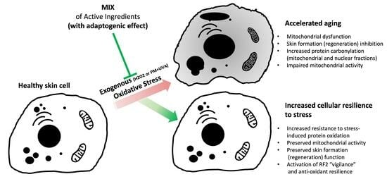
(1) Background: Skin undergoes constant changes, providing capabilities to repair and renovate its constituents once damaged and a fundamental shield to contrast environmental stress. Nevertheless, environmental stressors may overcome the skin’s protective potential inducing premature aging and accelerating the appearance of anaesthetic age-related skin aspects. Ultraviolet radiation (UVR) and pollutants (particulate matters, PAHs) contribute to skin aging and functional decline inducing harmful oxidative modifications of macromolecules and stress-related skin disorders. Innovative approaches to preserve skin are needed. (2) Methods: Skin keratinocytes were treated (or not) with a combination of ingredients (Lactobacillus plantarum extract, Withania somnifera root extract and Terminalia ferdinandiana fruit extract; “MIX”) in the presence or absence of stress (oxidative stress or pollution). The effects of the MIX adaptogen technology on (a) cellular resilience, (b) the regulation of cellular functions and (c) regeneration of skin were disclosed through expression proteomics and bioinformatics analyses first, and then through focused evaluations of protein carbonylation as a hallmark of oxidative stress’ deleterious impact and mitochondrial activity. (3) Results: The deleterious impact of stressors was evidenced, as well as the beneficial effects of the MIX through (a) mitochondrial activity preservation, (b) the “vigilance” of the NRF2 pathway activation, (c) NADPH production and protein homeostasis improvements, (d) preserving skin regeneration function and I the contrasting stress-induced oxidation (carbonylation) of mitochondrial and nuclear proteins. (4) Conclusions: The effects of the MIX on increasing cell adaptability and resilience under stress suggested a beneficial contribution in precision cosmetics and healthy human skin by acting as an adaptogen, an innovative approach that may be employed to improve resistance to harmful stress with a potential favourable impact on skin homeostasis.

Graphical Abstract

1. Introduction

Skin is the largest human organ providing a fundamental shield to contrast environmental stress. Cutaneous aging is a continuous, complex and cumulative biological process influenced by intrinsic (mainly genetic) and extrinsic factors. At midlife, these factors contribute to approximately 50% of alterations in skin function [1].
Physiologically, skin undergoes constant changes, with the capability to repair and renovate its constituents once damaged. Although the skin maintains a functional biological barrier against external aggressions, environmental stressors may overcome the skin’s protective potential. Besides environmental factors, biological and chronological aging also hamper the skin’s abilities to maintain homeostasis and repair, contributing to the aging process. Among environmental factors, ultraviolet radiation (UVR) is a primary element of skin impairment, inducing oxidative stress and harmful genetic modifications. Other factors, such as pollutants (particulate matters, volatile organic compounds (VOCs) and polycyclic aromatic hydrocarbons (PAHs)), also contribute to skin aging and decline the functionality of the skin [2,3,4,5,6,7].
Oxidative stress has been identified as a common and prevalent mechanism for cellular tissue damage and dysfunctions in an ample variety of age-related diseases and skin disorders, revealing, additionally, its interplay with inflammatory processes at both the tissue and organismal levels [8,9]. A major mechanism by which ambient PM exerts its detrimental effects is through the generation of oxidative stress. In particular, polycyclic aromatic hydrocarbons (PAHs) adsorbed on the surface of suspended particulate matter (PM) in air of urban areas can activate xenobiotic metabolism, ultimately inducing the production of reactive oxygen species (ROS) through the formation of quinone derivates and superoxide anion radicals. PAH metabolites contribute to the excessive generation of reactive oxygen species (ROS), causing oxidative stress. In addition, UV irradiation damages skin through the photosensitized production of ROS, mainly driven by UV-A (320–400 nm), which can penetrate to the deeper sections of the epidermis, reaching the dermis. Furthermore, several PAHs can induce strong oxidative stress and UV-A exposure and consequent photoactivation.
Among the oxidative modification of macromolecules, protein carbonylation has become a comprehensive warning and major hallmark of severe oxidative damage, protein dysfunction and stress-related disorders [10], being a harmful irreversible modification. This modification at the molecular level plays havoc with cellular/tissue functions in the aging organism, increasing alongside the organisms’ life [11].
Carbonylation can negatively affect, or totally abrogate, protein catalytic functions and may trigger the formation of potentially cytotoxic protein aggregates.
The nuclear factor erythroid-2-related factor 2 (Nrf2)-mediated antioxidant response pathway plays a central role in the cellular reduction–oxidation homeostasis, promoting a youthful cellular phenotype. This evolutionarily conserved pathway acts firstly as the transcription of cytoprotective genes, resulting in cellular cryoprotection and in the physiological cellular resilience to stress exposures (antioxidant, prosurvival, anti-inflammatory and macromolecular damage repair) [12,13].
Another well-known cellular mechanism implicated in cellular resilience and having a prosurvival role is the production of NADPH. In this pathway, glucose-6-phosphate dehydrogenase (G6PD) has a key role assuring the production of ribose and reducing equivalent nicotinamide adenine dinucleotide phosphate (NADPH), being vital components for the synthesis of many biological building blocks, such as nucleic and fatty acids. NADPH is well known for its remarkable role in the maintenance of antioxidant defences promoting health and life-span [14,15].
Moreover, protein carbonylation has been spotted as a potential mechanism contributing to mitochondrial dysfunction [16]. Mitochondria can produce up to 95% of all adenosine triphosphate (ATP) in the cell, sustain critical metabolic processes [17,18,19,20] and play a crucial role in inducing cell differentiation, sustaining proliferation, maintaining cell survival and triggering apoptosis, key processes for the development and maintenance of skin homeostasis and health [21,22,23]. Mitochondria are highly susceptible to detrimental environmental factors [24,25,26,27], harming its function and its genetic patrimony, resulting in mitochondrial DNA (mtDNA) deletions, perturbing the electron flow and energy production [28,29,30]. Mitochondrial dysfunction increases as age progresses. Additionally, wound healing and hair growth are part of the physiological functions of the skin, where mitochondria support optimal regeneration and differentiation, respectively.
Playing an important role in cellular metabolism and cell life-span, mitochondria assure the generation of cellular “energy storage” molecules (adenosine triphosphate) through oxidative phosphorylation (OXPHOS). Under unfavourable conditions, reactive oxygen species (ROS) are generated during OXPHOS as a by-product of aerobic respiration. Perturbations within the OXPHOS complexes generate more reactive oxygen species (ROS), less adenosine triphosphate and the mismanagement of mitochondrial dynamics with the consequent accumulation of oxidative damage. Moreover, the lower concentration of adenosine triphosphate restricts the activity of mitochondrial ATP-dependent proteases, which are less efficient or even not able to degrade all carbonylated proteins generated from the elevated ROS level. Reactive oxygen species can react and modify DNA, RNA, carbohydrates, proteins and lipids, triggering cellular and tissular dysfunction and promoting aging [31,32].
Biological adaptation refers to the ability of living organisms to adjust their biological functions to suit their external environment. This concept was first introduced by Laborit in 1976 [33], who proposed that the ability of a living being to survive and reproduce is based on its capacity to adapt to external conditions. In other words, biological adaptation involves the successful adjustment of an organism’s physiological processes to changes in its environment, enabling it to survive and thrive [34]. The human body is continuously exposed to a range of environmental stressors, such as pollution, temperature fluctuations, UV radiation, tobacco smoke and screen light [35]. The skin, acting as a barrier between the internal body and the external environment, also plays a critical role in the adaptation of humans to these stressors.
While the cosmetic industry has largely focused on developing products that enhance the appearance of the skin, there is growing interest in identifying ingredients that can also support the skin’s natural functions and ability to adapt to internal and external stressors. Adaptogens, in particular, have gained attention for their potential to help the skin maintain a healthy balance and resilience.
The definition of adaptogens has constantly evolved sinIe it was coined [36]. The dictionary definition is as follows: “natural substances used in herbal medicine to normalize and regulate the systems of the body”.
The increase in research and scientific evidence to understand the pharmacological and molecular mechanisms of action of these active ingredients has demonstrated that the term adaptogen is related to a physiological process, the adaptation to environmental challenges, which is a multistep process that includes diverse mechanisms of extracellular and intracellular interactions [36,37,38,39].
Adaptogens are mediators that help plants to persist in stress conditions, defined also as substances causing the “state of unspecific resistance” of an organism. The term adaptogens comprises families of herbal and natural products able to promote the adaptability and survival of living organisms under stress conditions, resulting in a decreased sensitivity to stressors and/or prolonged phase of resistance (stimulatory effect). These compounds are mild stress mimetics at low doses, activating adaptive stress-response signalling pathways to cope with severe stress and have been used in traditional or alternative medicines for preventing premature aging and to maintain good health and vitality [36,37,38,39,40,41,42].
If the beneficial properties of adaptogens have been studied often with oral intake, some studies using topical applications in nonhuman models have also proven their efficacy, notably with ashwagandha [43,44,45].
It is important to note that certain antioxidant treatments, if used incorrectly, can have negative effects on the skin that are just as harmful as those caused by stress. For instance, treatments that are too concentrated or applied at the wrong time can have adverse effects on the skin. Additionally, some treatments may target the wrong biological pathways, leading to unintended consequences.
To avoid such deleterious effects, it is crucial to adopt an approach that is tailored to the skin’s unique needs, ensuring that antioxidant treatments provide maximum benefits without causing any harm.
Preserving skin cells from oxidative damage and mitochondrial damage can help mitigate the cumulative injuries caused by environmental factors. Innovative approaches contrasting environmental aggressions that accelerate the aging process may employ compounds resulting in adaptogenic effects.
A mix of active ingredients was selected based on the understanding that the skin’s ability to adapt to stressors is facilitated by its three primary functions: protection, regulation and regeneration. These functions work together to maintain the skin’s health and resilience. The mix of ingredients includes:
-
An extract of Withania somnifera roots (or Ashwagandha), an adaptogen plant with a long history of use in Ayurvedic medicine for promoting longevity and slowing aging;
-
A ferment of Lactobacillus plantarum, known for its beneficial effects on the skin’s barrier function;
-
A superfruit extract of the Kakadu plum (Terminalia ferdinandiana), which is the richest natural source of vitamin C.
Together, these active ingredients were thoughtfully combined to create a technology (“Adapto.GN Smart Technology”) that can help increase the skin’s ability to adapt to modern life stressors of the current generation, GN.
By supporting the skin, the active ingredients can help optimize and enhance the skin’s functions.
The objective of our investigation was firstly focused on the characterizations of the effects of a mix compounds (Lactobacillus plantarum extract, Withania somnifera root extract and Terminalia ferdinandiana fruit extract) on human keratinocytes exposed to stress conditions (urban pollution and oxidative stress) through an expression proteomics phenotypical analysis. The obtained observations then inspired further evaluations on the potentially beneficial effects of the mix of compounds (1) on contrasting stress-induced protein carbonylation as a biomarker of oxidative damage and (2) the beneficial effects against stress exposure mitochondrial injury resulting in preserved mitochondrial function.

2. Materials and Methods

2.1. Cell Culture and Treatments

Human primary keratinocytes were seeded in multiwell plates, cultured in an SFM medium (Gibco, Strasburg, France) and incubated in optimal conditions at 37 °C, a humified atmosphere and 5% CO2. The stress exposures consisted of (1) 30 min of contact with a solution of 100 µM H2O2 or (2) contact with particulate matters (1 µg/cm2, 1 h of contact; Ref. ERM-CZ100; The item (ERM-CZ100) is supplied and certified by the “European Reference Materials” from the Joint Research Centre of the European Commission in Geel, Belgium, and is distributed by Sigma-Aldrich, Merck. European Reference Material with certified content in PAHs and the presence of >25% of PM2.5-like particles on cumulative distribution), followed by UV-A irradiation (dose of 2 J/cm2 at a wavelength peak of 365 nm). A fresh solution of active ingredients (the MIX (Adapto.GN technology TM)), composed of Lactobacillus plantarum extract 0.5%, Withania somnifera root extract 1.0% and Terminalia ferdinandiana fruit extract 0.4%, was obtained through the direct solubilization of each ingredient in the culture medium. The MIX solution was incubated with the cells for 72 h of contact prior to stress exposure or in basal conditions (in the absence of stress). The active mix solution was previously evaluated on cellular viability with an MTS (tetrazolium inner salt) viability assay, excluding cytotoxic effects at the tested concentrations.

2.2. Expression Proteomics Analysis and Ingenuity Pathway Analysis (IPA)

Right after the treatments and/or stress exposure to H2O2, the cells were harvested, snap-frozen in liquid nitrogen and stored at −80 °C until analyses. The cellular pellets (n = 3 replicates per experimental group) were submitted to protein extraction by using a buffer solution of 6M Urea, 2M Thiourea, 1% SDS (sodium dodecyl sulphate) and 1% deoxycholate (Sigma-Aldrich, Merck, Darmstadt, Germany). The protein quantification was then realized by using the Bradford assay [46], and the profiles and qualities of protein extractions were evaluated using SDS-PAGE. Fifty micrograms of proteins from each sample was typically digested and desalted through anion-exchange liquid chromatography (LC). A nanoscale LC was effectuated on a C18 column (UltiMateTM 3000 RSLCnano System; Thermo Fisher Scientific, Illkirch-Graffenstaden, France). Mass spectrometry (MS) analyses were performed on Tribrid Eclipse (Thermo Fisher Scientific) coupled to an Orbitrap-HCD-Ion Trap mass spectrometer (Thermo Fisher Scientific). Protein identifications and relative quantifications were conducted by using Protein Discoverer 2.4.0 software, MASCOT and SEQUEST on the SwissProt human protein database. Intergroup comparisons and statistics were performed by using PERSEUS software. The protein abundance ratio values were then converted to fold-change values, where the negative inverse (−1/x) was taken for values between 0 and 1 (e.g., an abundance ratio of 0.5 was converted in fold change −2), while values greater than 1 were not affected. Graphs and a principal component analysis (PCA) were generated with Prism (GraphPad software version 10, San Diego, CA, USA). The PCA was calculated using all protein abundance values (Log2) on PC1 and PC2. The data mining analysis of identified and grouped proteins in relation with their biological functions was then realized using IPA (Ingenuity Pathway Analysis, Qiagen, Courtaboeuf, France). Proteins from the dataset that were associated with biological functions in the Ingenuity Knowledge Base were considered for the analysis (minimum threshold of selection: >1.3-fold change of upregulation or <−1.3 of downregulation and p-value < 0.1). Right-tailed Fisher’s exact test was used to calculate a p-value determining the probability that each biological function, canonical pathway or upstream regulator assigned to that specific subdataset was due to chance alone. The functional analysis was restricted to human taxon and skin functions (focused on keratinocytes, fibroblasts, adipocytes, melanocytes, epidermis, dermis and skin over the Ingenuity Knowledge Base reference set) [47].

2.3. Mitochondrial Protein and Nuclear Protein (Histone-Enriched) Fraction Preparation

Right after the treatments and/or stress exposure to H2O2, the cells were harvested, snap-frozen in liquid nitrogen and stored at −80 °C until analyses. The cellular pellets (n = 5 replicates per group; per protein fraction) were independently processed and subjected to protein extraction using an adapted method based on a sequential lysis of cell membranes by increasing the detergent strength of the lysis buffers [48]. Reducing conditions were used in the overall process of extraction/enrichment in order to avoid unwanted oxidative reactions. The collected supernatants concerning organelle proteins (mitochondrial) or nuclear (histone enriched) proteins were precipitated with the trichloroacetic acid (TCA)/acetone method and then suspended in OxiProteomics buffer for solubilization. The quantification of the extracted proteins was performed using the Bradford method and the samples were equally distributed for a protein carbonylation level analysis.

2.4. Protein Carbonylation Analysed

The evaluation was performed on 5 replicates per experimental group, per each analysis: (1) organelles/mitochondrial proteins; (2) nuclear histone-enriched proteins. Right after the stress exposure, the cells were harvested, snap-frozen in liquid nitrogen and stored at −80 °C. At the moment of the analysis, the carbonylated proteins were extracted from each sample using an optimized lysis buffer and then labelled with a specific fluorescent probe [49]. After labelling, the proteins were resolved onto 4–20% gradient SDS-PAGE, then fixed to the gel and evidenced with differential fluorescence scanning at an emission wavelength of 647 nm. The total proteins were poststained with SyproRubyTM (Invitrogen, Strasbourg, France). Digital image acquisitions of carbonyl and the total proteins were performed using the iBright system (Thermo Fisher Scientific). Image processing and the densitometric analysis were performed using “ImageJ” software [50].
The signal of the carbonylated proteins (fluorescence units) was normalized with respect to the signal obtained with the total proteins for each sample to obtain the carbonyl score for each sample: carbonyl score (sample X) = carbonylated proteins (fluorescence signal; sample X)/total proteins (fluorescence signal; sample X). The statistics were obtained by using GraphPad Prism software.

2.5. Mitochondrial Function Analysis

The analysis was performed on n = 5 replicates per experimental group. Right after the stress exposures (H2O2 or pollution) and/or cell treatments with the active MIX, the mitochondrial function evaluation was conducted through an in situ treatment of cells with a Mitotracker® probe, following the provider’s instructions (Red CMXRos; Invitrogen). The control group did not receive any treatment or exposure to stress. The image collection of specific fluorescence signals was performed with the epi-fluorescence imaging system (EVOS M5000; Thermo fisher). The image analysis was effectuated with ImageJ software [50]. The levels of fluorescent probe accumulation in active mitochondria were obtained by integrating the specific intensity of a signal normalized on the surface of the evaluation and N of the nuclei.

3. Results and Discussion

3.1. MS (Mass Spectrometry)-Based Proteomic Analysis

The effects of a combination of active ingredients (MIX) were evaluated on skin cells (human primary keratinocytes) exposed (or not) to oxidative stress (H2O2) through a proteomic analysis. This approach allowed us to describe and characterize the changes in the expression of thousands of proteins supporting a better understanding of the beneficial protection against stress. Through a MS (mass spectrometry)-based proteomic analysis, we investigated the association between biological pathways, phenotypes, skin cell treatments and exposure to stress supporting precision cosmetics and promoting a healthy human skin.
In our study, “upregulated” and “downregulated” proteins referred to the proteins that showed an increase or decrease in their expression levels, respectively, when comparing two or more conditions. To quantify these changes in the protein expression, we employed a metric called ‘fold change’. Fold change is a ratio that represents how much a quantity has changed between an initial and a subsequent measurement. In our context, a fold change greater than one indicated an increase or upregulation in protein expression, while a fold change of less than one indicated a decrease or downregulation. For instance, a fold change of 2 would suggest that the protein expression doubled (upregulated) in the subsequent measurement compared to the initial one, whereas a fold change of 0.5 would indicate that the expression halved (downregulated).
More than 2400 proteins were identified and quantified per the experimental conditions (Figure 1). Among them, proteins with fold-change expression ratios versus the control group higher or lower than the control group or stress group (+1.3- or −1.3-fold change values) were considered, respectively, as upregulated (in red) or downregulated (in green, Figure 1).
The biplot representation (Figure 2 showed a homogenous positioning of the vectors (loadings, red dots) of each replicate belonging from the same group (generating clusters), and a relative difference in the positioning between the groups (correlation in this space) describing both intragroup (replicates n = 3) homogeneity and intergroup differential positioning.
The following results were evaluated according to skin functions: (1) regulation, (2) regeneration and (3) cellular protection/resilience. Among the identified proteins, the expression levels of “ADP/ATP translocase 1”, an antiporter that mediates the import of ADP (adenosine diphosphate) into the mitochondrial matrix for ATP (adenosine triphosphate) synthesis and the export of ATP out to fuel the cell resulted affected by stress (H2O2) exposure (downregulated; fold change −2.09, Table 1), were assessed. The expression of ANT1 carrying mutations caused a wide range of mitochondrial abnormalities clinically characterized by exercise intolerance, ptosis and muscle weakness [51]. Adenine nucleotide translocases (ANTs) are highly abundant proteins in mitochondria, comprising up to 1% of the total mitochondrial protein content [52]. In normal physiological conditions, the consumption of oxygen by mitochondria has been closely linked to the production of ATP. The proper functioning of ANT is crucial for maintaining healthy mitochondrial activity. One of the primary functions of ANT is to facilitate the exchange of ATP and ADP across the inner mitochondrial membrane, which is necessary for both ATP synthesis and the maintenance of a normal mitochondrial membrane potential (Δψm). Additionally, ANT also serves as an uncoupler, which is important for protecting mitochondria from excessive reactive oxygen species (ROS) production that can occur due to an elevated Δψm. These dual roles of ANT are critical for preserving mitochondrial health and function [53]. The pretreatment of cells with the MIX (MIX + Stress group) resulted in its significant upregulation (+2.37-fold change) when compared to the stressed group, contrasting the impact of stress exposure and preserving the levels of a key protein involved in mitochondrial activity.
Moreover, polymerase delta-interacting protein 3 contributed to an enhanced translational efficiency of spliced over nonspliced mRNAs. This protein recruited activated ribosomal protein S6 kinase beta-1 I/RPS6KB1 to the newly synthesized mRNA antiporter [54]. Oxidative stress (H2O2) negatively affected the expression of polymerase delta-interacting protein 3 (fold change variation versus control −2.63, Table 1), while the presence of the active MIX significantly contrasted the stress-induced variation (fold change +2.43 versus stress), suggesting a potentially improved and preserved protein homeostasis.
The structural maintenance of chromosome protein 3 is a central component of a complex required for chromosome cohesion during the cell cycle, which is coupled to DNA replication and involved in DNA repair [55], resulted in a significant downregulation (−6.13-fold change variation, Table 1) upon stress. The presence of the MIX of active ingredients (MIX + Stress) resulted in its significant upregulation (+5.53-fold change) when compared to the stressed group, preserving this important component of genetic maintenance and stability from the impact of oxidative stress exposure.
Interestingly, the levels of ADP/ATP translocase 1, polymerase delta-interacting protein 3 and structural maintenance of chromosome protein 3 were not significantly affected when the MIX was in contact with the keratinocytes in the basal conditions (absence of stress).
Then, the omics (proteome) dataset was analysed using IPA (Ingenuity Pathway Analysis; Qiagen) software to determine the biological significance of the observed experimental changes in the protein expression. Based on the integration of single protein expression results, the biological functions relevant to the skin expected to be activated or inhibited, given the observed protein expression profile, were identified as (a) major canonical pathways implicated in key aspects of cellular homeostasis (regulatory functions), (b) activated or inhibited upstream regulators (regulatory and protective functions, and (c) whether significant downstream biological processes increased or decreased (regeneration and resilience; Table 2).
The oxidative phosphorylation pathway, a key process in active mitochondria [56], was activated by the treatments with the combination of the MIX in the presence or absence of stress. This effect was not attended with the only exposure to stress (H2O2). This beneficial effect on mitochondria resulting from the activation of oxidative phosphorylation pathway was related to mitochondrial energy generation and production.
A predicted activation of the upstream regulator NRF2 was disclosed upon treatment with the MIX in the presence of stress underlying the effects on the regulation and protection of skin functions increasing cellular resilience to stress. In the nucleus, Nrf2 bound to the antioxidant response element (ARE) in the upstream promoter region of antioxidant genes and initiated their transcription, playing a vital role in response to oxidative stress, cytoprotection, being a recognized guardian of health span and a gatekeeper of species longevity [57]. The presence of the MIX in stress conditions showed beneficial effects on the “vigilance” of the Nrf2 signalling pathway resulting from activated antioxidant mechanisms (observed significant upregulation of five key proteins: the upregulation of phosphogluconate dehydrogenase [58], a key enzyme that produces NADPH, (PDG, fold change +1.48 vs. Ctrl), glucose-6-phosphate dehydrogenase [59] (G2PD; +1.39), malic enzyme 1 [60] (ME1; +1.39), transketolase [61] (TKT; +1.39), which connects the pentose–phosphate pathway to glycolysis and transaldolase 1 [62] (TALDO1; +1.39), an important enzyme for the balance of metabolites in the pentose–phosphate pathway. To note, transaldolase deficiency is associated with cutis laxa/wrinkled skin.
In basal conditions, the MIX treatment also showed a significant upregulation of PDG (+1.35), G2PD (+1.40) and malic enzyme 1 (ME1; +1.47). G2PD is responsible for the first step in the pentose–phosphate pathway, which, in a series of chemical reactions, converts glucose (a type of sugar found in most carbohydrates) to another sugar, ribose-5-phosphate. A dysfunction in the pentose–phosphate pathway can lead to a reduced production of NADPH (nicotinamide adenine dinucleotide phosphate) [59]. ME1 is a cytosolic protein that catalyses the conversion of malate to pyruvate, while concomitantly generating NADPH from NADP [60]. NADPH is one of the major reducing powers required for lipid production and protecting the cell against oxidative stress [63].
The biofunction formation (regeneration) of skin was predicted as slightly inhibited upon the basal treatment with the MIX (Z-score −1.091), being highly inhibited upon the exposure to the stress (Z-score −1.715), while not being affected in the presence of both the MIX and stress. These behaviours were aligned with the adaptogenic effects of the MIX, which exhibited slightly negative effects on the regeneration of the skin at basal conditions (mild stress-mimicking effects), but activated the adaptive stress response to protect and cope with the highly deleterious effects of stress, thus, maintaining a functional regeneration function of the skin comparable to the reference control conditions (not exposed to stress).
To note, among the differentially expressed proteins belonging to the so-called ”formation of skin regeneration”, Cornifin-A (SPRR1A) was upregulated upon the MIX treatment of the cells in the absence (basal conditions; fold change +6.37 vs. Ctrl) and in the presence of stress (fold change +10.12 vs. Ctrl), but not in the presence of stress only (−1.18 vs. Ctrl). Cornifin-A is a structural constituent of the skin epidermis that can participate widely in the construction of cell envelopes in cornifying epithelia characterized by either increased thickness or a requirement for extreme flexibility [64].

3.2. Carbonylation of Proteins

In order to confirm these promising results focused on key cellular functions, the effects of the MIX were evaluated on skin cells (primary keratinocytes) exposed (or not) to oxidative stress (H2O2) or pollution stress exposure (PM + UVA irradiation) through (a) a targeted evaluation of protein carbonylation levels of nuclear (histone-enriched) proteins and mitochondrial fraction and (b) a functional evaluation of mitochondrial activity. These investigations aimed to disclose the potential of the MIX to protect skin cells from stress-induced deleterious effects.
Carbonylated proteins (Oxi-Proteome [7,65]) were quantified upon detection, while the mitochondrial function was assessed by evaluating the membrane potential as a key indicator of mitochondrial activity through the accumulation of a selective fluorophore in active mitochondria. Increased levels of carbonylated (damaged) proteins were observed both in mitochondria and nuclear fraction upon H2O2 or pollution (PM + UVA) insults (Figure 3). In addition, the mitochondrial proteome fraction showed a higher susceptibility to oxidative damage than the nuclear proteome fraction for pollution stress exposure. The presence of the combination of active ingredients (MIX) showed a significant preservation of mitochondria-enriched proteome and nuclear (histones enriched) proteome from both H2O2-induced oxidative and pollution-induced damages, preventing the accumulation of carbonylated (oxidatively damaged) proteins. The alone presence of the MIX did not show any significant changes on the mitochondrial protein carbonylation basal levels. Interestingly, the presence of the MIX in basal conditions increased the carbonylation (p < 0.01) levels of nuclear proteins and showed protection upon stress conditions, again, in line with the action as stress mimetics in basal conditions and activating the adaptive stress response signalling pathways to contrast the deleterious effects of stress.

3.3. Mitochondrial Activity

Decreased levels of selective probe accumulation in the function of the mitochondrial membrane potential (essential for mitochondrial activity) were observed upon stress exposure (Figure 4). During the incubation with cells, the specific fluorescent probe passively diffused across the plasma membrane and selectively accumulated in active mitochondria in relation to their membrane potential, an essential component of mitochondrial function. The process in which the potential difference across the mitochondrial membrane was reduced is defined as mitochondrial depolarization, and it reflects a loss of mitochondrial function or activity. Upon both stress exposures (H2O2 or pollution), a significant decreased membrane potential was observed (reduced cumulation of the fluorescent probe in active mitochondria). The presence of the MIX showed a significant cellular resilience resulting in the protection of mitochondrial activity in the presence of pollution stress (PM + UVA; Figure 4).

4. Conclusions

Taken together, these results suggested that the presence of the MIX of active ingredients (Lactobacillus plantarum extract, Withania somnifera root extract and Terminalia ferdinandiana fruit extract) improved the response to negative stress consequences through (1) preserving mitochondrial activity, (2) activating the “vigilance” of NRF2 pathway, (3) improving NADPH production and protein homeostasis and (4) preserving skin regeneration under pollution exposure by boosting cellular antioxidant mechanisms.
Moreover, the MIX showed a significant increase in cellular resilience resulting in the protection of both mitochondria-enriched proteome and nuclear (histones enriched) proteome from H2O2- and pollution-induced oxidative damage (carbonylation).
Both effects indicated that the MIX acted as an adaptogen, improving the cellular resistance to harmful stresses with a potential favourable impact on skin health homeostasis.
Based on the obtained results on carbonylation levels of nuclear proteins, a mode of action such as eustressors (i.e., good stressors) or as mild stress mimetics was evidenced in the basal conditions, associated then to a stress-protective response against both stressors (H2O2 and pollution), confirming their action as adaptogens.
This study showed the effects of the MIX on increasing cell adaptability and resilience under stress, thus, as adaptogen technology, suggesting a beneficial contribution in precision cosmetics and healthy human skin targeting the adverse consequences of environmental stress.

Author Contributions

A.C. coguided, conducted the analysis, interpreted the results and wrote the paper. L.B. coguided, interpreted the results and wrote the paper. C.R., C.G., S.C., A.B., S.L., R.R., R.A. and M.A.B. coguided, supervised the study and read the paper. A.G. coguided, interpreted the results, wrote and reviewed the paper. All authors have read and agreed to the published version of the manuscript.

Funding

This research received no external funding.

Institutional Review Board Statement

Not applicable.

Informed Consent Statement

The study has been conducted complying strictly with the ethical rules for the donation and use of human samples for research use only.

Data Availability Statement

Data are contained within the article.

Acknowledgments

We thank Philippe Bastien (L’Oréal R&I) for the insightful discussions on statistics.

Conflicts of Interest

The authors belongs to either L’OREAL groups (Charline Ruaux, Celeste Grossgold, Suzy Levoy, Rawad Abdayem, Romain Roumiguiere, Stephanie Cheilian, Anne Bouchara, Audrey Gueniche), Oxiproteomics company (Andrea Cavagnino, Martin Baraibar) or Cilia company (Lionel Breton). The authors declare that the research was conducted in the absence of any commercial or financial relationships that could be construed as a potential conflict of interest. The funders had no role in the design of the study; in the collection, analyses or interpretation of data; in the writing of the manuscript; or in the decision to publish the results. The authors declare no conflict of interest.

References

  1. WHO. Guidelines on Hand Hygiene in Health Care: First Global Patient Safety Challenge Clean Care is Safer Care; World Health Organization—WHO: Geneva, Switzerland, 2009. [Google Scholar]
  2. Fussell, J.C.; Kelly, F.J. Oxidative contribution of air pollution to extrinsic skin ageing. Free Radic. Biol. Med. 2020, 151, 111–122. [Google Scholar] [CrossRef] [PubMed]
  3. Slominski, A.T.; Zmijewski, M.A.; Skobowiat, C.; Zbytek, B.; Slominski, R.M.; Steketee, J.D. Sensing the environment: Regulation of local and global homeostasis by the ski’s neuroendocrine system. Adv. Anat. Embryol. Cell Biol. 2012, 212, 1–115. [Google Scholar]
  4. Birch-Machin, M.A.; Russell, E.V.; Latimer, J.A. Mitochondrial DNA damage as a biomarker for ultraviolet radiation exposure and oxidative stress. Br. J. Dermatol. 2013, 169 (Suppl. 2), 9–14. [Google Scholar] [CrossRef] [PubMed]
  5. Hudson, L.; Bowman, A.; Rashdan, E.; Birch-Machin, M.A. Mitochondrial damage and ageing using skin as a model organ. Maturitas 2016, 93, 34–40. [Google Scholar] [CrossRef]
  6. Dezest, M.; Le Bechec, M.; Chavatte, L.; Desauziers, V.; Chaput, B.; Grolleau, J.L.; Descargues, P.; Nizard, C.; Schnebert, S.; Lacombe, S.; et al. Oxidative damage and impairment of protein quality control systems in keratinocytes exposed to a volatile organic compounds cocktail. Sci. Rep. 2017, 7, 10707. [Google Scholar] [CrossRef]
  7. Cavagnino, A.; Bobier, A.; Baraibar, M. The skin Oxi-Proteome as a molecular signature of exposome stress. H&PC Today 2019, 14, 4. [Google Scholar]
  8. Zuo, L.; Prather, E.R.; Stetskiv, M.; Garrison, D.E.; Meade, J.R.; Peace, T.I.; Zhou, T. Inflammaging and Oxidative Stress in Human Diseases: From Molecular Mechanisms to Novel Treatments. Int J Mol Sci. 2019, 20, 4472. [Google Scholar] [CrossRef]
  9. Fedorova, M.; Bollineni, R.C.; Hoffmann, R. Protein carbonylation as a major hallmark of oxidative damage: Update of analytical strategies. Mass Spectrom. Rev. 2014, 33, 79–97. [Google Scholar] [CrossRef]
  10. Nyström, T. Role of oxidative carbonylation in protein quality control and senescence. EMBO J. 2005, 24, 1311–1317. [Google Scholar] [CrossRef]
  11. Levine, R.L. Carbonyl modified proteins in cellular regulation, aging, and disease. Free Radic. Biol. Med. 2002, 32, 790–796. [Google Scholar] [CrossRef]
  12. Zhang, H. Oxidative stress response and Nrf2 signaling in aging. Free Radic. Biol. Med. 2015, 88 Pt B, 314–336. [Google Scholar] [CrossRef]
  13. Lavigne, E.G.; Cavagnino, A.; Steinschneider, R.; Breton, L.; Baraibar, M.A.; Jäger, S. Oxidative damage prevention in human skin and sensory neurons by a salicylic acid derivative. Free Radic. Biol. Med. 2022, 181, 98–104. [Google Scholar] [CrossRef] [PubMed]
  14. Yang, H.C. What has passed is prolog: New cellular and physiological roles of G6PD. Free Radic. Res. 2016, 50, 1047–1064. [Google Scholar] [CrossRef]
  15. Nobrega-Pereira, S. G6PD protects from oxidative damage and improves healthspan in mice. Nat. Commun. 2016, 7, 10894. [Google Scholar] [CrossRef] [PubMed]
  16. Frohnert, B.I.; Bernlohr, D.A. Protein carbonylation, mitochondrial dysfunction, and insulin resistance. Adv. Nutr. 2013, 4, 157–163. [Google Scholar] [CrossRef] [PubMed]
  17. McCormack, J.G.; Halestrap, A.P.; Denton, R.M. Role of calcium ions in regulation of mammalian intramitochondrial metabolism. Physiol Rev. 1990, 70, 391–425. [Google Scholar] [CrossRef]
  18. He, W.; Newman, J.C.; Wang, M.Z.; Ho, L.; Verdin, E. Mitochondrial sirtuins: Regulators of protein acylation and metabolism. Trends Endocrinol. Metab. 2012, 23, 467–476. [Google Scholar] [CrossRef]
  19. Tzameli, I. The evolving role of mitochondria in metabolism. Trends Endocrinol. Metab. 2012, 23, 417–419. [Google Scholar] [CrossRef]
  20. Birsoy, K.; Wang, T.; Chen, W.W.; Freinkman, E.; Abu-Remaileh, M.; Sabatini, D.M. An Essential Role of the Mitochondrial Electron Transport Chain in Cell Proliferation Is to Enable Aspartate Synthesis. Cell 2015, 162, 540–551. [Google Scholar] [CrossRef]
  21. Vakifahmetoglu-Norberg, H.; Ouchida, A.T.; Norberg, E. The role of mitochondria in metabolism and cell death. Biochem. Biophys. Res. Commun. 2017, 482, 426–431. [Google Scholar] [CrossRef]
  22. Noguchi, M.; Kasahara, A. Mitochondrial dynamics coordinate cell differentiation. Biochem. Biophys. Res. Commun. 2018, 500, 59–64. [Google Scholar] [CrossRef]
  23. Chandel, N.S. Mitochondria as signaling organelles. BMC Biol. 2014, 12, 34. [Google Scholar] [CrossRef]
  24. Slominski, A.T.; Zmijewski, M.A.; Semak, I.; Kim, T.K.; Janjetovic, Z.; Slominski, R.M.; Zmijewski, J.W. Melatonin, mitochondria, and the skin. Cell Mol. Life Sci. 2017, 74, 3913–3925. [Google Scholar] [CrossRef] [PubMed]
  25. Panich, U.; Sittithumcharee, G.; Rathviboon, N.; Jirawatnotai, S. Ultraviolet Radiation-Induced Skin Aging: The Role of DNA Damage and Oxidative Stress in Epidermal Stem Cell Damage Mediated Skin Aging. Stem. Cells Int. 2016, 2016, 7370642. [Google Scholar] [CrossRef] [PubMed]
  26. Victorelli, S.; Passos, J.F. Reactive Oxygen Species Detection in Senescent Cells. Methods Mol. Biol. 2019, 1896, 21–29. [Google Scholar]
  27. Bocheva, G.; Slominski, R.M.; Slominski, A.T. Neuroendocrine Aspects of Skin Aging. Int. J. Mol. Sci. 2019, 20, 2798. [Google Scholar] [CrossRef] [PubMed]
  28. Krutmann, J.; Schroeder, P. Role of mitochondria in photoaging of human skin: The defective powerhouse model. J. Investig. Dermatol. Symp. Proc. 2009, 14, 44–49. [Google Scholar] [CrossRef]
  29. Naidoo, K.; Hanna, R.; Birch-Machin, M.A. What is the role of mitochondrial dysfunction in skin photoaging? Exp. Dermatol. 2018, 27, 124–128. [Google Scholar] [CrossRef]
  30. Cabrera, F.; Ortega, M.; Velarde, F.; Parra, E.; Gallardo, S.; Barba, D.; Soto, L.; Peña, G.; Pedroza, L.A.; Jorgensen, C.; et al. Primary allogeneic mitochondrial mix (PAMM) transfer/transplant by MitoCeption to address damage in PBMCs caused by ultraviolet radiation. BMC Biotechnol. 2019, 19, 42. [Google Scholar] [CrossRef]
  31. Moller, I.M. Plant mitochondria and oxidative stress: Electron Transport, NADPH Turnover, and Metabolism of Reactive Oxygen Species. Annu. Rev. Plant Physiol. Plant Mol. Biol. 2001, 52, 561–591. [Google Scholar] [CrossRef]
  32. Sreedhar, A.; Aguilera-Aguirre, L.; Singh, K.K. Mitochondria in skin health, aging, and disease. Cell Death Dis. 2020, 11, 444. [Google Scholar] [CrossRef] [PubMed]
  33. Laborit, H. Éloge de la Fuite; Robert Laffont: Paris, France, 1976. [Google Scholar]
  34. Simonet, G. Le concept d’adaptation: Polysémie interdisciplinaire et implication pour les changements climatiques. Nat. Sci. Sociétés 2009, 17, 392–401. [Google Scholar] [CrossRef]
  35. Passeron, T.; Krutmann, J.; Andersen, M.L.; Katta, R.; Zouboulis, C. Clinical and biological impact of the exposome on the skin. J. Eur. Acad. Dermatol. Venereol. 2020, 34 (Suppl. 4), 4–25. [Google Scholar] [CrossRef] [PubMed]
  36. Panossian, A.G.; Efferth, T.; Shikov, A.N.; Pozharitskaya, O.N.; Kuchta, K.; Mukherjee, P.K.; Banerjee, S.; Heinrich, M.; Wu, W.; Guo, D.A.; et al. Evolution of the adaptogenic concept from traditional use to medical systems: Pharmacology of stress- and aging-related diseases. Med. Res. Rev. 2021, 41, 630–703. [Google Scholar] [CrossRef]
  37. Panossian, A.; Wikman, G.; Wagner, H. Plant adaptogens III. Earlier and more recent aspects and concepts on their mode of action. Phytomedicine 1999, 6, 287–300. [Google Scholar] [CrossRef]
  38. Panossian, A.; Gabrielian, E.; Wagner, H. On the mechanism of action of plant adaptogens with particular reference to cucurbitacin R diglucoside. Phytomedicine 1999, 6, 147–155. [Google Scholar] [CrossRef]
  39. Panossian, A. Understanding adaptogenic activity: Specificity of the pharmacological action of adaptogens and other phytochemicals. Ann. N. Y. Acad. Sci. 2017, 1401, 49–64. [Google Scholar] [CrossRef]
  40. Panossian, A.; Seo, E.J.; Efferth, T. Novel molecular mechanisms for the adaptogenic effects of herbal extracts on isolated brain cells using systems biology. Phytomedicine 2018, 50, 257–284. [Google Scholar] [CrossRef]
  41. Lazarev, N.V. General and specific in action of pharmacological agents. Farmacol. Toxicol. 1958, 21, 81–86. [Google Scholar]
  42. Lazarev, N.V.; Ljublina, E.I.; Ljublina, M.A. State of nonspecific resistance. Patol. Fiziol. Exp. Terapia. 1959, 3, 16–21. [Google Scholar]
  43. Yadav, B.; Bajaj, A.; Saxena, M.; Saxena, A.K. In Vitro Anticancer Activity of the Root, Stem and Leaves of Withania Somnifera against Various Human Cancer Cell Lines. Indian J. Pharm. Sci. 2010, 72, 659–663. [Google Scholar] [CrossRef]
  44. Ilayperuma, I.; Ratnasooriya, W.D.; Weerasooriya, T.R. Effect of Withania somnifera root extract on the sexual behaviour of male rats. Asian J. Androl. 2002, 4, 295–298. [Google Scholar] [PubMed]
  45. Saleem, S.; Muhammad, G.; Hussain, M.A.; Altaf, M.; Bukhari, S.N.A. Withania somnifera L.: Insights into the phytochemical profile, therapeutic potential, clinical trials, and future prospective. Iran. J. Basic Med. Sci. 2020, 23, 1501–1526. [Google Scholar] [PubMed]
  46. Bradford, M.M. A rapid and sensitive method for the quantitation of microgram quantities of protein utilizing the principle of protein-dye binding. Anal. Biochem. 1976, 72, 248–254. [Google Scholar] [CrossRef] [PubMed]
  47. Krämer, A.; Green, J.; Pollard, J., Jr.; Tugendreich, S. Causal analysis approaches in Ingenuity Pathway Analysis. Bioinformatics 2014, 30, 523–530. [Google Scholar] [CrossRef]
  48. Baghirova, S.; Hughes, B.G.; Hendzel, M.J.; Schulz, R. Sequential fractionation and isolation of subcellular proteins from tissue or cultured cells. MethodsX 2015, 2, 440–445. [Google Scholar] [CrossRef]
  49. Baraibar, M.A.; Ladouce, R.; Friguet, B. Proteomic quantification and identification of carbonylated proteins upon oxidative stress and during cellular aging. J. Proteom. 2013, 92, 63–70. [Google Scholar] [CrossRef]
  50. Schneider, C.A.; Rasband, W.S.; Eliceiri, K.W. NIH Image to ImageJ: 25 years of image analysis. Nat. Methods 2012, 9, 671–675. [Google Scholar] [CrossRef]
  51. Kawamata, H.; Tiranti, V.; Magrané, J.; Chinopoulos, C.; Manfredi, G. adPEO mutations in ANT1 impair ADP-ATP translocation in muscle mitochondria. Hum. Mol. Genet. 2011, 20, 2964–2974. [Google Scholar] [CrossRef]
  52. Brand, M.D.; Pakay, J.L.; Ocloo, A.; Kokoszka, J.; Wallace, D.C.; Brookes, P.S.; Cornwall, E.J. The basal proton conductance of mitochondria depends on adenine nucleotide translocase content. Biochem. J. 2005, 392 Pt 2, 353–362. [Google Scholar] [CrossRef]
  53. Kim, E.H.; Koh, E.H.; Park, J.Y.; Lee, K.U. Adenine nucleotide translocator as a regulator of mitochondrial function: Implication in the pathogenesis of metabolic syndrome. Korean Diabetes J. 2010, 34, 146–153. [Google Scholar] [CrossRef] [PubMed]
  54. Folco, E.G.; Lee, C.S.; Dufu, K.; Yamazaki, T.; Reed, R. The proteins PDIP3 and ZC11A associate with the human TREX complex in an ATP-dependent manner and function in mRNA export. PLoS ONE 2012, 7, e43804. [Google Scholar] [CrossRef] [PubMed]
  55. Sumara, I.; Vorlaufer, E.; Gieffers, C.; Peters, B.H.; Peters, J.M. Characterization of vertebrate cohesin complexes and their regulation in prophase. J. Cell Biol. 2000, 151, 749–762. [Google Scholar] [CrossRef]
  56. Vercellino, I.; Sazanov, L.A. The assembly, regulation and function of the mitochondrial respiratory chain. Nat. Rev. Mol. Cell Biol. 2022, 23, 141–161. [Google Scholar] [CrossRef] [PubMed]
  57. Matsumaru, D.; Motohashi, H. The KEAP1-NRF2 System in Healthy Aging and Longevity. Antioxidants 2021, 10, 1929. [Google Scholar] [CrossRef]
  58. Chayen, J.; Howat, D.W.; Bitensky, L. Cellular biochemistry of glucose 6-phosphate and 6-phosphogluconate dehydrogenase activities. Cell Biochem. Funct. 1986, 4, 249–253. [Google Scholar] [CrossRef] [PubMed]
  59. Stanton, R.C. Glucose-6-phosphate dehydrogenase, NADPH, and cell survival. IUBMB Life 2012, 64, 362–369. [Google Scholar] [CrossRef]
  60. White, K.; Someya, S. The roles of NADPH and isocitrate dehydrogenase in cochlear mitochondrial antioxidant defense and aging. Hear. Res. 2023, 427, 108659. [Google Scholar] [CrossRef]
  61. Kochetov, G.A.; Solovjeva, O.N. Structure and functioning mechanism of transketolase. Biochim. Biophys. Acta 2014, 1844, 1608–1618. [Google Scholar] [CrossRef]
  62. Moriyama, T.; Tanaka, S.; Nakayama, Y.; Fukumoto, M.; Tsujimura, K.; Yamada, K.; Bamba, T.; Yoneda, Y.; Fukusaki, E.; Oka, M. Two isoforms of TALDO1 generated by alternative translational initiation show differential nucleocytoplasmic distribution to regulate the global metabolic network. Sci. Rep. 2016, 6, 34648. [Google Scholar] [CrossRef]
  63. Xu, M.; Ding, L.; Liang, J.; Yang, X.; Liu, Y.; Wang, Y.; Ding, M.; Huang, X. NAD kinase sustains lipogenesis and mitochondrial metabolismthrough fatty acid synthesis. Cell Rep. 2021, 37, 110157. [Google Scholar] [CrossRef] [PubMed]
  64. Rajagopalan, P.; Jain, A.P.; Nanjappa, V.; Patel, K.; Mangalaparthi, K.K.; Babu, N.; Cavusoglu, N.; Roy, N.; Soeur, J.; Breton, L.; et al. Proteome-wide changes in primary skin keratinocytes exposed to diesel particulate extract-A role for antioxidants in skin health. J. Dermatol. Sci. 2019, 96, 114–124. [Google Scholar] [CrossRef] [PubMed]
  65. Lourenço Dos Santos, S.; Baraibar, M.A.; Lundberg, S.; Eeg-Olofsson, O.; Larsson, L.; Friguet, B. Oxidative proteome alterations during skeletal muscle ageing. Redox Biol. 2015, 5, 267–274. [Google Scholar] [CrossRef] [PubMed]
Figure 1. Stacked bar representation of significantly differentially expressed proteins in treated conditions versus “Ctrl” (basal conditions) or versus “Stress”. The number of upregulated proteins with fold change expression ratios higher than 1.3 are represented in the red bar, while the number of proteins downregulated are shown in green (fold change ratio lower than −1.3). The total number of identified proteins in the experimental dataset is shown as a white bar (cumulated distribution = 2432 proteins).
Cosmetics 10 00155 g001
Figure 2. The results of the PCA were visualized though a biplot scaled representation of the loadings (each replicate for experimental group) and PC scores. Proportion of variance PC1 96.26%, PC2 0.81%.
Cosmetics 10 00155 g002
Figure 3. (a) Protein carbonylation of the mitochondrial purified fraction and (b) of the nuclear (histone-enriched) fraction. The levels of protein carbonylation for each experimental condition are reported as vertical bar graph representation of the mean and standard deviation. *** p-value < 0.001 ** p-value < 0.01 * p-value < 0.05 § p-value < 0.1—ANOVA and Tukey’s post hoc multicomparison statistical analyses.
Cosmetics 10 00155 g003
Figure 4. (a) Mitochondrial activity. The levels of fluorescent probe (Mitrotracker®) accumulation in active mitochondria are reported as vertical bar representation of mean values for each experimental group +/− standard deviation from the mean. The intensity of fluorescence was normalized with the number of nuclei per each analysed image (detection and quantification from DAPI nuclear staining collected images). (b) In situ visualization of mitochondrial polarization (yellow), functional mitochondria accumulate more efficiently the probe (high intensity of the signal, yellow). Mitochondria injury and depolarization led to lower levels of probe accumulation (decreased fluorescence signal). The specific fluorescence emission signal for the fluorescent probe included in active mitochondria was obtained by using different and specific excitation and emission wavelengths (Ex: 542/20 Em: 593/40). *** p-Value < 0.001 n.s.—not significantly different—ANOVA and Tukey’s post hoc multi-comparisons statistical analyses.
Cosmetics 10 00155 g004
Table 1. Subset of proteins significantly (p < 0.05) up- or downregulated in “Stress” (compared to control) and in “Mix + Stress” condition (compared to stress). Only the proteins showing a relative fold change (vs. ctrl) >1.3 in the group STRESS were included. The fold change ratios were presented as negative values for downregulation and positive values for upregulation.
Accession
Number
DescriptionMIX (in Basal Conditions)Stress (H2O2)MIX + Stress
Fold Change
(vs. Ctrl)
p-ValueFold Change
(vs. Ctrl)
p-ValueFold Change
(vs. Stress)
p-Value
P12235ADP/ATP translocase 1+1.140.525−2.090.039+2.370.007
Q9BY77Polymerase delta-interacting protein 3−1.080.780−2.630.007+2.430.012
Q9UQE7Structural maintenance of chromosome protein 3−1.870.416−6.130.006+5.530.020
Table 2. Relevant canonical pathway, upstream analysis and biofunction analysis originated from the comparison between the conditions “MIX (basal conditions)” or “Stress” or “MIX + Stress” versus the condition “Ctrl”. A positive z-score indicated a predicted activation and a negative z-score indicated a predicted inactivation of the enriched pathway or biofunction in relation to the group of binary comparison. NaN, a z-core, could not be calculated. The overlap p-value identified pathways or biological functions that were statistically significantly overlapping between the dataset proteins and the proteins that were included in the specific pathway or biological function.
AnalysisMIX (in Basal Condition)
(vs. Ctrl)
Stress (H2O2)
(vs. Ctrl)
MIX + Stress
(vs. Ctrl)
z-Scorep-Valuez-Scorep-Valuez-Scorep-Value
Canonical Pathway
Oxidative Phosphorylation
2.309<0.001NaN0.482.4490.035
Upstream Analysis
Nuclear factor, erythroid 2 like 2 (NRF2)
NaN<0.001NaN/2.236<0.001
Biofunction
Formation (Regeneration) of the skin
−1.091<0.001−1.715<0.001NaN<0.001
Disclaimer/Publisher’s Note: The statements, opinions and data contained in all publications are solely those of the individual author(s) and contributor(s) and not of MDPI and/or the editor(s). MDPI and/or the editor(s) disclaim responsibility for any injury to people or property resulting from any ideas, methods, instructions or products referred to in the content.