Article

Intracellular Ca2+-Mediated Mechanisms for the Pacemaker Depolarization of the Mouse and Guinea Pig Sinus Node Tissue

Department of Pharmacology, Faculty of Pharmaceutical Sciences, Toho University, 2-2-1 Miyama Funabashi, Chiba 274-8510, Japan
*
Author to whom correspondence should be addressed.
Biomolecules 2022, 12(3), 377; https://doi.org/10.3390/biom12030377
Submission received: 30 November 2021 / Revised: 24 January 2022 / Accepted: 11 February 2022 / Published: 28 February 2022
(This article belongs to the Special Issue Molecular Pathogenesis of Cardiac Arrhythmia)

Abstract

:
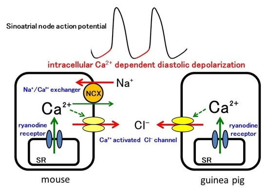
Intracellular Ca2+-mediated mechanisms for pacemaker depolarization were studied in sinus node tissue preparations from mice and guinea pigs. Microelectrode recordings revealed that the sinus node of the mouse, which had a higher beating rate, had a steeper slope of the pacemaker depolarization than that of the guinea pig. BAPTA and ryanodine, agents that interfere with intracellular Ca2+, significantly decreased the slope of the pacemaker depolarization in both species. In contrast, SEA0400, a specific inhibitor of the Na+-Ca2+ exchanger (NCX), as well as change to low Na+ extracellular solution, significantly decreased the slope in the mouse, but not in the guinea pig. Niflumic acid, a blocker of the Ca2+ activated Cl channel, decreased the slope in both species. Confocal microscopy revealed the presence of spontaneous Ca2+ oscillations during the interval between Ca2+ transients; such phenomenon was more pronounced in the mouse than in the guinea pig. Thus, although intracellular Ca2+-mediated mechanisms were involved in the pacemaker depolarization of the sinus node in both species, the NCX current was involved in the mouse but not in the guinea pig.

Graphical Abstract

1. Introduction

The contraction of the myocardium is driven by the action potential originating in the sinus node, the cardiac pacemaker. The action potential of the sinus node has a characteristic pacemaker depolarization phase (phase 4 depolarization) in the membrane voltage range of −60–−40 mV which leads the membrane potential to the threshold level for the rapid action potential upstroke (phase 0 depolarization). The pacemaker depolarization is formed by multiple inward membrane currents including the hyperpolarization-activated current (If), L-type and T-type Ca2+ currents, and the sustained inward current (Ist) [1,2,3,4,5]. The combination of membrane currents involved in the pacemaker depolarization appears to be different among animal species. For example, the If current manifests in sinus node cells of most species including mice, guinea pigs, and rabbits, while it was masked by the inwardly rectifying K+ current (IK1) in rats and monkeys [6]. The current density and function of the T-type Ca2+ current was reported to be larger in the sinus nodes from smaller animals [3,7].
It has been postulated that the pacemaker depolarization is also influenced by intracellular Ca2+. The Ca2+ released from the sarcoplasmic reticulum (SR) causes the Na+-Ca2+ exchanger (NCX) to generate a depolarizing current and accelerate the pacemaker depolarization. This mechanism was referred to as the Ca2+ clock and was considered to function in coordination with the ion channels of the cell membrane (membrane clock) to form the pacemaker depolarization [8,9]. However, the related experimental results appear to vary among researchers, and definitive conclusions have not been reached for the involvement of intracellular Ca2+-mediated mechanisms or the precise pacemaking mechanisms in each animal species. For example, in the guinea pig sinus node, some researchers emphasize the involvement of the Ca2+-clock while others reported negative results [4,9,10].
One of the possible reasons for this discrepancy is the use of isolated sinus node cells. The sinus node consists of cardiomyocytes with different electrophysiological properties [5,11], and these cells work as a functional syncytium to form the pacemaker depolarization. Thus, in a strict sense, none of the cells isolated from the sinus node region exactly represent the sinus node pacemaker. Thus, it is of value to obtain information on the function of the sinus node as a whole. In the present study, we intended to clarify the involvement of intracellular Ca2+-mediated mechanisms in the pacemaker depolarization of the mouse and guinea pig sinus node using tissue preparations. We performed standard microelectrode measurements of action potentials with sinus node tissue preparations and applied selective pharmacological agents.

2. Materials and Methods

Standard glass microelectrode experiments were performed with isolated sinus node tissue from the mouse and the guinea pig as described previously [2,3]. Microelectrode penetrations into the sinus node region were made from the epicardial surface. The extracellular solution was of the following composition (mM concentration): NaCl 118.4, KCl 4.7, CaCl2 2.5, MgSO4 1.2, KH2PO4 1.2, NaHCO3 24.9, and glucose 11.1 (pH 7.4), and the solution was gassed with 95% O2-5% CO2 and maintained at 36 ± 0.5 °C. The low Na+ extracellular solution was prepared with the equimolar substitution of NaCl with LiCl so that the final Na+ concentration was 70 mM. Change to low Na+ solution and return to normal solution were performed with a flow-switching device which enabled change of the extracellular solution within approximately 1 s. To chelate intracellular Ca2+ with O,O’-Bis (2-aminophenyl) ethyleneglycol-N,N,N′,N′-tetraacetic acid (BAPTA), its cell-permeable tetraacetoxymethyl ester (BAPTA-AM) was applied to the preparations at a final concentration of 300 μM.
The action potential parameters measured were firing rate, cycle length, maximum diastolic potential, threshold potential, the slope of the pacemaker depolarization (slope), maximum rate of rise of the action potential upstroke (maximum rate of rise; (dV/dt)max), peak potential, and duration at 50% repolarization (APD50). To obtain the slope value, the middle portion of the pacemaker depolarization phase was fitted by a straight line; the curved regions close to the maximum diastolic potential and threshold potential were not included in the fitting. The spontaneous firing of the sinus node tissue preparations was well maintained; the change in firing rate at 10 and 30 min after the addition of vehicle was less than 1% and 2% of the basal value, respectively.
For the analysis of intracellular Ca2+ movements, the sinus node tissue preparations were incubated with a Ca2+ indicator (Cal-590 AM) at 10 μM for 1 h at 37 °C in the following composition: NaCl 143, KCl 4.7, MgCl2 1.0, NaH2PO4 0.33, glucose 5.5, and HEPES 5 (mM). The recording chamber, the bottom of which was a coverslip, was placed on the stage of an inverted microscope. Preparations were placed at the bottom of the recording chamber epicardial surface down. The normal extracellular solution (NaCl 143, KCl 4.7, MgCl2 1.0, CaCl2 1.8, NaH2PO4 0.33, glucose 5.5, and HEPES 5 (mM) maintained at 32 °C) was continuously perfused. Confocal microscopic analyses of the Ca2+ oscillations in the myocardial layer were performed with a rapid scanning confocal microscope A1R (Nikon, Tokyo, Japan). The emission 570–620 nm on excitation at 561 nm was detected for Cal-590 fluorescence, and the scanning was performed at a speed of frame/16.9–33.8 ms. The fluorescent intensity of Cal-590 at each time point was normalized against the basal intensity.
BAPTA-AM (Tokyo Chemical Industry, Tokyo, Japan), ryanodine (Wako Pure Chemical Industries, Osaka, Japan), SEA0400 (synthesized in our faculty), niflumic acid (Sigma-Aldrich, St. Louis, MO, USA), and Cal-590 AM (Cosmo Bio, Tokyo, Japan) were dissolved in dimethyl sulfoxide (DMSO). All data were expressed as the mean ± standard error of the mean (S.E.M). Data were analyzed by the paired t-test. A p value less than 0.05 was considered statistically significant.

3. Results

Microelectrode recordings showed that the sinus node of the mouse, which had a higher firing rate, had a steeper slope of the pacemaker depolarization and a shorter action potential duration than that of the guinea pig. There was no difference in the maximum diastolic potential and threshold potential between the mouse and guinea pig; the maximum diastolic potential was about −60 mV, and the threshold potential was about −40 mV in both species.
In the mouse sinus node, chelation of intracellular Ca2+ with BAPTA (300 μM) induced significant decreases in the firing rate and the slope of the pacemaker depolarization (Figure 1 and Table 1). BAPTA had no significant effect on other parameters in the mouse. In the guinea pig sinus node, BAPTA induced significant decreases in the firing rate and the slope of the pacemaker depolarization (Figure 1 and Table 1). BAPTA shifted the maximum diastolic potential and the threshold potential towards depolarized potentials in the guinea pig.
In the mouse and guinea pig sinus node, ryanodine (0.1 μM) significantly decreased the firing rate and the slope of the pacemaker depolarization (Figure 1 and Table 1). Ryanodine had no significant effect on other parameters in both species.
In the mouse sinus node, SEA0400 (1 and 10 μM) decreased the firing rate and the slope of the pacemaker depolarization (Figure 2 and Table 2). SEA0400 shifted the maximum diastolic potential and the threshold potential towards depolarized potentials and decreased the maximum rate of rise and the peak potential in the mouse. Further, when the effect of SEA0400 on the beating rate of the mouse right atria was examined after the beating rate was decreased with carbachol, SEA0400 significantly decreased the beating rate; the beating rate in the presence of 0.3 μM carbachol before and after the application of SEA0400 (1 μM) was 234.1 ± 10.7 and 163.8 ± 29.1 (n = 6), respectively.
In the guinea pig sinus node, SEA0400 had no effect on the action potential waveform (Figure 2 and Table 2). Further, when the effect of SEA0400 on the beating rate of the guinea pig right atria was examined after the beating rate was increased with noradrenaline, SEA0400 had no effect on the beating rate; the beating rate in the presence of 3 μM noradrenaline before and after the application of SEA0400 (1 μM) was 328.4 ± 8.9 and 324.6 ± 8.9 (n = 5), respectively.
To support the results obtained with SEA0400, we intended to reduce the inward NCX current with low Na+ extracellular solution. In the mouse sinus node, changing the extracellular solution to low Na+ solution significantly decreased the firing rate and the slope of the pacemaker depolarization (Figure 3 and Table 3). The low Na+ solution shifted the maximum diastolic potential and the threshold potential towards depolarized potentials and decreased the maximum rate of rise and the peak potential. In the guinea pig, changing the extracellular solution to low Na+ solution had no significant effect on the firing rate and the slope of the pacemaker depolarization. The low Na+ solution did not affect any of the action potential parameters (Figure 3 and Table 3).
In the mouse sinus node, niflumic acid (30 μM), a blocker of the Ca2+-activated Cl channel, significantly decreased the firing rate and the slope of the pacemaker depolarization (Figure 4 and Table 4). Niflumic acid had no significant effect on other parameters in the mouse. In the guinea pig sinus node, niflumic acid significantly decreased the firing rate and the slope of the pacemaker depolarization (Figure 4 and Table 4). The action potential duration was shortened by niflumic acid in the guinea pig.
To analyze intracellular Ca2+ movements, the sinus node tissue preparations loaded with the Ca2+ sensitive fluoroprobe, Cal-590, were observed with a rapid scanning confocal microscope. In both species, Ca2+ transients, synchronized elevations of Ca2+ fluorescence throughout the myocardial tissue, were observed at a constant frequency. In mouse sinus node, spontaneous Ca2+ oscillations, non-synchronized local rises in Ca2+ fluorescence, were observed in roughly one third of the cells (Figure 5). Such Ca2+ oscillations were less frequently observed in the guinea pig (Figure 6).
In the mouse sinus node, SEA0400 (1 μM) markedly reduced the firing frequency of spontaneous Ca2+ transients (Figure 5); the frequency before and 10 min after the addition of 1 μM SEA0400 was 185.8 ± 24.1 and 110.3 ± 23.2 bpm (n = 5; p < 0.05), respectively. SEA0400 caused an increase in basal Ca2+ concentration; the basal Ca2+ fluorescence at 10 min after the addition of 1 μM SEA0400 was 110.3 ± 8.7% (n = 5) of the value before application. SEA0400 had no apparent effect on the local Ca2+ oscillations.
In the guinea pig sinus node, SEA0400 had no effect on the firing frequency of spontaneous Ca2+ transients (Figure 6); the frequency before and 10 min after the addition of 1 μM SEA0400 was 157.2 ± 5.9 and 158.2 ± 8.5 bpm (n = 5), respectively. SEA0400 caused an increase in basal Ca2+ concentration; the basal Ca2+ fluorescence at 10 min after the addition of 1 μM SEA0400 was 112.0 ± 4.5% (n = 5) of the value before application.

4. Discussion

In the present study, we intended to clarify the involvement of intracellular Ca2+-mediated mechanisms in the pacemaker depolarization of the mouse and guinea pig sinus node. We applied standard microelectrode techniques on tissue preparations and used selective pharmacological agents. Compared to isolated cardiomyocytes, myocardial tissue preparations may have disadvantages such as less efficient delivery of oxygen and pharmacological agents to the cells of interest. On the other hand, they have advantages such as absence of cell isolation damage and cell selection bias. Above all, tissue preparations enable the observation of the sinus node as a whole working as a functional syncytium.
First of all, we examined whether or not intracellular Ca2+-mediated mechanisms are involved in the pacemaking of the sinus node. Treatment of the sinus node tissue preparations with either BAPTA or ryanodine, agents that interfere with intracellular Ca2+, decreased the firing rate and the slope of the pacemaker depolarization both in the mouse and guinea pig. These results indicated that Ca2+ released from the SR through the ryanodine receptor contributes to the formation of the pacemaker depolarization of the sinus node in both species. As both BAPTA and ryanodine may have a broad effect on intracellular Ca2+-mediated events, their effects may also reflect changes other than the instantaneous reduction of intracellular Ca2+ [4]. Thus, we intended to clarify the transporters that convert intracellular Ca2+ into the pacemaker depolarization.
The Na+/Ca2+ exchanger (NCX) is the major pathway for transsarcolemal Ca2+ extrusion from the cytoplasm; it pumps out one Ca2+ ion in exchange for three Na+ ions (forward mode NCX). It has been postulated that the net inward current that occurs in this process contributes to the pacemaker depolarization of the sinus node. To examine this hypothesis in sinus node tissue preparations, we used SEA0400, a potent and selective inhibitor of NCX. In voltage-clamped guinea pig ventricular myocytes, 1 μM SEA0400 inhibited the NCX current by more than 80%, with no effect on the sodium current, L-type calcium current, delayed rectifier potassium current, and the inwardly rectifying potassium current [12]. The effectiveness of SEA0400 on tissue preparations was also confirmed in the mouse and guinea pig myocardium [13,14,15].
In the mouse sinus node, the slope of the pacemaker depolarization, as well as the firing rate of action potentials and Ca2+ transients, was decreased by SEA0400. Similar results were obtained with low Na+ extracellular solution. Thus, the NCX current activated by the Ca2+ released from the SR contributes to the pacemaker depolarization in mouse sinus node. These results were consistent with the observation that the firing frequency of the sinus node was lower in atrial-specific NCX knock out mice than in wild type mice [16]. In contrast, in the guinea pig sinus node, treatment with SEA0400, as well as low Na+ extracellular solution, affected neither the firing rate nor the slope of the pacemaker depolarization. SEA0400 caused a small increase in the basal Ca2+ concentration. These results indicated that NCX contributes to transarcolemmal Ca2+ efflux but does not play an essential role in the pacemaker depolarization of the guinea pig sinus node. This lack of SEA0400 effect in the guinea pig sinus node was not due to its lower firing rate because SEA0400 was ineffective in the guinea pig right atria even after its beating rate was increased by noradrenaline to a level similar to that of the mouse, and effective in the mouse right atria even after its beating rate was decreased by carbachol to a level similar to that of the guinea pig.
The effects of SEA0400 and low Na+ on the maximum rate of rise and the peak potential could be explained by their effect on the pacemaker depolarization. SEA0400 and low Na+ markedly reduced the slope of pacemaker depolarization in the mouse sinus node. This means that the membrane potential is kept in a partially depolarized potential range close to the threshold potential for a longer time, which may cause increased inactivation of the L-type Ca2+ channel leading to a reduced maximum rate of rise and peak potential. In contrast, in the guinea pig sinus node, in which the slope of the pacemaker depolarization was unchanged, neither the maximum rate of rise nor the peak potential was affected.
The pacemaker depolarization is considered to be formed by several inward currents such as the hyperpolarization-activated current (If), L-type and T-type Ca2+ currents, and the sustained inward current (Ist) [1,2,3,4,5]. In addition, the deactivation of IK is dominant in the early phase of pacemaker depolarization in the guinea pig sinus node [4,6]. The NCX current in the sinus node was detected both in the mouse and guinea pig; the current density was not different between the two species under voltage-clamp conditions [17,18]. Since the NCX currents observed were relatively small (<5 pA/pF), they may possibly be masked by other membrane currents. As the NCX current density in the intact sinus node is affected by the intracellular supply of Ca2+ to the NCX, it is possible that some difference in intracellular Ca2+ handling properties causes a difference in NCX current density in the spontaneously firing sinus node. In fact, spontaneous Ca2+ oscillations were observed during the interval between Ca2+ transients in sinus node tissue preparations, which was more pronounced in the mouse than in the guinea pig. This appears to be consistent with the observation in isolated sinus node cardiomyocytes that the number of Ca2+ sparks occurring during the pacemaker depolarization was larger in the mouse than in the guinea pig [9,19].
Concerning the sinus node of other animal species, it was reported that SEA0400 had no effect on the heart rate in the isolated rabbit heart [20] and in anesthetized dogs [21]. To the best of our knowledge, there is no information on the effect of SEA0400 on the human sinus node. Concerning ectopic pacemakers, we observed in the guinea pig pulmonary vein that both SEA0400 and ryanodine inhibited the spontaneous electrical activity [22]. Ca2+ sparks occurred during the interval between Ca2+ transients, and the frequency of Ca2+ spark firing increased abruptly just before the onset of Ca2+ transients [23]. These results indicated that the NCX current activated by Ca2+ released from the SR was involved in the pacemaker depolarization of the pulmonary vein myocardium. Thus, the contribution of NCX to myocardial pacemaking varies among animal species and type of myocardium.
To obtain further information on the Ca2+-mediated pacemaking mechanisms of the sinus node, we examined the effect of niflumic acid, a blocker of the Ca2+-activated Cl channel, which was identified in sinus node cells [24,25]. As the estimated reversal potential for Cl ions in cardiomyocytes was about −30 mV [26], activation of the Ca2+-activated Cl channel could generate a depolarizing current during the pacemaker depolarization. Niflumic acid reduced the firing rate and the slope of pacemaker depolarization in the sinus node of both mouse and guinea pig. These results indicated that the inward current through the Cl channel activated by Ca2+ released from the SR contributes to the pacemaker depolarization of the sinus node both in the mouse and guinea pig.
The mouse has a high heart rate (400 bpm>), and its sinus node has a steeper slope of pacemaker depolarization among experimental animal species. The present study showed that the Ca2+-activated Cl current (IClCa) was involved in the pacemaker depolarization of both mouse and guinea pig, but the NCX current contributed to cardiac pacemaking only in the mouse. Using similar experimental techniques, we previously showed that the contribution of the T-type Ca2+ channel to cardiac pacemaking differs among animal species; the effects of R-(−)-efonidipine, which is a selective T-type Ca2+ channel inhibitor, was prominent in the mouse, small but significant in the guinea pig, and was not observed in the rabbit [2]. Voltage gated Na+ channels, which are considered not to play a role in pacemaker activity in the majority of mammalian species including the guinea pig [10], was reported to play a certain role in the pacemaking of the mouse sinus node [27]. The present and earlier studies revealed that the guinea pig sinus node depends on ICaL, Ist, If, and IClCa, but not INCX for its pacemaking [4,5,11]. In the mouse sinus node, additional currents including the ICaT, INCX, and INa also contribute to pacemaking [2,7,16,27]. Thus, the mouse sinus node appears to have a greater variety of ionic mechanisms to maintain its high firing rate.

5. Conclusions

Intracellular Ca2+-mediated mechanisms were involved in the pacemaker depolarization of the sinus node both in the mouse and guinea pig. The NCX current was involved in the mouse but not in the guinea pig. This difference could partly explain the higher firing rate of the mouse sinus node.

Author Contributions

Conceptualization, I.N. and H.T.; methodology, I.N. and H.T.; software, S.H.; formal analysis, I.N. and R.O.; investigation, R.O., K.J. and A.F.; resources, H.T.; data curation, I.N.; writing—original draft preparation, I.N.; writing—review and editing, H.T.; visualization, I.N.; supervision, H.T.; project administration, I.N. and H.T.; funding acquisition, I.N., S.H. and H.T. All authors have read and agreed to the published version of the manuscript.

Funding

This research was funded by JSPS KAKENHI, Grant Numbers 20K07299, 20K16013 and 20K07091.

Institutional Review Board Statement

The study was approved by the Ethics Committee of Toho University (21-55-362; March 2021) and conducted in accordance with the “Guiding Principles for the Care and Use of Laboratory Animals” approved by The Japanese Pharmacological Society.

Informed Consent Statement

Not applicable.

Data Availability Statement

Not applicable.

Conflicts of Interest

The authors declare no conflict of interest.

References

  1. DiFrancesco, D. The role of the funny current in pacemaker activity. Circ. Res. 2010, 106, 434–446. [Google Scholar] [CrossRef] [PubMed] [Green Version]
  2. Tanaka, H.; Komikado, C.; Namekata, I.; Nakamura, H.; Suzuki, M.; Tsuneoka, Y.; Shigenobu, K.; Takahara, A. Species difference in the contribution of T-type calcium current to cardiac pacemaking as revealed by R(−)-efonidipine. J. Pharmacol. Sci. 2008, 107, 99–102. [Google Scholar] [CrossRef] [PubMed] [Green Version]
  3. Tanaka, H.; Namekata, I.; Ogawa, T.; Tsuneoka, Y.; Komikado, C.; Takahara, A.; Iida-Tanaka, N.; Izumi-Nakaseko, H.; Tsuru, H.; Adachi-Akahane, S. Effects of S(+)-efonidipine on the rabbit sinus node action potential and calcium channel subunits CaV 1.2, CaV 1.3 and CaV 3.1. Eur. J. Pharmacol. 2010, 649, 263–267. [Google Scholar] [CrossRef] [PubMed]
  4. Himeno, Y.; Toyoda, F.; Satoh, H.; Amano, A.; Cha, C.Y.; Matsuura, H.; Noma, A. Minor contribution of cytosolic Ca2+ transients to the pacemaker rhythm in guinea pig sinoatrial node cells. Am. J. Physiol. Heart Circ. Physiol. 2011, 300, 251–261. [Google Scholar] [CrossRef] [PubMed] [Green Version]
  5. Toyoda, F.; Ding, W.G.; Matsuura, H. Heterogeneous functional expression of the sustained inward Na+ current in guinea pig sinoatrial node cells. Pflug. Arch. 2018, 470, 481–490. [Google Scholar] [CrossRef]
  6. Satoh, H. Sino-atrial nodal cells of mammalian hearts: Ionic currents and gene expression of pacemaker ionic channels. J. Smooth Muscle Res. 2003, 39, 175–193. [Google Scholar] [CrossRef] [Green Version]
  7. Ono, K.; Iijima, T. Cardiac T-type Ca2+ channels in the heart. J. Mol. Cell. Cardiol. 2010, 48, 65–70. [Google Scholar] [CrossRef]
  8. Maltsev, V.A.; Vinogradova, T.M.; Lakatta, E.G. The emergence of a general theory of the initiation and strength of the heartbeat. J. Pharmacol. Sci. 2006, 100, 338–369. [Google Scholar] [CrossRef] [Green Version]
  9. Sirenko, S.G.; Yang, D.; Maltseva, L.A.; Kim, M.S.; Lakatta, E.G.; Maltsev, V.A. Spontaneous, local diastolic subsarcolemmal calcium releases in single, isolated guinea-pig sinoatrial nodal cells. PLoS ONE 2017, 12, e0185222. [Google Scholar] [CrossRef] [Green Version]
  10. Sanders, L.; Rakovic, S.; Lowe, M.; Mattick, P.A.; Terrar, D.A. Fundamental importance of Na+-Ca2+ exchange for the pacemaking mechanism in guinea-pig sino-atrial node. J. Physiol. 2006, 571, 639–649. [Google Scholar] [CrossRef]
  11. Mangoni, M.E.; Nargeot, J. Properties of the hyperpolarization-activated current (If) in isolated mouse sino-atrial cells. Cardiovasc. Res. 2001, 52, 51–64. [Google Scholar] [CrossRef] [Green Version]
  12. Tanaka, H.; Nishimaru, K.; Aikawa, T.; Hirayama, W.; Tanaka, Y.; Shigenobu, K. Effect of SEA0400, a novel inhibitor of sodium-calcium exchanger, on myocardial ionic currents. Br. J. Pharmacol. 2002, 135, 1096–1100. [Google Scholar] [CrossRef] [PubMed] [Green Version]
  13. Tanaka, H.; Namekata, I.; Takeda, K.; Kazama, A.; Shimizu, Y.; Moriwaki, R.; Hirayama, W.; Sato, A.; Kawanishi, T.; Shigenobu, K. Unique excitation-contraction characteristics of mouse myocardium as revealed by SEA0400, a specific inhibitor of Na+-Ca2+ exchanger. Naunyn Schmiedebergs Arch. Pharmacol. 2005, 371, 526–534. [Google Scholar] [CrossRef] [PubMed]
  14. Tanaka, H.; Shimada, H.; Namekata, I.; Kawanishi, T.; Iida-Tanaka, N.; Shigenobu, K. Involvement of the Na+/Ca2+ exchanger in ouabain-induced inotropy and arrhythmogenesis in guinea-pig myocardium as revealed by SEA0400. J. Pharmacol. Sci. 2007, 103, 241–246. [Google Scholar] [CrossRef] [Green Version]
  15. Namekata, I.; Nakamura, H.; Shimada, H.; Tanaka, H.; Shigenobu, K. Cardioprotection without cardiosuppression by SEA0400, a novel inhibitor of Na+-Ca2+ exchanger, during ischemia and reperfusion in guinea-pig myocardium. Life Sci. 2005, 77, 312–324. [Google Scholar] [CrossRef]
  16. Torrente, A.G.; Zhang, R.; Zaini, A.; Giani, J.F.; Kang, J.; Lamp, S.T.; Philipson, K.D.; Goldhaber, J.I. Burst pacemaker activity of the sinoatrial node in sodium-calcium exchanger knockout mice. Proc. Natl. Acad. Sci. USA 2015, 112, 9769–9774. [Google Scholar] [CrossRef] [Green Version]
  17. Gao, Z.; Rasmussen, T.P.; Li, Y.; Kutschke, W.; Koval, O.M.; Wu, Y.; Wu, Y.; Hall, D.D.; Joiner, M.L.; Wu, X.Q.; et al. Genetic inhibition of Na+-Ca2+ exchanger current disables fight or flight sinoatrial node activity without affecting resting heart rate. Circ. Res. 2013, 112, 309–317. [Google Scholar] [CrossRef] [Green Version]
  18. Kojima, A.; Ito, Y.; Kitagawa, H.; Matsuura, H.; Nosaka, S. Direct negative chronotropic action of desflurane on sinoatrial node pacemaker activity in the guinea pig heart. Anesthesiology 2014, 120, 1400–1413. [Google Scholar] [CrossRef] [Green Version]
  19. Chen, B.; Wu, Y.; Mohler, P.J.; Anderson, M.E.; Song, L.S. Local control of Ca2+-induced Ca2+ release in mouse sinoatrial node cells. J. Mol. Cell. Cardiol. 2009, 47, 706–715. [Google Scholar] [CrossRef] [Green Version]
  20. Magee, W.P.; Deshmukh, G.; Deninno, M.P.; Sutt, J.C.; Chapman, J.G.; Tracey, W.R. Differing cardioprotective efficacy of the Na+/Ca2+ exchanger inhibitors SEA0400 and KB-R7943. Am. J. Physiol. Heart Circ. Physiol. 2003, 284, 903–910. [Google Scholar] [CrossRef] [Green Version]
  21. Takahashi, T.; Takahashi, K.; Onishi, M.; Suzuki, T.; Tanaka, Y.; Ota, T.; Yoshida, S.; Nakaike, S.; Matsuda, T.; Baba, A. Effects of SEA0400, a novel inhibitor of the Na+/Ca2+ exchanger, on myocardial stunning in anesthetized dogs. Eur. J. Pharmacol. 2004, 505, 163–168. [Google Scholar] [CrossRef] [PubMed]
  22. Namekata, I.; Tsuneoka, Y.; Takahara, A.; Shimada, H.; Sugimoto, T.; Takeda, K.; Nagaharu, M.; Shigenobu, K.; Kawanishi, T.; Tanaka, H. Involvement of the Na+/Ca2+ exchanger in the automaticity of guinea-pig pulmonary vein myocardium as revealed by SEA0400. J. Pharmacol. Sci. 2009, 110, 111–116. [Google Scholar] [CrossRef] [PubMed] [Green Version]
  23. Namekata, I.; Tanaka, Y.; Ohmori, T.; Tsuneoka, Y.; Hamaguchi, S.; Tanaka, H.; Tanaka, H. Cell morphology and early-phase Ca2+ transients of guinea-pig pulmonary vein cardiomyocytes compared with atrial and ventricular cardiomyocytes. Bioimages 2019, 27, 1–12. [Google Scholar]
  24. Verkerk, A.O.; Wilders, R.; Zegers, J.G.; van Borren, M.M.; Ravesloot, J.H.; Verheijck, E.E. Ca2+-activated Cl current in rabbit sinoatrial node cells. J. Physiol. 2002, 540, 105–117. [Google Scholar] [CrossRef] [PubMed]
  25. Mao, Z.; Wang, Y.; Peng, H.; He, F.; Zhu, L.; Huang, H.; Huang, X.; Lu, X.; Tan, X. A newly identified missense mutation in CLCA2 is associated with autosomal dominant cardiac conduction block. Gene 2019, 714, 143990. [Google Scholar] [CrossRef]
  26. Turner, D.; Kang, C.; Mesirca, P.; Hong, J.; Mangoni, M.E.; Glukhov, A.V.; Sah, R. Electrophysiological and Molecular Mechanisms of Sinoatrial node mechanosensitivity. Front. Cardiovasc. Med. 2021, 8, 662410. [Google Scholar] [CrossRef]
  27. Lei, M.; Jones, S.A.; Liu, J.; Lancaster, M.K.; Fung, S.S.; Dobrzynski, H.; Camelliti, P.; Maier, S.K.; Noble, D.; Boyett, M.R. Requirement of neuronal- and cardiac-type sodium channels for murine sinoatrial node pacemaking. J. Physiol. 2004, 559, 835–843. [Google Scholar] [CrossRef]
Figure 1. Effects of BAPTA and ryanodine on the sinus node action potential of the mouse and guinea pig. Typical traces before (black lines) and after (red lines) application of 300 μM BAPTA (upper) and 0.1 μM ryanodine (lower).
Biomolecules 12 00377 g001
Figure 2. Effect of SEA0400 on the sinus node action potential of the mouse and guinea pig. Typical traces before (black lines) and after (red lines) application of 1 μM (upper) and 10 μM (lower).
Biomolecules 12 00377 g002
Figure 3. Effect of low Na+ solution on the sinus node action potential of the mouse and guinea pig. Typical traces before (black lines) and after (red lines) rapid change of the extracellular solution to low Na+ solution.
Biomolecules 12 00377 g003
Figure 4. Effect of niflumic acid on the sinus node action potential of the mouse and guinea pig. Typical traces before (black lines) and after (red lines) application of 30 μM niflumic acid.
Biomolecules 12 00377 g004
Figure 5. Effect of SEA0400 on spontaneously occurring Ca2+ transients in the mouse sinus node tissue. (A) Typical x-y images of the myocardium loaded with Cal-590 before (ac) and 10 min after the addition of 1μM SEA0400 (df). Panels c and f are the images at the peak of the Ca2+ transient. Note that Ca2+ oscillations were observed during the interval between the Ca2+ transients (arrows). (B) Time course of the changes in fluorescence before addition (ac) and 10 min after the addition of 1 μM SEA0400 (df). Time course of the fluorescence intensity quantified in the whole field of view (a,d), cell 1 (b,e), and cell 2 (c,f) as shown in panel (A)/(c).
Biomolecules 12 00377 g005
Figure 6. Effect of SEA0400 on spontaneously occurring Ca2+ transients in the guinea pig sinus node tissue. (A) Typical x-y images of the myocardium loaded with Cal-590 before (ac) and 10 min after the addition of 1μM SEA0400 (df). Panels c and f are the images at the peak of the Ca2+ transient. Note that no Ca2+ oscillation was observed during the interval between the Ca2+ transients (a,b,d,e). (B) Time course of the changes in fluorescence before addition (ac) and 10 min after the addition of 1 μM SEA0400 (df). Time course of the fluorescence intensity quantified in the whole field of view (a,d), cell 1 (b,e), and cell 2 (c,f) as shown in panel (A)/(c).
Biomolecules 12 00377 g006
Table 1. Action potential parameters before and after application of BAPTA and ryanodine.
BAPTARyanodine
MouseGuinea PigMouseGuinea Pig
Firing rate (bpm)423.7 ± 10.1241.4 ± 16.1453.6 ± 10.0248.7 ± 11.7
396.0 ± 6.4 *213.1 ± 12.2 *414.3 ± 13.9 *237.4 ± 12.6 *
Cycle length (ms)141.9 ± 3.4254.2 ± 17.0132.5 ± 2.9241.3 ± 13.4
151.7 ± 2.4 *286.7 ± 17.2 *145.5 ± 4.7 *256.7 ± 13.6 *
Maximum diastolic potential (mV)−60.6 ± 1.4−62.9 ± 1.2−58.8 ± 1.2−63.2 ± 1.3
−60.0 ± 1.7−58.9 ± 2.1 *−55.9 ± 1.7−60.9 ± 1.7
Slope of pacemaker depolarization (mV/s)192.0 ± 26.2147.4 ± 14.7223.5 ± 25.8138.2 ± 10.4
130.0 ± 17.7 *104.8 ± 9.8 *110.0 ± 24.5 *119.2 ± 9.1 *
Threshold potential (mV)−50.4 ± 1.6−49.0 ± 1.6−46.9 ± 1.9−52.6 ± 1.9
−51.6 ± 1.5−46.4 ± 2.1 *−45.8 ± 3.0−49.1 ± 2.3
Maximum rate of rise (V/s)10.3 ± 2.69.0 ± 2.28.7 ± 2.718.7 ± 9.4
9.0 ± 2.28.1 ± 1.97.8 ± 2.215.6 ± 7.8
Peak potential (mV)0.9 ± 2.518.9 ± 1.14.6 ± 1.218.3 ± 1.7
−0.9 ± 3.217.7 ± 1.54.2 ± 1.017.8 ± 1.6
Duration at 50% repolarization (ms)30.3 ± 1.781.8 ± 2.028.3 ± 0.975.4 ± 5.9
30.2 ± 1.485.6 ± 6.328.9 ± 1.477.4 ± 6.4
Upper and lower values in each row indicate the parameters obtained before and after (10 min for BAPTA and 5 min for ryanodine) the application of agents, respectively. Values indicate the mean ± S.E.M. from 5–6 preparations. Asterisks indicate significant difference from corresponding values before the application of agents as evaluated by the paired t-test (p < 0.05).
Table 2. Action potential parameters before and after application of SEA0400.
SEA0400 (1 μM)SEA0400 (10 μM)
MouseGuinea PigMouseGuinea Pig
Firing rate (bpm)449.7 ± 24.9239.0 ± 9.2428.2 ± 27.2239.4 ± 8.9
396.8 ± 29.0 *240.1 ± 9.2363.8 ± 17.7 *237.4 ± 11.6
Cycle length (ms)135.2 ± 8.1253.0 ± 10.1142.1 ± 8.0252.0 ± 9.0
155.0 ± 12.2 *251.8 ± 9.7166.4 ± 7.6 *255.1 ± 12.3
Maximum diastolic potential (mV)−55.8 ± 1.8−62.6 ± 2.5−59.2 ± 1.0−61.7 ± 2.1
−51.7 ± 1.9 *−62.1 ± 2.4−55.9 ± 1.3 *−59.1 ± 3.0
Slope of pacemaker depolarization (mV/s)217.0 ± 22.2148.3 ± 16.2215.4 ± 26.6172.5 ± 6.0
173.2 ± 22.9 *150.8 ± 13.8161.0 ± 20.9 *177.0 ± 16.3
Threshold potential (mV)−46.0 ± 1.2−49.8 ± 2.7−48.6 ± 1.2−48.3 ± 2.5
−41.9 ± 1.4 *−48.7 ± 2.5−45.5 ± 1.3 *−43.9 ± 3.1
Maximum rate of rise (V/s)6.7 ± 0.67.0 ± 1.57.9 ± 1.65.8 ± 1.5
5.6 ± 0.7 *7.7 ± 2.24.5 ± 0.8 *4.8 ± 1.1
Peak potential (mV)4.8 ± 0.712.0 ± 2.14.7 ± 1.611.9 ± 1.6
−0.5 ± 1.3 *12.8 ± 1.8−11.2 ± 3.0 *12.0 ± 1.6
Duration at 50% repolarization (ms)33.5 ± 2.180.4 ± 5.431.3 ± 2.285.4 ± 6.2
35.5 ± 1.579.8 ± 4.436.2 ± 2.283.6 ± 5.3
Upper and lower values in each row indicate the parameters obtained before and after (10 min) the application of agents, respectively. Values indicate the mean ± S.E.M. from 5–6 preparations. Asterisks indicate significant difference from corresponding values before the application of agents as evaluated by the paired t-test (p < 0.05).
Table 3. Action potential parameters before and after the rapid change of the extracellular solution to low Na+ solution.
Low Na+
MouseGuinea Pig
Firing rate (bpm)498.4 ± 17.1232.7 ± 9.5
455.5 ± 20.5 *232.7 ± 9.9
Cycle length (ms)121.1 ± 4.0260.2 ± 12.0
133.1 ± 5.9 *260.5 ± 12.6
Maximum diastolic potential (mV)−60.4 ± 1.1−63.2 ± 0.9
−56.9 ± 0.9 *−64.0 ± 1.6
Slope of pacemaker depolarization (mV/s)251.4 ± 25.3166.3 ± 17.4
206.7 ± 30.7 *187.3 ± 20.4
Threshold potential (mV)−49.8 ± 1.1−48.2 ± 1.1
−47.6 ± 1.0 *−48.1 ± 1.8
Maximum rate of rise (V/s)8.6 ± 1.14.0 ± 0.7
5.6 ± 0.6 *3.6 ± 0.6 *
Peak potential (mV)1.9 ± 1.311.0 ± 1.2
−5.1 ± 2.4 *8.3 ± 2.4
Duration at 50% repolarization (ms)28.7 ± 1.389.3 ± 3.5
32.2 ± 1.3 *88.8 ± 4.2
Upper and lower values in each row indicate the parameters obtained before and after (1 min) the application of low Na+ solution. Values indicate the mean ± S.E.M. from 6 preparations. Asterisks indicate significant difference from corresponding values before the application of agent as evaluated by the paired t-test (p < 0.05).
Table 4. Action potential parameters before and after application of niflumic acid.
Niflumic Acid
MouseGuinea Pig
Firing rate (bpm)444.5 ± 18.1236.6 ± 14.1
418.4 ± 21.0 *205.0 ± 12.5 *
Cycle length (ms)136.1 ± 5.5258.4 ± 15.9
145.3 ± 7.6 *298.0 ± 17.7 *
Maximum diastolic potential (mV)−61.0 ± 1.1−62.7 ± 3.1
−60.6 ± 1.8−61.8 ± 2.8
Slope of pacemaker depolarization (mV/s)221.5 ± 40.9152.2 ± 20.2
173.8 ± 35.7 *104.8 ± 12.4 *
Threshold potential (mV)−50.5 ± 1.7−48.8 ± 3.2
−51.7 ± 2.5−49.0 ± 3.5
Maximum rate of rise (V/s)14.5 ± 6.58.4 ± 2.1
14.7 ± 6.86.9 ± 1.3
Peak potential (mV)5.8 ± 0.819.4 ± 4.0
5.2 ± 1.617.8 ± 3.4
Duration at 50% repolarization (ms)31.3 ± 1.278.5 ± 2.6
32.1 ± 1.074.3 ± 3.2 *
Upper and lower values in each row indicate the parameters obtained before and after (10 min) the application of niflumic acid. Values indicate the mean ± S.E.M. from 6 preparations. Asterisks indicate significant difference from corresponding values before the application of agent as evaluated by the paired t-test (p < 0.05).
Publisher’s Note: MDPI stays neutral with regard to jurisdictional claims in published maps and institutional affiliations.