1. Introduction
The
Plectranthus genus (Lamiaceae) comprises nearly 350 different species with a vast distribution and is recognized for its rich bio- and ethnobotanical diversity [
1,
2,
3] (see
Supplementary Materials, Figure S1). Its antibacterial, antifungal, antiviral, and antiproliferative potential have been well-described in literature [
2]. These characteristics contribute to its popular use in traditional medicine, mainly through herbal preparations, thus having major importance in the provision of primary health care [
3,
4,
5].
Cancer is still one of the major leading causes of death worldwide and the success of anticancer therapies is dependent on the development of multidrug resistance (MDR) [
5,
6]. Plants have long been used for the treatment of cancer and there are notable anticancer plant-derived drugs used in clinical practice, namely vinca alkaloids (vinblastine and vincristine) and paclitaxel [
7,
8]. In addition, several plant-derived products, namely terpenoids, and extracts have been described for their capability to inhibit P-glycoprotein (P-gp), and thus modulate MDR [
9].
Terpenoids are also considered as responsible for the antimicrobial properties attributed to the aforementioned genus [
3]. The increasing resistance to antibiotics and rising range of infections have become a public health-concerning issue [
10].
Diterpenes are frequently found in the
Plectranthus genus and are compounds of high interest, considering their wide spectrum of biological activity, characterized by antimicrobial and anti-inflamatory activity, but also, anticancer properties [
11]. The majority of diterpene metabolites isolated from
Plectranthus spp. belong to abietane, kaurane, and labdane classes [
11]. The more prominent functional groups are phenol or quinone diterpenes belonging to royleanone, coleon, or parvifloron abietanes, which have been shown to be potent anticancer agent classes [
12,
13]. It is known that the major component of the essential oil of
P. madagascariensis is the diterpene 6,7 dehydroroyleanone, which is easily identified due to its characteristic orange reddish color [
14,
15] and has already shown cytotoxic properties [
16,
17]. On the other hand, it is also ascribed antitumoral activity by a number of other
Plectranthus species such as
P. amboinicus,
P. barbatus, and
P. hadiensis [
2], so the screening of their extracts is a significant achievement. Other previous studies showed that natural compounds with antitumor activity, isolated from
Plectranthus, induce apoptosis by caspases activation. The discovery of natural tumor-promoting phorbol esters acting as protein kinase C (PKC) activators led to a new interest in the role of these proteins, but also, and more importantly, to new studies focusing on diterpenes as PKC modulators. It is known that some of the PKC isoforms are implied in tumoral regression, while others are involved in tumoral invasion, so modulating its activity is an important issue [
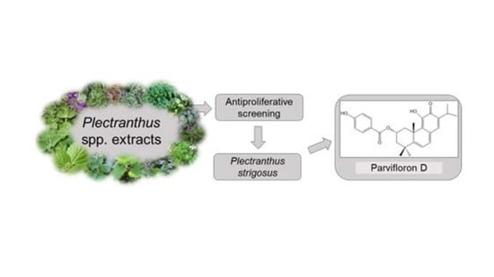
18]. Having this in mind, the present study was aimed at screening the antimicrobial and cytotoxic properties of several
Plectranthus spp. extracts. These goals were accomplished by comparing the efficacy of 31 plant extracts and identifying the bioactive component responsible for the extracts activity.
2. Experimental
2.1. Plant Material
Plectranthus spp. (
Lamiaceae family) were cultivated in Instituto Superior de Agronomia Campus (Lisbon University), from seeds provided by the herbarium of the Botanical Garden of Lisbon, Portugal. The air dried and powdered whole plants were selected based on their ethnopharmacological features:
Plectranthus aliciae (Codd) van Jaarsv. and Edwards,
Plectranthus amboinicus (Lour.) Spreng.,
Plectranthus barbatus Andrews,
P. ecklonii Benth.,
P. fruticosus L’Hér.,
Plectranthus hadiensis (Forssk.) Schweinf. ex Spreng. var.
hadiensis, P. hereroensis Engl.,
Plectranthus japonicus Koidz.,
Plectranthus madagascariensis (Pers.) Benth. var.
madagascariensis,
P. madagascariensis (Pers.) Benth var. ‘Lynne’,
Plectranthus malvinus van Jaarsv. and T.J. Edwards,
Plectranthus oertendahlii Th. Fr.,
Plectranthus reflexus van Jaarsv. and T.J. Edwards,
Plectranthus stylesii Edwards,
P. strigosus Benth. ex E.Mey., and
Plectranthus zuluensis T. Cooke. The plant names were verified with the Index Kewensis [
19]. The plants were grown in Parque Botânico da Tapada da Ajuda from cuttings provided through the Kirstenbosch National Botanical Gardens, South Africa; were collected between 2007 and 2008, always in June and September; and voucher specimens were deposited in the Herbarium “João de Carvalho e Vasconcellos” of the “Instituto Superior de Agronomia”, Lisboa (LISI), Portugal [
20].
Traditional uses of the mentioned plants and their voucher numbers are displayed in
Table S1.
2.2. Extraction Procedure of Plectranthus spp.
Thirty-one extracts were prepared from sixteen different
Plectranthus species. The air-dried leaves of each plant were milled in a kitchen mill and weighed. The extraction procedure was carried out in triplicate in an ultrasonic (US) bath (Bandelin SONOREX RK 510H, Berlin, Germany) for 60 min at 25 °C—using acetone, ethanol, and/or methanol—in a concentration of 10% (
w/
v). The suspensions were then filtered using Filtrer Laurent—Prat demas 150 mm (Couze-St-Front, France). After each filtration, the solvent was removed in a rotary evaporator (Sigma-Aldrich, IKA HBR 4 basic heating bath, Essen, Germany) at 40 °C. After submitting the suspensions to ultrasounds three times, the final extracts were weighed, and the yield of extraction determined. The percentage yield for each extract was determined (
Figure S2).
2.3. Microorganisms Used and Growth Conditions
Bacterial strains Enterococcus faecalis (ATCC 29212), Escherichia coli (ATCC 25922), Pseudomonas aeruginosa (ATCC 27853), and Staphylococcus aureus (ATCC 25923), and yeast strains Candida albicans (ATCC 10231) and Saccharomyces cerevisiae (ATCC 9763) were chosen based on their clinical and pharmacological importance. In addition, S. cerevisiae was added due to its general toxicity screening value. The tested bacteria were precultivated on Mueller Hinton agar and yeasts in Sabouraud agar (Biokar diagnostics, Allonne, France). Cultures of microorganisms were grown at 37 °C (30 °C for the yeasts) for 24 h and were maintained on respective agar slants at 4 °C.
2.4. Antimicrobial Screening Assays
2.4.1. Well Diffusion Method
The extracts were reconstituted in DMSO—Dimethyl Sulfoxide—to a 1 mg/mL concentration as well as the stock solutions of reference antibiotics (vancomycin and norfloxacin, Gram-positive and -negative, respectively). Nystatin was used as the positive control for the yeasts. Afterwards, Petri dishes containing 20 mL of solid Mueller–Hinton culture medium (Sabouraud for the yeasts) were inoculated with 100 μL of microorganism suspension (correspondent to a 0.5 McFarland standard solution). The inocula were evenly harvested on the medium surface using a sterile swab [
20]. Wells of approximately 5 mm in diameter were punched aseptically in the medium, with a sterile glass Pasteur pipette and 50 μL of each extract and antimicrobial agent were introduced into the well [
20,
21]. The solvent DMSO was used as negative control. Plates were then incubated at 37 °C (30 °C for the yeasts) for 24 h. The antimicrobial activity was then evaluated by measuring the diameter (mm) of the inhibition zone formed around the wells and the results compared to positive and negative controls [
20]. This method was tested at least in triplicate.
2.4.2. Determination of Minimum Inhibitory Concentration
Only extracts that showed inhibitory activity on the well diffusion method were tested for this method, so only the bacteria were active. After screening the antimicrobial activity of the plant extracts, the microplate broth microdilution method was performed for those that were considered active (inhibition zone > 10 mm). As such, 100 μL of liquid Mueller–Hilton medium was distributed in each well of a 96-well plate. The first well of each row contained 100 μL of extract, the positive control, or negative control solutions (1 mg/mL concentration). Serial dilutions were made to 1:2 proportion between each row of wells (1.95–500 μg/mL range). Finally, 10 μL of bacterial suspension of 0.5 McFarland density were added to each well. Plates were then incubated at 37 °C for 24 h, allowing the bacterial growth. The latter was measured with an absorbance microplate reader (Thermo Scientific Multiskan FC, Loughborough, UK) set to 620 nm. All assays were carried out in triplicate for each tested microorganism [
20].
2.4.3. Determination of Minimal Bactericidal Concentration
After concluding the minimum inhibitory concentration (MIC) assay, the minimal bactericidal concentration (MBC) assay was done accordingly to the agar dilution method. Thus, a swab from each well where no growth was observed—containing the tested extract concentration series—was plated on a Mueller Hinton Agar [
21,
22,
23]. The Petri plates were then incubated for 24 h at 37 °C, for further evaluation. The lowest concentration that revealed no visible bacterial growth after subculturing was taken as its MBC. Only the bacteria showed inhibition growth.
2.5. Cell Lines and Reagents
2.5.1. Chemicals
MTT (3-(4,5-dimethyl-2-thiazolyl)-2, 5-diphenyl-2H-tetrazolium bromide), rhodamine 123 (Rho123), and dimethylsulfoxide (DMSO) were acquired from Sigma-Aldrich Chemie GmbH. Fetal bovine serum (FBS), RPMI 1640 medium (Roswell Park Memorial Institut), DMEM (Dulbecco’s Modified Eagle Medium), penicillin–streptomycin solution, antibiotic–antimycotic solution, l-glutamine, and trypsin/EDTA were purchased from PAA (Vienna, Austria).
2.5.2. Cell Culture Maintenance
Human colon (HCT116) and breast (MCF-7) adenocarcinoma, and non-small cell lung carcinoma (NCI-H460) cell lines were purchase from ATCC. All cancer cells were cultured in RPMI-1640 medium with ultraglutamine (Lonza, VWR, Carnaxide, Portugal), and supplemented with 10% fetal bovine serum (FBS; Gibco, Alfagene, Carcavelos, Portugal). Cells were maintained at 37 °C in a humidified atmosphere of 5% CO2.
Sensitive non-small cell lung carcinoma NCI-H460, colorectal carcinoma DLD1 cell lines, and normal human embryonal bronchial fibroblasts MRC-5 were purchased from American Type Culture Collection (Rockville, MD, USA).
NCI-H460 and DLD1 cells were maintained in RPMI 1640 medium containing 10% heat in activated FBS, 2 mM
l-glutamine, 4.5 g/L glucose, 10,000 U/mL penicillin, 10 mg/mL streptomycin, and 25 mg/mL amphotericin B solution at 37 °C in a humidified 5% CO2 atmosphere. NCI-H460/R cells were originally selected from NCI-H460 cells and cultured in a medium containing 100 nM doxorubicin (DOX) [
23]. DLD1-TxR cells were selected by continuous exposure to stepwise increasing concentrations of paclitaxel (PTX) from DLD1 cells [
24].
MRC-5 cells were cultured in DMEM supplemented with 10% FBS, 4 g/L glucose, l-glutamine (2 mM), and 5000 U/mL penicillin, 5 mg/mL streptomycin solution at 37 °C in a humidified 5% CO2 atmosphere.
All cell lines were subcultured at 72 h intervals using 0.25% trypsin/EDTA and seeded into a fresh medium at the density of 8000 cells/cm2.
2.6. Preliminary Evaluation of General Toxicity on Artemia salina Model
Aiming at the evaluation of the preliminary toxicity of the selected
Plectranthus spp. extracts, a lethality test on
Artemia salina L. was carried out [
25]. The general toxicity on
A. salina was tested according to the method of Zhang et al. 2012 [
26], with slight alterations.
Brine shrimp cysts (obtained from JBL GmbH and Co. KG, D-67141 Neuhofen Germany) were hatched in artificial sea water with a concentration of 35 g/L. A container with two connected chambers with a simple handmade communicating vessel was used for brine shrimp hatching. An air pump inserted in the container ensured a regular air flow supply, thus saturating the artificial sea water for successful hatching. The cysts were then incubated for 48 h at 24 °C. Each extract was tested at a concentration of 100 ppm. To do so, ten to fifteen nauplii were transferred into wells of 24-well plates containing artificial sea water, and 100 μL of each extract was added to the wells (final volume per well: 1 mL). After 24 h exposure to the extracts (24 °C), the number of dead nauplii (mortality rate (%)) was determined (Equation (1)). Also, LC
50 (Lethal Concentration, 50%) values (μg/mL) were calculated for the most toxic extracts. DMSO was used as the solvent and was kept at 10% (
v/
v) in all samples tested.
2.7. Cytotoxicity Screening
2.8. Liquid Chromatography–Mass Spectrometry Analysis
Liquid chromatography–mass spectrometry (LC–MS)/MS analyses were performed on a Waters Alliance 2695 high performance liquid chromatography (HPLC) separation module connected to a Micromass Quattro Micro triple Quadropole (Waters, Dublin, Ireland) Mass Spectrometer equipped with a Waters electrospray ionization (ESI) source. Mass spectrometer was operated in positive mode and spectra were recorded in the m/z 60 to 800 Da range. In order to optimize detection conditions, a standard solution of parvifloron D was infused in the mass spectrometer and capillary and cone voltages, and collision energy were tuned to maximize (recursor ion > product ion) transition signal. ESI capillary voltage was optimized to 3 kV, cone voltage was set on 30 V. Source and desolvation temperature were adjusted to 120 °C and 350 °C, respectively. High purity nitrogen (N2) was used as drying gas and as a nebulizing gas. Ultra-high purity argon (Ar) was used as collision gas. A reversed phase WatersTM Atlantis C-18 (Waters, Milford, MA, USA; 5 μm × 2.1 mm × 150 mm) was used, with an injection volume of 15 μL. The mobile phase consisted in 0.5% formic acid in Milli-Q water (eluent A) and acetonitrile (eluent B). A flow rate of 0.2 mL/min was used, with the following gradient program: 0–15 min from 60% to 20% A, 15–20 min at 20% A, 20–30 min 60% A. MassLynx software (Waters, version 4.1) was used for data acquisition and processing.
The quantification of the identified compound was carried out in an Agilent Technologies 1200 Infinity Series system with diode array detector (DAD; Agilent, Santa Clara, CA, USA), equipped with a Zorbax XDB-C18, (250 × 4.0 mm i.d., 5μm) column, from Merck and ChemStation Software (Hewlett-Packard, Alto Palo, CA, USA). Each extract was analyzed (after 20 μL injection) and a gradient elution mixture composed of solution A (methanol), solution B (acetonitrile), and solution C (0.3% trichloroacetic acid in water) was used as follows: 0 min, 15% A, 5% B, and 80% C; 20 min, 70% A, 30% B, and 0% C; 25 min, 70% A, 30% B, and 0% C; and 28 min, 15% A, 5% B, and 80% C. The flow rate was set at 1 mL.min−1. For quantification and identification purposes, the compounds present in the samples were compared with calibration standards. Compound identification was based on ultraviolet (UV) spectra comparison and retention time. The time of analysis was of 28 min, including the stabilization of the RP-18 column. All analyses were performed in triplicate.
2.9. Anticancer Effects of the Identified Compound
The anticancer properties of the parvifloron D present in the most active extract were assessed through the use of a model system of NCI-H460 and its resistant counterpart NCI-H460/R, along with normal human embryonal bronchial fibroblasts MRC-5. MTT test was applied after 72 h incubation with diterpenes, applied within a range of concentrations 2.5–50 μM.
2.9.1. Cell Viability Assessed by MTT Assay
MTT assay is based on the reduction of 3-(4, 5-dimethyl-2-thizolyl)-2,5-diphenyl-2H-tetrazolium bromide into formazan dye by active mitochondria of living cells. Briefly, cells grown in 25 cm2 tissue flasks were trypsinized, seeded in 96-well tissue culture plates (2,000 cells/well), and incubated overnight in 100 mL of appropriate medium. After 24 h, cells were treated with increasing concentrations of parvifloron D (2.5–50 μM). All treatments lasted 72 h. At the end of treatment period, 100 mL of MTT solution (1 mg/mL) was added to each well and plates were incubated at 37 °C for 4 h. Formazan product was dissolved in 200 mL DMSO. The absorbance of obtained dye was measured at 540 nm using an automatic microplate reader (LKB 5060-006 Micro Plate Reader, Vienna, Austria).
Relative cell growth rate (CG) was determined according to the following equitation:
where A is absorbance. IC
50 value was defined as concentration of each drug that inhibited cell growth by 50%. IC
50 was calculated by nonlinear regression analysis using log (inhibitor) vs. normalized response in GraphPad Prism 6 software.
2.9.2. Assay for P-gp Inhibiting Activity
The function of P-gp was analyzed by flow-cytometry utilizing the ability of its substrate Rho123 to emit fluorescence. Studies were carried out with parvifloron D. Dex-VER was used as a positive control. The MDR cells (NCI-H460/R) were grown to 80% confluence in 75 cm2 flasks, trypsinized, and resuspended in 5 mL centrifuge tubes in a Rho123-containing medium (5 μM Rho123). The cells were treated with Dex-VER and parvifloron D (5 μM) and incubated at 37 °C in 5% CO2 for 30 mmL of cold phosphate-buffered saline, and analyzed usinin. The samples were washed twice, resuspended in 1.0 g CyFlow Space flow cytometer (Partec, Münster, Germany). The mean fluorescence intensity (MFI) of Rho123 was assessed on fluorescence channel 1. At least 10,000 events were assayed for each sample.
2.9.3. Effects of Compounds on Microtubule
The effect of the identified compound on interphase on microtubules, mitotic spindles, nuclear morphology, and cell cycle was assessed. In order to do so, human lung carcinoma A549 cells were incubated for 20 h with serial concentrations of the diterpene ranging from 10 nM to 200 μM.
The DNA content was analyzed by flow cytometry using a BD FACSVerseTM cytometer (BD Biosciences, San Jose, CA, USA). Cells were collected and centrifuged at 500× g, washed with phosphate buffer saline (PBS), and resuspended in 50 μL of PBS. Following dropwise addition of 1 mL of ice-cold 75% ethanol, fixed cells were stored at −20 °C for 1 h. Samples were then centrifuged at 500× g and washed with PBS before resuspension in 1 mL of PBS containing 50 μg/mL propidium iodide and 100 μg/mL RNase A, and incubation for 1 h at 37 °C in the dark. The percentage of cells with decreased DNA staining, composed of apoptotic cells resulting from either fragmentation or decreased chromatin, was counted (minimum of 10,000 cells per experimental condition). Cell debris was excluded from analysis by selective gating based on anterior and right angle scattering.
3. Results and Discussion
The acetonic, ethanolic, and/or methanolic extracts of several
Plectranthus species (
Figure S1) were evaluated concerning their antimicrobial activity through the well diffusion method and further MIC and MBC determination. Furthermore, in order to evaluate their cytotoxic properties, toxicity assays were performed on
Artemia salina L. and different cancer cell lines.
All 31 extracts were screened at a fixed concentration (1 mg/mL). Overall, five extracts demonstrated inhibitory activities against either or both
Enterococcus faecalis (ATCC 29212) and
Staphylococcus aureus (ATCC 25923) bacterial strains (
Table S2). All other extracts did not display antibacterial effects, showing inhibition zones lower than 10 mm (data not shown). The extracts prepared with acetone revealed better antimicrobial activity, particularly against Gram-positive bacteria.
A significant inhibition zone was recorded for the acetonic extracts of P. aliciae and P. madagascariensis var. “Lynne”, for both Gram-positive bacteria used (E.faecalis and S. aureus), with results similar to those of positive control. The remaining active extracts, however, showed selectivity only against S. aureus.
Regarding the results obtained on the well diffusion test, only Gram-positive bacteria were taken into account on these assays. Thus, in order to assess the lowest growth inhibitory concentration of the most promising
Plectranthus spp. extracts, minimum inhibitory concentration values (MIC) were determined. Their bacteriostatic and bactericidal properties were also evaluated (
Table 1) MIC values ranged from 15.6 to 125 μg/mL, while MBC values ranged from >62.5 to 250 μg/mL.
Comparing the MBC and MIC values of the selected extracts, and having in mind that a bactericidal effect is associated to a MBC no more than four times the MIC value [
21], it is possible to conclude that the extracts are mainly bactericidal rather than bacteriostatic.
The preliminary evaluation of toxicity on
Artemia salina is a fairly easy and inexpensive method and is particularly interesting when testing plant extracts as it can be used as a prescreen of their potential antitumor activities This assay also helps in gaining a preliminary understanding of which extracts may deserve additional testing. All 31 extracts were tested (data not shown), but only five of them exhibited toxic effects with values ranging from 13.62 to 110.76 μg/mL (
Table 2). Curiously, these five acetonic extracts were the same that induced
S. aureus growth inhibition. The lowest LC
50, and consequently, the highest toxic potential, was attributed, once again, to the activity of
P. strigosus. On the contrary, the least toxic extract was attributed to
P. stylesii.
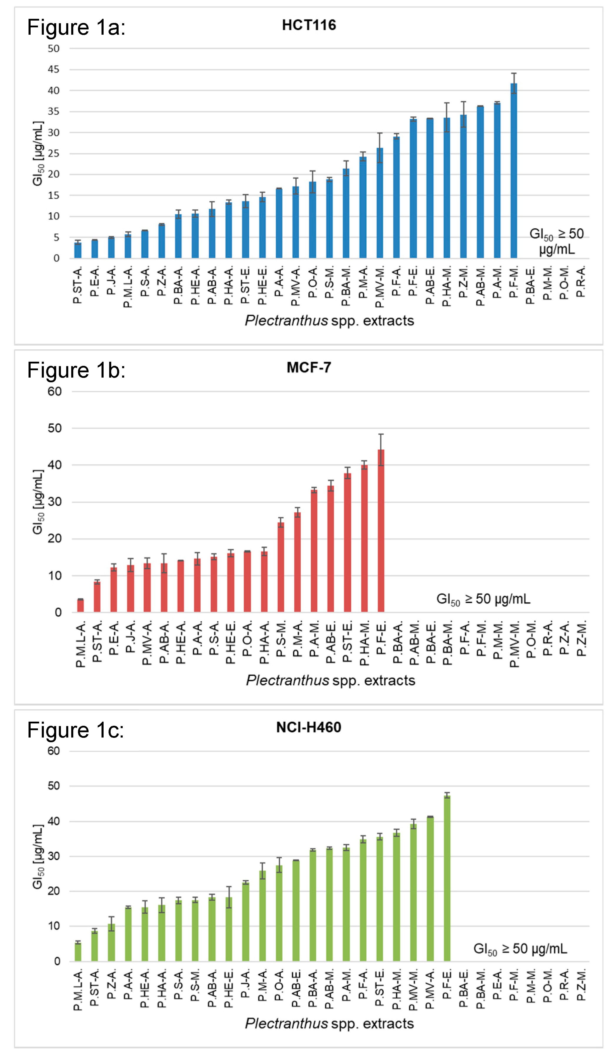
The GI
50 values were also determined for each of the 31 extracts, in HCT116, MCF-7, and NCI-H460 cell lines, and the observed reduction in the cell growth was compared to the growth of cells treated with DMSO only (set as 100% growth). The results are summarized in
Figure 1 below. The data bars not shown refer to a GI
50 value equal or higher than 50 μg/mL.
According to the results, acetonic extracts revealed better cytotoxic effects. Five of these—P. aliciae, P. japonicus, P. madagascariensis var. “Lynne”, P. stylesii, and P. strigosus—have consistently exhibited a broad cytotoxic potential. When analyzing GI50 on the three tested cell lines, values ranged between 3.47 to 16.67 μg/mL, showing effective growth inhibition against specific cell lines.
P. strigosus acetonic extract displayed the highest overall anticancer potential, given its growth inhibitory activities towards HCT116 (GI50 = 3.78 ± 0.49 μg/mL), MCF-7 (GI50 = 8.35 ± 0.57μg/mL), and NCI-H460 (GI50 = 8.75 ± 0.70 μg/mL). On the other hand, the highest inhibitory growth activities on MCF-7 and NCI-H460 cell lines were attributed to the effect of P. madagascariensis var. “Lynne” (GI50 = 3.47 ± 0.15μg/mL and 5.39 ± 0.48, respectively).
The extract with the lowest anticancer potential was P. reflexus methanolic extract, for which GI50 was not reached in the screened range in all tested cell lines (GI50 ≥ 50 μg/mL).
Overall, Plectranthus spp. extracts recorded a broader cytotoxic activity against HCT116, given that 27 out of the 31 extracts tested have GI50 lower than 50 μg/mL.
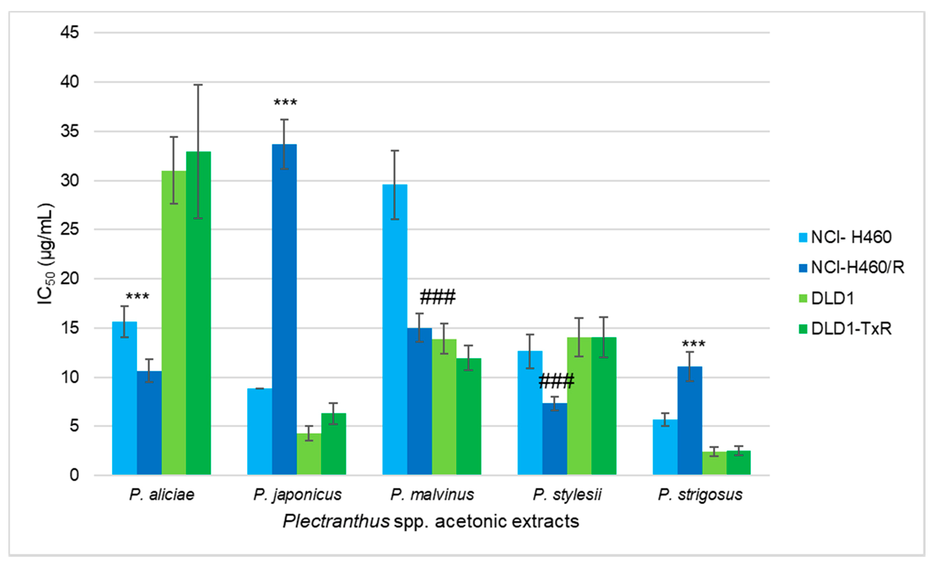
According to the results obtained in the previous screening (
Figure 1), the most promising
Plectranthus spp. extracts were tested in MDR cell lines with P-gp overexpression: NCI-H460/R (non-small cell lung carcinoma cell line) and DLD1-TxR (colorectal adenocarcinoma cell line). The effects of the five selected acetonic extracts were assessed by SRB assay after 72 h, and their IC
50 values were determined (
Figure 2).
P. strigosus acetonic extract exerted an overall best effect in both non-small cell lung carcinoma and colorectal adenocarcinoma cells, which showed IC
50 values of 5.64 μg/mL (NCI-H460), 11.09 μg/mL (NCI-H460/R), 2.41 μg/mL (DLD1), and 2.51 μg/mL (DLD1-TxR).
All five extracts exerted similar results towards colorectal adenocarcinoma cells (DLD1 and DLD1-TxR). Thus, their efficacy might not depend on the presence of a MDR phenotype. Acetonic extracts from P. aliciae, P. japonicas, and P. strigosus were less active against non-small cell lung carcinoma MDR cells (NCI-H460/R) in comparison with their sensitive counterparts (NCI-H460). Interestingly, resistant NCI-H460/R were more sensitive to extracts from P. malvinus and P. stylesii showing collateral sensitivity, the phenomenon when MDR cells are more vulnerable than corresponding sensitive cells.
In order to assess the chemical composition of the most active extract (
P. strigosus acetonic extract), and in an attempt to identify the compounds responsible for the referred activity, a combination of HPLC–DAD (diode array detector) –MS techniques were carried out. The HPLC–MS analysis showed two main peaks at 19.8 and 22.0 min, which revealed a UV spectrum characteristic of quinone methide diterpenes [
29]. The ESI mass spectra of the compounds pointed to the presence of parvifloron E or F (peak at 19.8 min,
m/
z 451 [M + H]
+) and parvifloron D (peak at 22 min,
m/
z 435 [M + H]
+). The presence of parvifloron D was confirmed through MS/MS analysis by comparison with a standard compound. MS/MS spectra showed product ions at
m/
z 297 (base peak) and
m/
z 279, corresponding to the loss of the ester moiety ([M-C
7H
5O
3 + H]
+) and ester moiety and H
2O ([M-C
7H
5O
3-H
2O + H]
+), see
Supplementary Materials, Figure S3. The 19.8 min compound displayed a similar fragmentation pattern corresponding to the same diterpenic scaffold, with one more hydroxyl group on the aromatic ring substituent. Therefore, this compound was tentatively identified as parvifloron F. The quantification of parvifloron D was performed through HPLC–DAD/UV and was found to be present in the acetonic extract of
P. strigosus with a relative concentration of 9%.
Given its antitumor properties [
30], the presence of the latter may explain, to a certain extent, the foreseen toxic properties of
P. strigosus acetonic extract. In order to further explore its cytotoxicity, additional studies were performed on this compound.
Afterwards, the preliminary toxicity evaluation of parvifloron D on Artemia salina L. resulted in a 76.6% mortality rate on this species after 24 h incubation.
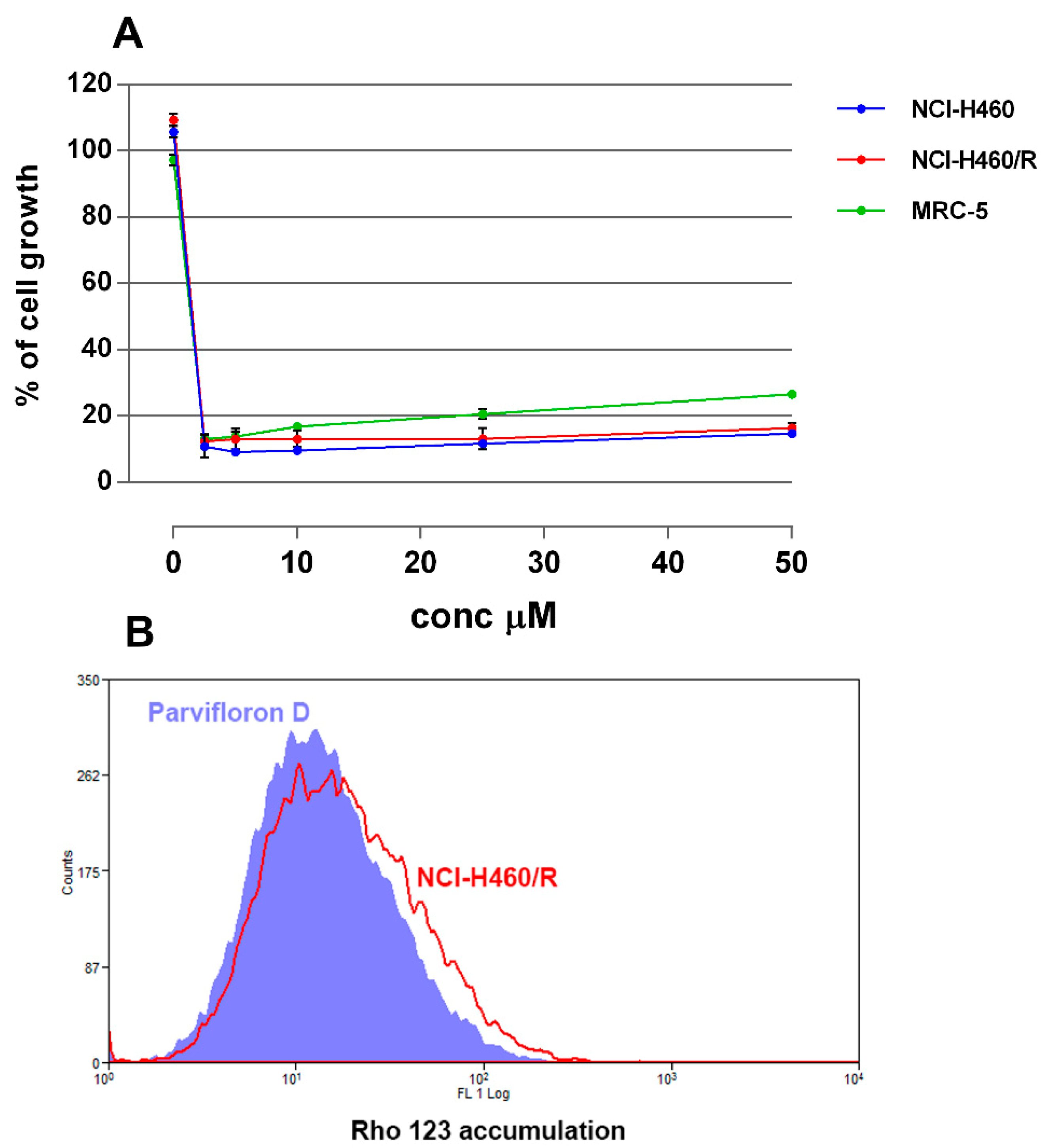
In addition, its cytotoxic activity was further evaluated on NCI-H460 and NCI-H460/R cell lines. To determine whether parvifloron D is selective towards cancer cells, the cytotoxic effect in normal embryonic bronchial fibroblasts (MRC-5) was also evaluated.
As expected, this diterpene demonstrated high cytoxicity and exerted a similar activity in all three cell lines, with IC
50 ranging from 1.7 to 1.9 μM. Interestingly, it also showed the same efficacy in sensitive and MDR cancer cells, which implies that parvifloron D is able to evade P-gp efflux activity (
Figure 3A). However, it was equally active against normal cells (
Figure 3A). Parvifloron D was also tested regarding its potential to inhibit P-gp activity in NCI-H460/R cells. Unfortunately, parvifloron D was not able to increase the Rhodamine 123 accumulation in P-gp overexpressing cells (
Figure 3B). Considering its high cytotoxic potential, further examinations of parvifloron D should be performed. Its mechanism in cancer and normal cells, with respect to the application of lower concentrations and different time scheduling, are necessary to clarify whether this compound should be developed as an anticancer agent.
In order to check whether this abietane diterpene was a microtubule stabilizer agent, A549 cell morphologies in the IC50 culture plates were examined with an inverted microscope, after 20 h incubation with parvifloron D. Paclitaxel was used as control tubulin specific drug. All microtubule targeting drugs, stabilizers, or depolymerizers in contact with A549 cell lines induced the appearance of round cells (mitotic cells) that detach from the plate. However, parvifloron D only displayed a cytotoxic effect rather than cytostatic, with no mitotic blockage, practically ruling out an action on mitotic spindle microtubules (data not shown). These results suggest that the antibacterial and anticancer activities of the acetonic extract of P. strigosus are attributed to the presence of parvifloron D. These results correlate to the representative HPLC–DAD/UV (λ = 254 nm) chromatogram of acetonic extract of P. strigosus and UV DAD spectra of peaks. The prepared acetonic extract of P. strigosus (c = 0.67 mg/mL) was revealed to have Parvifloron D (9% (w/w) in its composition.

 ,
, 


