Article

Separation and Determination of Some of the Main Cholesterol-Related Compounds in Blood by Gas Chromatography-Mass Spectrometry (Selected Ion Monitoring Mode)

by
Lucia Valverde-Som
1,*,
Alegría Carrasco-Pancorbo
1,
Saleta Sierra
2,
Soraya Santana
2,
Cristina Ruiz-Samblás
1,
Natalia Navas
1,
Javier S. Burgos
2 and
Luis Cuadros-Rodríguez
1
1
Department of Analytical Chemistry, Faculty of Science, University of Granada, C/Fuentenueva s/n, E-18071 Granada, Spain
2
Neuron Bio, P.T.S. Granada, C/Avicena 4, E-18016 Granada, Spain
*
Author to whom correspondence should be addressed.
Separations 2018, 5(1), 17; https://doi.org/10.3390/separations5010017
Submission received: 22 September 2017 / Revised: 12 February 2018 / Accepted: 27 February 2018 / Published: 5 March 2018

Abstract

:
Oxysterols are metabolites produced in the first step of cholesterol metabolism, which is related to neurodegenerative disorder. They can be detected by testing blood, plasma, serum, or cerebrospinal fluid. In this study, some cholesterol precursors and oxysterols were determined by gas chromatography coupled to mass spectrometry. The selected cholesterol-related compounds were desmosterol, lathosterol, lanosterol, 7α-hydroxycholesterol, 7β-hydroxycholesterol, 24(S)-hydroxycholesterol, 25-hydroxycholesterol, 7-ketocholesterol, and 27-hydroxycholesterol. A powerful method was developed and validated considering various analytical parameters, such as linearity index, detection and quantification limits, selectivity and matrix effect, precision (repeatability), and trueness (recovery factor) for each cholesterol-related compound. 7α-hydroxycholesterol, 7β-hydroxycholesterol, and desmosterol exhibited the lowest detection and quantification limits, with 0.01 and 0.03 μg/mL, respectively, in the three cases. 7-ketocholesterol and lathosterol showed matrix effect percentages between 95.5% and 104.8%, respectively (demonstrating a negligible matrix effect), and very satisfactory repeatability values (i.e., overall performance of the method). Next, the method was applied to the analysis of a very interesting selection of mouse plasma samples (9 plasma extracts of non-transgenic and transgenic mice that had been fed different diets). Although the number of samples was limited, the current study led to some biologically relevant conclusions regarding brain cholesterol metabolism.

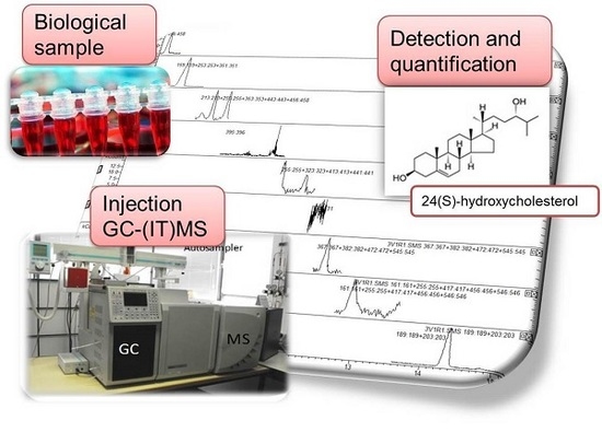
Graphical Abstract

1. Introduction

Oxysterols play a crucial role as (a) regulators of expression of genes involved in lipid and sterol biosynthesis; (b) substrates for the formation of bile acids; and (c) mediators of reverse cholesterol transport, through which excess cholesterol is returned to the liver for excretion [1,2]. However, an overabundance of oxysterols can cause several diseases, such as cardiovascular disease, retina degeneration, inflammatory bowel disease, atherosclerosis, and neurodegenerative disorders [3,4,5].
Oxysterols can be found in several chemical forms and are enzymatically produced in the first steps of cholesterol (Chol) metabolism [6,7]. For example, Alzheimer’s disease (AD) pathology is initiated (or accelerated) by a deregulation of the metabolism of Chol, sphingolipids, and fatty acids; in fact, hypercholesterolemia is an important risk factor for AD [8,9]. These metabolites are present in blood (serum or plasma), cerebrospinal fluid and brain tissues. Desmosterol, lanosterol, and lathosterol are the precursors of Chol synthesis. Chol is metabolized into side chain oxysterols (24(S)-hydroxycholesterol, 25-hydroxycholesterol, and 27-hydroxycholesterol) during its hepatic conversion [10] and transported into the brain by apolipoprotein E. Under normal conditions, no exchange of cholesterol occurs over the blood-brain barrier. However, Chol can be transported to the human brain in the form of a side-chain oxidized oxysterol such as 24(S)-hydroxycholesterol and 27-hydroxycholesterol [5,11,12]. As a consequence, the presence of these oxysterols has been related to neurological disorders and such oxysterols have been proposed as potential AD biomarkers [9,13].
Over the last decades, significant scientific developments and cutting-edge technologies have enabled a technological revolution in molecular biological methods. Metabolomics is a field that deals with the study of metabolites, which are the final products of cell regulation processes due to responses to genetic and environmental changes in biological systems [14].
Different approaches can be used in metabolomics [15]. Metabolite targeted analysis, for example, involves the detection, identification, and quantification of a single metabolite or a small group of specific metabolites of particular importance. The comprehensive characterization of a group of metabolites in an organism, tissue or cell implies the use of advanced analytical techniques. Oxysterols have been determined so far by liquid chromatography (LC) or gas chromatography (GC), mainly coupled to mass spectrometry (MS) and also by nuclear magnetic resonance (NMR) [6,16]. MS and NMR have a great potential to select proper biomarkers in order to diagnose associated diseases [13,17].
To determine the oxysterols present in biological fluids and tissues, there are different ways to carry out the sample preparation that obviously depend on the matrix, analyte/s and technique/s to be used. Blood plasma has been separated from blood serum by using ethylenediaminetetraacetic acid (EDTA) and further centrifugating the blood sample [18,19]. Next, sterols can be extracted from blood plasma by alkaline hydrolysis. This leads to the degradation of lipids such as phospholipids and triglycerides and avoids their potential interferences. Finally, solid phase extraction (SPE) has been utilized to separate sterols from other substances [18,20].
A derivatization reaction is usually required prior to GC analysis in order to increase the volatility of oxysterols; N,O-bis(trimethylsilyl)trifluoracetamide/trimethylchloroside (BSTFA:TMCS) is a very widely used derivatization reagent. However, N-methyl-N-(trimethylsilyl)-trifluoroacetamide-1,4-dithioerythritol-trimethyliodosilane (MSTFA:DTE:TMIS) has been proposed as one of the most efficient derivatization reagents based on its specificity/sensitivity ratio in GC-MS [21,22].
LC is recognized as a chromatographic technique that does not require a previous derivatization of the samples. Yet, in the specific case of determining oxysterols, the quantification can be more difficult if the analytes are not derivatized [22]. The use of N,N-dimethyl-glycine, picolinyl esters, and Girard P reagents has been described in LC-MS [23,24]. The derivatized compounds are more polar than the native sterols, which improves the ionization in the MS system. In fact, the number of papers describing LC-MS analytical methodologies for oxysterol determination in biological matrices has lately increased [25,26,27].
Traditionally, the instrumental analysis of sterols and related compounds in plasma has used GC and GC-MS [18]. Although GC and its use coupled to MS have some limitations, these techniques are widely used for sterol analysis due to their chromatographic resolving capacity, their robustness, and the relatively low cost of acquiring and operating the instruments [6,19,20,28]. GC-MS with electron ionization provides spectra rich in structural information, allowing the determination of unknown compounds [29]. By contrast, LC-MS requires Girard P derivatizations and exploring the mass-load ratio with the MS3 mode in order to obtain structural information and identify unexpected oxysterols [27]. The use of MS in tandem (MSn) can improve the sensitivity of the methods [30,31], but can also entail some methodological difficulties. A review conducted by Griffiths and Wang in 2009 [32] explained the biological importance of oxysterols and various relevant samples of brain and body fluids analyzed with GC-MS and LC-MS/MS. In 2016, both authors with other colleagues presented a review of MS-based methods for oxysterol analysis paying particular attention to LC-MS [27].
The aim of the present study was to develop a reliable and fit-for-purpose bioanalytical method for the determination of some Chol-related compounds (selected as potential biomarkers of AD by the biotechnology-based company Neuron Bio) in mouse plasma from six APPswe mice and three non-transgenic littermate mice of both sexes that were fed different diets from the age of 9 months to the age of 15 months. The analytical method was developed by using GC-MS in selected ion monitoring (SIM) mode. An ion trap (IT) mass analyzer was used to obtain the m/z signal of the analytes under study (7α-hydroxycholesterol (7αOHChol), desmosterol (Desm), lathosterol (Latho), 7β-hydroxycholesterol (7βOHChol), lanosterol (Lano), 24(S)-hydroxycholesterol (24OHChol), 25-hydroxycholesterol (25OHChol), 7-ketocholesterol (7KetoChol), and 27-hydroxycholesterol (27OHChol)) applying an MS method with 9 different segments. In other words, we defined specific segments or analytical windows with different MS conditions in order to selectively determine each compound. This strategy enabled us to detect and quantify Chol-related compounds with a similar structure in mouse plasma. The potential interfering effect produced by high concentrations of cholesterol was avoided by applying the SIM strategy.

2. Materials and Methods

2.1. Chemicals and Reagents

(HPLC)-grade methanol and ethyl acetate (EA) were provided by Panreac, Spain. BSTFA (N,O-bis(trimethylsilyl)trifluoracetamide) with 1% TMCS (trimethylchloroside) was obtained from Sigma-Aldrich, St. Louis, MO, USA, as a derivatization reagent. Solid standards of analytes were supplied by Enzo Life Sciences, Farmingdale, NY, USA (24OHChol, >98%), MP Biomedicals, Illkirch-Graffenstaden, Strasbourg, France (25OHChol, >98%), Sigma-Aldrich, St. Louis, MO, USA (Chol, ≥99%; Latho, ≥93%; Desm, ≥84%; and 7KetoChol, ≥90%), and Avanti Polar Lipids, Alabaster, AL, USA (Lano, >99%; 7αOHChol, >99%; 7βOHChol, >99%; and 27OHChol, >99%). In addition, 98% purity betulin supplied by Avanti Polar Lipids, Alabaster, AL, USA, was used as internal standard (IS). All these chemical compounds as well their corresponding solutions were stored in the dark at −20 °C.
Betulin was chosen as internal standard because it is not a naturally occurring sterol in blood. This analyte has been also used as IS in other similar applications [33,34].

2.2. Standard Solutions

A multi-standard stock solution containing 100 μg/mL of each analyte (Chol, Desm, Lano, Latho, 7KetoOH, 7αOHChol, 7βOHChol, 24OHChol, 25OHChol, and 27OHChol) was prepared in methanol weighing the appropriate amount of the solid pure standards. A 100 μg/mL stock standard solution of betulin was also prepared in methanol.

2.3. Samples and Sample Preparation

The study was performed with two sample groups: (i) mice blood plasma as test samples and (ii) goat blood serum as quality control samples.
Each extract was prepared applying the following procedure: 200 μL of sample were poured into a glass tube along with 40 μL of internal standard and 0.5 mL of EA. The tube was shaken in a vortex mixer (1 min), sonicated (50 W) for 15 min, and centrifuged for 5 min at 10,000 rpm (approx. 8400 g). Subsequently, 100 μL of the organic phase were transferred to an Eppendorf vial and stored in the dark at −20 °C until the analysis was performed.
The final concentration of IS was 40 μg/mL in all the extracts. The solvent was evaporated with N2 and an aliquot of 50 μL of derivatization reagent was added after that. The reaction mixture was left at room temperature for 1 h (this was previously optimized by our team and it was proved that the total derivatization of the compounds of interest was achieved). In every case, 50 μL of the derivatized sample extract were transferred to a chromatographic vial containing a 250 μL insert.
(i)
Mouse plasma as test samples
Six 9-month-old APPswe (transgenic) mice and three 9-month-old non-transgenic (Non-Tg) littermate mice of both sexes were used. They were kept in temperature- and light-controlled rooms in Neuron Bio facilities. Three APPswe mice were fed with NST0037 (a novel statin developed by Neuron Bio (Granada, Spain) [35])—enriched diet until the age of 15 months; the rest—three APPswe and three Non-Tg littermate mice—were fed with a control diet from until the same age. In the rest of the paper, each type of mice will be denoted by using letters A, B and C: A are non-transgenic mice, Non-Tg; B are transgenic mice, APPswe; and C are transgenic mice, APPswe, fed with NST0037.
The 15-month-old mice were anesthetized with sodium pentobarbital (300 mg/kg); their blood was collected and the plasma was obtained and frozen at −80 °C.
(ii)
Goat serum as quality control samples
The set of quality control samples was composed of 5 extracts of the blood serum of a single goat in EA. Each extract (300 μL) was prepared in the same way as the test samples, starting from 600 μL of blood sample. As indicated above, the extracts were stored in the dark at −20 °C until the analysis took place. It should be noted that the sample treatment removes the proteins; thus, extracts from matrices of plasma and serum are quite similar and serum samples can be used as representative examples of plasma samples for an effective analytical quality control.
Quality control (QC) standard solutions were prepared by adding 0.5, 1.0, 2.5, and 5.0 μg/mL of each analyte into goat serum extracts. In order to effectively monitor the analytical method, two types of QC samples were used: some were spiked before and others were spiked after the extraction.
An overall diagram showing the preparation of both sample groups (i.e., test samples and quality control samples) is shown in Figure 1.

2.4. GC-(EI/IT)MS Instrument and Method Conditions

All separations were performed by using a Varian GC 3800 gas chromatograph (Palo Alto, CA, USA) equipped with a split/splitless injector. The GC system was coupled to a Varian (IT)MS 4000 (ion trap) mass spectrometer (Palo Alto, CA, USA) equipped with an electron impact (EI) source. 1 μL of each sample was introduced into the GC equipment using a robotized autosampler module (CombiPal, CTC ANALYTICS, Zwingen, Switzerland). Splitless injection mode was selected and the injector temperature was held at 250 °C.
The analytical column was a capillary column coated with a 5% diphenyl-95% dimethyl polysiloxane stationary phase (DB-5MS; 30 m × 0.25 mm i.d. × 0.25 µm film thickness (Agilent Technologies J&W, Santa Clara, CA, USA). A silica deactivated pre-column (1 m × 0.25 mm i.d.) with press-fit connections (Agilent Technologies, Santa Clara, CA, USA) was used.
At the beginning of the runs, the column oven temperature was held at 200 °C for 0.5 min, then programmed to increase by 20 °C/min up to 300 °C and maintained at that value for 10 min, with a total run time of 15.50 min. Helium (99.995%) was used as the carrier gas and its flow rate was 1.2 mL/min. This method was a modification of a previously published method [21,36].
The MS conditions were as follows: the ion source temperature was held at 250 °C during the GC-MS runs. The transfer-line temperature was maintained at 280 °C. The electron energy was 70 eV and the emission current 10 µA. In full-scan mode, average spectra were acquired in the m/z range of 50–600 m/z and were recorded at a scan speed of 1.20 s. Scan control, data acquisition, and processing were performed using MS Workstation (Varian, Palo Alto, CA, USA) software data system, version 6.9.
SIM mode was selected and applied for monitoring different ions in each segment. Table 1 shows the different segments with their time range, m/z selected in each analytical window and the analyte of interest to be determined in each case. Peak identification was based on the comparison of retention times and MS signals with those obtained from the analysis of pure standards.

3. Results and Discussion

3.1. Optimization of Detection Conditions

The starting GC conditions were provided by Neuron Bio. Further GC-MS optimization was carried out in 5 stages to improve the starting conditions. In a first stage of the optimization, each analyte was individually injected in GC-MS (full scan mode) to determine its retention time and its characteristic m/z. The values obtained for each analyte and the IS are shown on Table 2. Regarding the m/z values, it should be noted that the MS signals appear in decreasing order of intensity.
After characterizing the individual behavior of each analyte, we analyzed a multi-standard solution (full scan mode); an example of the result is provided in Figure 2.
It is well known that the concentration of cholesterol is quite high in plasma samples; therefore, to simulate a real biological situation, we prepared a standard solution containing 2000 μg/mL of Chol (cholesterol is the most abundant sterol in human plasma with levels in the order of 1 to 3 mg/mL [20]) and 5 μg/mL of the rest of the analytes, which is enough to view the analytes with full scan mode. The result is illustrated in Figure 3, which shows that Desm (which elutes just after Chol) was properly detected even with very high concentration levels of Chol in the sample under study. However, Figure 3 also shows that when Chol was present at high concentrations, the signal of the other analytes was very low in comparison, which hampered their appropriate determination. Another issue related to Chol concentration is that the peak corresponding to 7α-hydroxycholesterol was hardly detectable. Thus, the next step of the optimization focused on improving the conditions of ion detection with MS through the use of SIM mode.
We selected SIM operational mode and created specific segments or analytical windows (Table 1) in order to better detect each analyte (using specific MS parameters and looking for the characteristic m/z signals). A multi-standard solution containing 10 μg/mL of each analyte was injected into the GC-MS system (SIM mode). The result is shown in Figure 4.
As stated before, IT was used as mass analyzer. Even though IT can be used to perform MS/MS experiments, the use of SIM mode was selective enough to properly detect and quantify the compounds under study.
As a result, 9 analytes of interest plus the IS were detected separately by using 9 specific segments. The segments were optimized and defined taking into account the information included in Table 2 (i.e., retention times and m/z responses). The transfer and detection parameters were optimized aiming for the maximum sensitivity with the highest possible resolution. SIM led to very promising results in terms of improvement of the signal-to-noise ratio (S/N), sensitivity, detection and quantification limits, and some other important analytical validation characteristics.
After optimizing the GC-MS conditions, some biological samples were injected to prove the suitability of the new methodology in relatively long sequences (including biological samples, which are sometimes considerably dirty).
After 25 analyses, the need of a pre-column was quite evident to reduce the noise, which increased when a great number of consecutive injections and chromatographic runs were carried out. A silica phase with the same internal diameter as the column (0.25 mm) was selected. The pre-column had 1 m length and was connected to the column with press-fit connections. Once the pre-column was installed the S/N remained constant. The information provided in Table 2 regarding retention time was achieved with the pre-column already connected to the system.

3.2. Validation of the Developed Method

Multi-standard solutions and QC samples were used to validate the method. Table 3 shows all the analytical parameters calculated during the validation of the developed method [37,38].
Multi-standard solutions were prepared at different concentration levels (0.1, 0.2, 0.5, 1.0, 2.5, and 5.0 μg/mL of each analyte) to build the calibration curves. Forty μg/mL of betulin (as for the biological samples) were added to each multi-standard solution. Fifty μL of the multi-standard solution were transferred to a vial containing a 250 μL insert. The solvent was then evaporated with N2, after which an aliquot of 50 μL of derivatization reagent was added. The reaction mixture was left at room temperature for 1 h.
Calibration curves, one for each Chol-related compound, were fitted by least-squares regression from relative area signal (area_Chol-related analyte/area_IS) vs. Chol-related analyte concentration. The intercept was estimated as zero for all the analytes. For this reason, the relative areas were directly proportional to the concentration of each analyte. Thus, it was possible to calculate a response factor (RF) for each compound, which was constant and independent of the concentration of each analyte; values are shown in Table 3. Each point of the calibration graph corresponded to the mean value of four independent injections.
Determination coefficients and linearity indices (LIN) [39] were calculated for each calibration curve to assess goodness of fit. LIN = 1 − (SDslope/slope) is included in Table 3 and was above 98% in all the cases, except for 25OHChol (96.5%), which had a slightly higher detection limit. Therefore, it was not possible to detect low concentrations.
The calibration curve, response factor, and linearity index were calculated considering the four replicates of each concentration level of the multi-standard solutions injected.
Limit of detection (LOD) and limit of quantification (LOQ) were determined considering the standard deviation calculated from six replicates of the lower sample concentration and multiplying it by 3.28 and 10, respectively. The 3.28 value is the 95%-quantile of the non-centrally t-distribution with ∞ degrees of freedom (α = β = 0.05) [40]. Detection and quantification limits for 25OHChol were slightly higher than for the rest of the analytes, which can be explained because only one m/z signal was characteristic for this analyte; therefore, its determination was not as selective as that of the others. 7αOHChol, 7βOHChol, and Desm exhibited the lowest detection and quantification limits, with 0.01 and 0.03 μg/mL, respectively, in three cases.
Spiked goat serum samples were prepared to test the matrix effect and control the repeatability of the overall analytical process. To verify the matrix effect, calibration curves were prepared by spiking goat serum after the extraction with the Chol-related compounds (samples codes: QC00-05, QC00-10, QC00-25, QC00-50) at different levels: 0.5, 1.0, 2.5, and 5.0 µg/mL. The slopes of these calibration curves were compared with those achieved by using external standard calibration. The native levels of analytes present in the goat blood serum sample (QC00) were previously determined in order to perform an effective blank correction. In QC00, the following compounds were found: 7αOHChol, 7βOHChol, 24OHChol, and 7KetoChol. The other Chol-related compounds were not detected (nd; below the detection limit). The area values found for this sample were considered in all the subsequent calculations and were subtracted from the values achieved after spiking with the known concentration levels to assess the matrix effect. Such concentration levels were QC00-05 (spiked with 0.5 μg/mL of multi-standard solution), QC00-10 (spiked with 1.0 μg/mL of multi-standard solution), QC00-25 (spiked with 2.5 μg/mL of multi-standard solution), and QC00-50 (spiked with 5.0 μg/mL of multi-standard solution). As shown on Table 3, matrix effect recoveries were found between 95.5% and 104.8% for 7KetoChol and Latho, respectively (100% was the value that would be achieved if the slopes of the external standard calibration and standard addition calibration curves were identical); in other words, the matrix effect was not observed in the current study.
Moreover, to provide an estimation of overall repeatability (including sample preparation and measurement), goat serum samples spiked with the Chol-related analytes under study—before the extraction—were prepared at the same levels as shown above (samples codes: QC 05, QC 10, QC 25, and QC 50). The standard deviation (SD) was calculated from four consecutive measurements; to estimate the repeatability of the overall process, SD was calculated from three independent goat serum samples that were extracted, injected, and analyzed separately. The average SD of the determined values is provided in Table 3 and was found to be within 0.059–0.181 for repeatability of the measurements and 0.166–0.294 for the overall process.
The same preparations were useful to assess the recovery factor. The theoretical added concentration did not exactly coincide with the concentration obtained after the extraction and the corresponding measurement. Given that no matrix effect was observed in the previous step, we concluded that the difference was due to the recovery (obviously lower than 100%). Recovery values were calculated and are shown in Table 3: they ranged from 65.8% to 97.8% for 27OHChol and 25OHChol, respectively. These recovery factors should be used to calculate the real concentration of each analyte in any biological sample. They were applied to obtain the quantitative values shown in Table 4.

3.3. Application of the Method to the Analysis of Biological Samples

Biological test samples were used for the application of the method. Each test sample was prepared and injected four times. Figure 5 shows an example of the results achieved for a mouse plasma sample (TA2) by using the SIM method with the 9 optimized segments.
In this example, all the selected analytes were found, with the exception of Lano (segment 4) and 25OHChol (segment 6); both analytes were indicated as not detected. In addition, some Chol-related compounds were detected but not quantified (they were below the LOQ). This was the case of Latho in samples TA1, TB1, TC2, and TC3 and 7KetoChol in samples TB1 and TC1. The rest of the analytes were found at concentration levels above their LOQs.
Quantitative results are summarized in Table 4, which includes the average concentration (μg/mL) of each analyte and the relative standard deviation (%RSD). The recovery factor was used to recalculate the real analyte concentration after extraction.
As stated above, there were three types of mice (A, B, and C) and three specimens in each group (1, 2, and 3). Samples belonging to the same group did not always show very similar results, what could be easily explained considering the fact that we studied different specimens.
Lano and 25OHChol were not detected in any sample. Desm was only detected in TA samples (TA1, TA2, and TA3) with concentration values ranging from 0.25 to 0.39 μg/mL, and in TB1, with a value of 0.19 μg/mL. Latho was found in the three specimens of Type A mice, with amounts of 0.39 and 0.46 μg/mL (TA3 and TA2); it was detected but not quantified in TA1. It was also found in two specimens of Type B mice (TB1 and TB2), with concentration values of 0.49 and 0.19 μg/mL respectively (the latter was a rough estimation), and in two Type C mice samples (estimated values—concentrations below LOQ—of 0.20 and 0.34 μg/mL—samples TC2 and TC3) (see Table 2). Overall, the rest of Chol-related compounds showed a decreasing trend in terms of concentration levels in mice A, B, and C.
Results showed that 7αOHChol was found at higher concentrations in Type A mice with the following values: 0.76, 0.22, and 0.70 μg/mL. In Type B samples, however, concentration values were 0.14, 0.42, and 0.11 μg/mL in the three specimens respectively. Type C mice showed the lowest values, ranging from 0.057 to 0.15 μg/mL. The situation was similar for 7βOHChol: the highest values were obtained for Type A mice (0.80, 0.41, and 0.76 μg/mL). 27OHChol and 24OHChol were found in all the samples. Their highest levels were determined in TA2 (0.87 μg/mL of 27OHChol), and TB2 (2.01 μg/mL of 24OHChol). As 24OHChol (also known as cerebrosterol) is a cholesterol catabolite produced almost exclusively in the brain, a reduction in its plasma levels after treatment with statins is regarded as an indirect marker of the inhibitory effect of these statins on brain cholesterol biosynthesis.
7KetoChol was found in all samples except for TB1 and TC1, in which it was detected but not quantified. In this case, its highest levels were determined in TA3 (2.99 μg/mL of 7KetoChol).
Overall, Type C mice showed a lower concentration of all Chol-related compounds, followed by Type B mice and Type A mice, which tended to exhibit the highest concentration values. These results are consistent with the fact that Type C mice were fed with statin. Several studies have pointed at a relationship between treatment with statins and a reduction of the risk of developing Alzheimer’s disease [41,42,43,44]. This can be explained considering that statins can help to reduce oxysterol levels, which affect amyloid beta production. Our findings indicate that a statin-rich diet can affect the brain cholesterol metabolism of mice to a certain extent; further studies are still needed to clarify whether the use of statins can be a useful strategy for treating the onset of clinical dementia, but promising results can already be found in the literature.

4. Conclusions

This study proves that GC-(IT)MS, working in SIM mode, is an analytical platform suitable to detect and quantify cholesterol precursors and oxysterols in mouse plasma samples. The high cholesterol concentration present in this biological fluid may impede the proper detection of the chromatographic peaks of some Chol-related compounds that were eluting close to the cholesterol peak; however, this potential issue was avoided by using the SIM mode. The use of a GC pre-column was another methodological improvement tested in this study. We demonstrated that it can easily reduce the background noise usually found in the biological samples; this strategy also extends the lifetime of the analytical column.
Other chromatography–MS methods have been reported in the literature, showing even better performance characteristics. Yet, this study proves that the proposed method is fit for monitoring the contents of some AD biomarkers in plasma samples from mammals that are used as experimental animals. Sample selection in this study was very special as it included non-transgenic and transgenic mice that were fed different diets. Overall, Type C mice (i.e., transgenic mice fed with statin) showed a lower concentration of every Chol-related compound; this fact may be explained considering that statins can help to reduce oxysterols levels, which affect amyloid beta production. We consider the results to be very promising, however, we are aware about some limitations of our study; several experiments are already under way to increase the number of samples analyzed and ensure that the observed variations in measurements are due to the treatment and not to potential population variability.

Acknowledgments

The authors are grateful for the interesting collaboration with the biotechnology-based company Neuron Bio. The research project was funded by CEI BioTic/University of Granada.

Ethical Conduct of Research

The experimental protocol was approved by the Neuron Bio Ethics Committee for Animal Research. Animal care was provided by qualified technicians supervised by veterinarians. Animals were treated in accordance with European legislation (Directive 2010/63/EU) and the International guidelines for ethical conduct in the care and use of experimental animals were applied throughout the study.

Author Contributions

All authors contributed to the bibliographic search for the introduction; Lucia Valverde-Som prepared the multi-standard stock solution, which was designed by Lucia Valverde-Som, Alegría Carrasco-Pancorbo, Natalia Navas and Luis Cuadros-Rodríguez; Saleta Sierra, Soraya Santana and Javier S. Burgos conceived and designed the extraction treatment; Saleta Sierra and Soraya Santana prepared the sample extraction; Lucia Valverde-Som, Alegría Carrasco-Pancorbo and Cristina Ruiz-Samblás optimized the GC-(IT)MS with SIM mode method; Lucia Valverde-Som performed the experiments and analyzed the data; Alegría Carrasco-Pancorbo and Luis Cuadros-Rodríguez guided the project; Lucia Valverde-Som, Alegría Carrasco-Pancorbo and Luis Cuadros-Rodríguez wrote the paper; all the authors revised the paper.

Conflicts of Interest

The authors declare no conflict of interest.

References

  1. Garenc, C.; Julien, P.; Levy, E. Oxysterols in biological systems: The gastrointestinal tract, liver, vascular wall and central nervous system. Free Radical Res. 2010, 44, 47–73. [Google Scholar] [CrossRef] [PubMed]
  2. Bjökhem, I.; Heverin, M.; Leoni, V.; Meaney, S.; Diczfalusy, U. Oxysterols and Alzheimer’s disease. Acta Neurol. Scand. 2006, 114, 43–49. [Google Scholar] [CrossRef] [PubMed]
  3. Terao, J. Cholesterol hydroperoxides and their degradation mechanism. In Lipid Hydroperoxide-Derived Modification of Biomolecules; Kato, Y., Ed.; Springer: London, UK, 2014; pp. 83–91. [Google Scholar]
  4. Mulder, M. Sterols in the central nervous system. Curr. Opin. Clin. Nutr. 2009, 12, 152–158. [Google Scholar] [CrossRef] [PubMed]
  5. Poli, G.; Biasi, F.; Leonarduzzi, G. Oxysterol in the pathogenesis of major chronic diseases. Redox Biol. 2013, 1, 125–130. [Google Scholar] [CrossRef] [PubMed]
  6. Griffiths, W.J.; Wang, Y. Analysis of oxysterol metabolomes. Biochim. Biophys. Acta 2011, 1811, 784–799. [Google Scholar]
  7. Griffiths, W.J.; Abdel-Khalik, J.; Yutuc, E.; Morgan, A.H.; Gilmore, I.; Hearn, T.; Wang, Y. Cholesterolomics: An update. Anal. Biochem. 2017, 524, 56–67. [Google Scholar] [CrossRef] [PubMed]
  8. Trushina, E.; Mielke, M.M. Recent advances in the application of metabolomics to Alzheimer’s Disease. Biochim. Biophys. Acta 2014, 1842, 1232–1239. [Google Scholar] [CrossRef] [PubMed]
  9. Leoni, V.; Caccia, C. Review. Oxysterols as biomarkers in neurodegenerative diseases. Chem. Phys. Lipids 2011, 164, 515–524. [Google Scholar] [CrossRef] [PubMed]
  10. Baila-Rueda, L.; Cenarro, A.; Cofán, M.; Orera, I.; Barcelo-Batllori, S.; Pocoví, M.; Ros, E.; Civeira, F.; Domeño, C. Simultaneous determination of oxysterols, phytosterols and cholesterol precursors by high performance liquid chromatography tándem mass spectrometry in human serum. Anal. Methods 2013, 5, 2249–2257. [Google Scholar] [CrossRef]
  11. Heverin, M.; Bogdanovic, N.; Lütjohann, D.; Bayer, T.; Pikuleva, I.; Bretillon, L.; Dicfalusy, U.; Winblad, B.; Bjökhem, I. Changes in the levels of cerebral and extracerebral sterols in the brain of patients with Alzheimer’s disease. J. Lipid Res. 2004, 45, 186–193. [Google Scholar] [CrossRef] [PubMed]
  12. Lim, W.L.F.; Martins, I.J.; Martins, R.N. The involvement of lipids in Alzheimer’s disease. J. Genet. Genom. 2014, 41, 261–274. [Google Scholar] [CrossRef] [PubMed]
  13. Kölsch, H.; Heun, R.; Kerksiek, A.; Bergmann, K.V.; Maier, W.; Lütjohann, D. Altered levels of plasma 24S- and 27-hydroxycholesterol in demented patients. Neurosci. Lett. 2004, 368, 303–308. [Google Scholar] [CrossRef] [PubMed]
  14. De la Luz-Hdez, K. Metabolomics and mammalian cell culture. In Metabolomics; Roessner, U., Ed.; InTech: Rijeka, Croatia, 2012; pp. 3–18. [Google Scholar]
  15. Nielsen, J. Metabolomics in functional genomics and systems biology. In Metabolome Analysis. An Introduction; Villas-Bôas, S.G., Roessner, U., Hansen, M.A.E., Smedsgaard, J., Nielsen, J., Eds.; John Wiley & Sons: Hoboken, NY, USA, 2007; pp. 3–14. [Google Scholar]
  16. Gouveia, M.J.; Brindley, P.J.; Santos, L.L.; Correia da Costa, J.M.; Gomes, P.; Vale, N. Mass spectrometry techniques in the survey of steroid metabolites as potential disease biomarkers: A review. Metabolis 2013, 62, 1206–1217. [Google Scholar] [CrossRef] [PubMed]
  17. Want, E.J.; Cravatt, B.F.; Siuzdak, G. The expanding role of mass spectrometry in metabolite profiling and characterization. ChemBioChem 2005, 6, 1941–1951. [Google Scholar] [CrossRef] [PubMed]
  18. Dzeletovic, S.; Breuer, O.; Lund, E.; Diczfalusy, U. Determination of cholesterol oxidation products in human plasma by isotope dilution-mass spectrometry. Anal. Biochem. 1995, 225, 73–80. [Google Scholar] [CrossRef] [PubMed]
  19. Ahmida, H.S.M.; Bertucci, P.; Franzò, L.; Massoud, R.; Cortese, C.; Lala, A.; Federici, G. Simultaneous determination of plasmatic phytosterols and cholesterol precursors using gas chromatography-mass spectrometry (GC-MS) with selective ion monitoring (SIM). J. Chromatogr. B 2006, 842, 43–47. [Google Scholar] [CrossRef] [PubMed]
  20. McDonald, J.G.; Smith, D.D.; Stiles, A.R.; Rusell, D.W. A comprehensive method for extraction and quantitative analysis of sterols and steroids from human plasma. J. Lipid Res. 2012, 53, 1399–1409. [Google Scholar] [CrossRef] [PubMed]
  21. Saraiva, D.; Semedo, R.; Castilho, M.C.; Silva, J.M.; Ramos, F. Selection of the derivatization reagent—The case of human blood colesterol, its precursors and phytosterols GC-MS analyses. J. Chromatogr. B 2011, 879, 3806–3811. [Google Scholar] [CrossRef] [PubMed]
  22. Andrade, I.; Santos, L.; Ramos, F. Advances in analytical methods to study cholesterol metabolism: The determination of serum noncholesterol sterol. Biomed. Chromatogr. 2013, 27, 1234–1242. [Google Scholar] [CrossRef] [PubMed]
  23. Griffiths, W.J.; Crick, P.J.; Wang, Y. Methods for oxysterol analysis: Past, present and future. Biochem. Pharmacol. 2013, 86, 3–14. [Google Scholar] [CrossRef] [PubMed]
  24. Karuna, R.; Christen, I.; Sailer, A.W.; Bitsch, F.; Zhang, J. Detection of dihydroxycholesterol in human plasma using HPLC-ESI-MS/MS. Steroids 2015, 99, 131–138. [Google Scholar] [CrossRef] [PubMed]
  25. Sugimoto, H.; Kakehi, M.; Satomi, Y.; Kamiguchi, H.; Jinno, F. Method development for the determination of 24S-hydroxycholesterol in human plasma without derivatization by high-performance liquid chromatography with tandem mass spectrometry in atmospheric pressure chemical ionization mode. J. Sep. Sci. 2015, 38, 3516–3524. [Google Scholar] [CrossRef] [PubMed]
  26. Pataj, Z.; Liebisch, G.; Schmitz, G.; Matysik, S. Quantification of oxysterols in human plasma and red blood cells by liquid chromatography high-resolution tandem mass spectrometry. J. Chromatogr. A 2016, 1439, 82–88. [Google Scholar] [CrossRef] [PubMed]
  27. Griffiths, W.J.; Abdel-Khalik, J.; Crick, P.J.; Yutuc, E.; Wang, Y. New methods for analysis of oxysterols and related compounds by LC-MS. J. Steroid Biochem. 2016, 162, 4–26. [Google Scholar] [CrossRef] [PubMed]
  28. Menéndez-Carreño, M.; García-Herreros, C.; Astiasarán, I.; Ansorena, D. Validation of a gas chromatography-mass spectrometry method for the analysis of sterol oxidation products in serum. J. Chromatogr. B 2008, 864, 61–68. [Google Scholar] [CrossRef] [PubMed]
  29. Schött, H.F.; Lütjohann, D. Validation of an isotope dilution gas chromatography-mass spectrometry method for combined analysis of oxysterols and oxyphytosterols in serum samples. Steroids 2015, 99, 139–150. [Google Scholar] [CrossRef] [PubMed]
  30. Matysik, S.; Klünemann, H.H.; Schmitz, G. Gas chromatography-tandem mass spectrometry method for the simultaneous determination of oxysterols, plant sterols, and cholesterol precursors. Clin. Chem. 2012, 58, 1557–1564. [Google Scholar] [CrossRef] [PubMed]
  31. Matysik, S.; Schmitz, G. Application of gas chromatography-triple quadrupole mass spectrometry to the determination of sterol components in biological samples in consideration of the ionization mode. Biochimie 2013, 95, 489–495. [Google Scholar] [CrossRef] [PubMed]
  32. Griffiths, W.J.; Wang, Y. Analysis of neurosterols by GC-MS and LC-MS/MS. J. Chromatogr. B 2009, 877, 2778–2805. [Google Scholar] [CrossRef] [PubMed]
  33. Narváez-Rivas, M.; Pham, A.J.; Schilling, M.W.; León-Camacho, M. A new SPE/GC-fid method for the determination of cholesterol oxidation products. Application to subcutaneous fat from Iberian dry-curred ham. Talanta 2014, 122, 58–62. [Google Scholar] [CrossRef] [PubMed]
  34. Toivo, J.; Piironen, V.; Kalo, P.; Varo, P. Gas chromatographic determination of major sterols in edible oils and fast using solid-phase extraction in simple preparation. Chromatographia 1998, 48, 745–750. [Google Scholar] [CrossRef]
  35. Campoy, S.; Sierra, S.; Suarez, B.; Ramos, M.C.; Velasco, J.; Burgos, J.S.; Adrio, J.L. Semisynthesis of novel monacolin J derivatives: Hypocholesterolemic and neuroprotective activities. J. Antibiot. 2010, 63, 499–505. [Google Scholar] [CrossRef] [PubMed]
  36. Yamazaki, R.; Nakagawa, S.; Tanabe, A.; Ikeuchi, T.; Miida, T.; Yamato, S. Determination of 24S-hydroxycholesterol in human cerebrospinal fluid by gas chromatography/mass spectrometry. Bunseki Kagaku 2008, 57, 707–713. [Google Scholar] [CrossRef]
  37. EURACHEM/CITAC Guide 2014. The Fitness for Purpose of Analytical Methods, 2nd ed.; 2014; Available online: www.eurachem.org (accessed on 10 September 2017).
  38. Thompson, M.; Ellison, S.R.; Wood, R. Harmonized Guidelines for Single Laboratory Validation of Methods of Analysis; IUPAC Technical Report 2002; International Union of Pure and Applied Chemistry: Research Triangle Park, NC, USA, 2002; Volume 74, pp. 835–855. [Google Scholar]
  39. Cuadros-Rodríguez, L.; García-Campaña, A.M.; Bosque-Sendra, J.M. Statistical Estimation of Linear Calibration Range. Anal. Lett. 1996, 29, 1231–1239. [Google Scholar] [CrossRef]
  40. International Organization for Standardization. Capability of Detection—Part 2: Methodology in the Linear Calibration Case; ISO 11843-2:2000; International Organization for Standardization: Geneva, Switzerland, 2000. [Google Scholar]
  41. Ostrowski, S.M.; Johnson, K.; Siefert; Shank, M.S.; Sironi, L.; Wolozin, B.; Landreth, G.E.; Ziady, A.G. Simvastain inhibits protein isoprenylation in the brain. Neuroscience 2016, 329, 264–274. [Google Scholar] [CrossRef] [PubMed]
  42. Sallustio, F.; Studer, V. Targeting new pharmacological approaches for Alzheimer’s disease: Potential for statins and phosphodiesterase inhibitors. CNS Neurol. Disord. 2016, 15, 647–659. [Google Scholar] [CrossRef]
  43. McGuinness, B.; Craig, D.; Bullock, R.; Malouf, R.; Passmore, P. Statins for the treatment of dementia. Cochrane DB. Syst. Rev. 2004, 7, 1–74. [Google Scholar]
  44. Refolo, L.M.; Pappolla, M.A.; LaFrancois, J.; Malester, B.; Schmidt, S.D.; Thomas-Bryant, T.; Tint, G.S.; Wang, R.; Mercken, M.; Pentanceska, S.S.; et al. A cholesterol-lowering drug reduces β-amyloid pathology in a transgenic mouse model of Alzheimer’s disease. Neurobiol. Dis. 2001, 8, 890–899. [Google Scholar] [CrossRef] [PubMed]
Figure 1. Diagram of the preparation of (a) test samples and (b) quality control samples. The sample code was indicated in every vial. The sample codes for the test samples (T) include information about the type of mice (A, B, or C) and the specimen number (1, 2, or 3). The codes of the quality control samples (QC) indicate the spiked concentration (50, 25, 10, 05, or 00) in μg/mL. (EA: ethyl acetate; IS: internal standard; BSTFA/TMS: silylation reagent).
Separations 05 00017 g001aSeparations 05 00017 g001b
Figure 2. Chromatogram of a multi-standard solution (20 μg/mL in terms of each standard) to determine the retention time and the m/z ions of each Chol-related compound. The letters used to refer to the peaks mean the following: (a) BSTFA/TMS (silylation reagent); (b) 7α-hydroxycholesterol; (c) cholesterol; (d) desmosterol; (e) lathosterol; (f) 7β-hydroxycholesterol; (g) lanosterol; (h) 24(S)-hydroxycholesterol; (i) 25-hydroxycholesterol; (j) 7-ketocholesterol; (k) 27-hydroxycholesterol; (l) betulin (IS). The same letter codes are used in the rest of the manuscript.
Separations 05 00017 g002
Figure 3. Chromatogram of a standard solution simulating blood cholesterol concentration (2000 μg/mL). Peak identification as in Figure 2.
Separations 05 00017 g003
Figure 4. Result of analyzing a mix of the analytes under study (plus IS) at a concentration of 10 μg/mL using the segments created. Compounds determined in each segment (segment numbers are shown on the left side of the figure): (1) 7α-hydroxycholesterol; (2) desmosterol; (3) lathosterol and 7β-hydroxycholesterol; (4) lanosterol; (5) 24(S)-hydroxycholesterol; (6) 25-hydroxycholesterol; (7) 7-ketocholesterol; (8) 27-hydroxycholesterol; and (9) betulin (IS). Letter codes are the same as in Figure 2.
Separations 05 00017 g004
Figure 5. Results of the analysis of the test sample (TA2) using the created segments to show the application of the developed method to the analysis of biological samples. Compounds determined in each segment and numbers of segments are the same as in Figure 4.
Separations 05 00017 g005
Table 1. Distribution of the segments used for the selective monitoring of each analyte.
SegmentTime Range (min)m/z SelectedAnalyte
14.00–8.98456–4587αOHChol
28.98–9.64159, 253, 351 Desm
39.64–10.20213, 255, 353, 443, 456–458Latho, 7βOHChol
410.20–11.31395–396Lano
511.31–11.99255, 323, 413, 44124OHChol
611.99–12.2513125OHChol
712.25–12.70367, 382, 472, 5457KetoChol
812.70–13.60161, 255, 417, 456, 54627OHChol
913.60–15.50189, 203betulin (IS)
Table 2. Retention time (tR) and characteristic m/z signals of cholesterol precursors and oxysterols (in decreasing order of intensity).
Standardm/ztR (min)
7α-hydroxycholesterol 456, 4578.73
cholesterol368, 329, 133, 353, 1459.14
desmosterol253, 351, 159, 4569.49
lathosterol255, 458, 213, 443, 3539.74
7β-hydroxycholesterol456, 4579.92
lanosterol395, 39610.68
24(S)-hydroxycholesterol145, 323, 413, 159, 441, 255, 54611.82
25-hydroxycholesterol131, 14512.17
7-ketocholesterol472, 367, 382, 545, 16112.40
27-hydroxycholesterol456, 417, 161, 546, 255, 441, 12913.06
betulin (IS)189, 203, 49614.47
Table 3. Analytical parameters obtained during the validation of the method.
CompoundRFLIN (%)LOD (µg/mL)LOQ (µg/mL)Recovery Factor (%)Matrix Effect (%)Repeatability
Consecutive MeasurementsOverall Process
7αOH Chol0.11598.20.010.0383.897.30.1060.200
Desm0.01398.70.010.0374.296.90.0730.166
Latho0.02598.00.120.3767.7104.80.0880.277
7βOH Chol0.10098.80.010.0367.0100.10.1590.271
Lano0.04098.60.020.0772.798.00.1480.170
24OH Chol0.005398.00.140.4471.096.50.1810.228
25OH Chol0.02296.50.351.0697.899.40.1460.294
7Keto Chol0.02097.90.070.2073.295.50.1140.197
27OH Chol0.02398.50.020.0765.897.70.0590.203
Table 4. Concentration levels found for each Chol-related compound in the test samples. The concentration was recalculated applying the appropriate recovery factor.
Data7αOH CholDesmLatho7βOH CholLano24OH Chol25OH Chol7Keto Chol27OH Chol
TA1Average (μg/mL)0.760.25d (0.13)0.80nd1.28nd2.920.39
RSD (%)5.5130.0833.670.89-14.75-3.9014.74
TA2Average (μg/mL)0.220.280.460.41nd1.06nd1.620.87
RSD (%)3.8031.1922.5918.89-8.50-8.1033.02
TA3Average (μg/mL)0.700.390.390.76nd0.99nd2.990.84
RSD (%)9.487.6127.309.16-11.98-9.1422.16
TB1Average (μg/mL)0.140.190.490.19nd0.76ndd (0.17)0.51
RSD (%)19.5330.1921.5921.76-17.01-27.2326.05
TB2Average (μg/mL)0.42ndd (0.19)0.35nd2.01nd1.700.42
RSD (%)0.24-30.1910.11-13.94-11.1710.57
TB3Average (μg/mL)0.11ndnd0.11nd1.04nd0.530.35
RSD (%)5.24--11.97-1.36-9.0028.14
TC1Average (μg/mL)0.061ndnd0.04nd0.72ndd (0.16)0.26
RSD (%)9.74--10.26-20.86-11.5623.14
TC2Average (μg/mL)0.15ndd (0.20)0.13nd0.85nd0.870.24
RSD (%)5.33-29.6722.95-19.91-2.0130.00
TC3Average (μg/mL)0.057ndd (0.34)d (0.016)nd0.98nd0.560.31
RSD (%)1.99-24.0921.72-19.98-21.5722.01
RSD: Relative standard deviation; nd: not detected; d: detected, but below LOQ (in parentheses, when the value is <LOQ, a rough estimation is given).