Communication

Antibiotic Activity of a Paraphaeosphaeria sporulosa-Produced Diketopiperazine against Salmonella enterica

1
Consiglio per la ricerca in agricoltura e l’economia agraria, Cerealicoltura e Colture Industriali. Via Torrino, 2; I-81100 Caserta, Italy
2
Istituto Zooprofilattico Sperimentale del Mezzogiorno, Via Salute, 2; Portici, 80055 Napoli, Italy
3
Cosvitec, scarl, Via G. Ferraris, 171, 80100 Napoli, Italy
4
Dipartimento di Agraria, Università degli Studi di Napoli Federico II. Via Università, 100; Portici, 80055 Napoli, Italy
5
Dipartimento di Farmacia, Università degli Studi di Napoli Federico II. Via Domenico Montesano, 49; 80131 Napoli, Italy
6
Dipartimento di Medicina Veterinaria e Produzioni Animali—Università degli Studi di Napoli Federico II. Via Federico Delpino, 1; 80137 Napoli, Italy
7
Consiglio Nazionale delle Ricerche, Istituto per la Protezione Sostenibile delle Piante, Via Università, 133, Portici, 80131 Napoli, Italy
*
Author to whom correspondence should be addressed.
J. Fungi 2020, 6(2), 83; https://doi.org/10.3390/jof6020083
Submission received: 25 May 2020 / Revised: 6 June 2020 / Accepted: 7 June 2020 / Published: 10 June 2020

Abstract

:
A diketopiperazine has been purified from a culture filtrate of the endophytic fungus Paraphaeosphaeria sporulosa, isolated from healthy tissues of strawberry plants in a survey of microbes as sources of anti-bacterial metabolites. Its structure has been determined by nuclear magnetic resonance (NMR) and liquid chromatography–mass spectrometry (LC–MS) analyses and was found to be identical to cyclo(L-Pro-L-Phe) purified from species of other fungal genera. This secondary metabolite has been selected following bioguided-assay fractionation against two strains of Salmonella enterica, the causal agent of bovine gastroenteritis. The diketopiperazine cyclo(L-Pro-L-Phe), isolated for the first time from Paraphaeosphaeria species, showed minimum inhibitory concentration (MIC) values of 71.3 and 78.6 μg/mL against the two S. enterica strains. This finding may be significant in limiting the use of synthetic antibiotics in animal husbandry and reducing the emergence of bacterial multidrug resistance. Further in vivo experiments of P. sporulosa diketopiperazines are important for the future application of these metabolites.

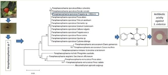
Graphical Abstract

1. Introduction

Animal infections caused by pathogenic bacteria are commonly treated with antibiotics. However, their massive and often improper use has led to a noticeable increase in microbial multidrug-resistance (MDR), considered a severe problem for global public health [1,2]. Antimicrobial resistance extends treatments and increases the risk of spreading the resistant microorganisms and an increase of health-care costs. In addition, the development of new antibiotics is restricted by economic and regulatory obstacles [3].
Overuse of drugs is also reported in animal husbandry, where antibiotics are used to prevent infection and as growth supplements [4,5]. In this field, previous reports showed that chronic treatments with a single drug can lead to MDR [6]. Many isolates belonging to the genus Salmonella—very common in cattle and considered one of the most relevant foodborne zoonoses—are resistant to multiple antibiotics [7,8]. The consumption of contaminated food of animal origin (eggs, meat, milk) may transfer resistant bacteria to humans, predisposing them for serious infections [8].
A very high number of new bioactive secondary metabolites have been reported in the literature in the last 20 years [9]. They are largely used in different areas, such as medicine, animal husbandry, environment and agriculture. Natural compounds are major sources of new active principles for drug formulates. All around the world, researchers investigate the biological properties of metabolites extracted mainly from plants, fungi and marine-derived organisms [10]. The number of bioactive molecules isolated from fungal species has increased since the early 1990s [9].
Endophytic fungi may colonize living plant tissue without causing any negative effects and are an important source of biologically active secondary metabolites. The molecules isolated from these fungi belong to different classes of natural compounds: isoprenoids, polyketides, amino acid derivatives, terpenoids, steroids, xanthones, chinones, phenols, isocumarines, benzopyranones, tetralones, cytochalasines and enniatines [11].
During a survey on fungi as a source of anti-bacterial metabolites, an endophytic fungus was isolated from the stem and crown of healthy strawberry plants. The objectives of the present study were to: (i) fully characterize this fungal isolate at the morphological and molecular levels; (ii) test the activity of the fungal culture filtrate and its organic extracts against two strains of S. enterica, the causal agent of bovine gastroenteritis; (iii) determine the half maximal inhibitory concentration (IC50) and minimum inhibitory concentration (MIC) of the resulting active fraction(s); and (iv) identify the bio-active compound(s) present in the active fraction(s).

2. Materials and Methods

2.1. Fungal Isolation and Morphological Characterization

Fungal isolations were performed from tissues (stem and crown) of healthy strawberry plants (Fragaria x ananassa) collected in April 2014 from various orchards located in Caserta province, Southern Italy. Tissue fragments (3–4 mm2) were excised with a surgical blade, dipped in 2% (vol/vol) NaClO for 30 s, rinsed twice in sterile distilled water and dried on sterile Whatman filter paper under a laminar flow hood. The samples were placed in 90 mm-diameter Petri dishes containing Potato Dextrose Agar (PDA, Conda, Madrid, Spain), amended with streptomycin sulphate (100 mg/L). The dishes were kept at 24 ± 2 °C in the dark. After 4–5 days of incubation, the fungal colonies emerging on the vegetable fragments were transferred on fresh PDA plates. Single-spore cultures were obtained by spreading 0.2 mL of a suspension containing 1 × 10³ conidia/mL, prepared from 7 days-old plates, on 1% water agar plates. Then, the germinated spores were transferred onto PDA dishes. The growing colonies were observed with an optical microscope to allow for a morphological characterization (colony diameter, colony color and degree of sporulation) (Olympus BH-2, Olympus America, Lake Success, NY, USA).

2.2. Molecular Characterization and Phylogenetic Analysis

The internal transcribed spacers (ITS) regions and the 5.8S gene, in the cluster of ribosomal genes, were sequenced for molecular characterization. Genomic DNA was extracted in triplicate with the DNeasy Plant Mini Kit (Qiagen, Hilden, Germany). Subsequently, an aliquot was PCR-amplified by using primers ITS4/ITS5 [12]. The reaction mixture contained: the DNA template (50 ng), 1× reaction buffer, 2 mM MgCl2, 200 µM of each dNTP, 400 nM of each primer and 2.5 U Taq DNA polymerase. PCR was performed in 38 cycles; each cycle consisted of three steps—denaturation at 95 °C (1 min), annealing at 55 °C (1 min) and extension at 72 °C (1 min)—followed by a final extension at 72 °C (10 min). Amplicons of the expected size were purified from agarose gel with the DNA Clean & Concentrator kit (Zymo Research, Orange, CA, USA), sequenced at Microgem (Naples, Italy) and then analyzed using the NCBI-BLASTn program [13]. The sequence of the isolate (named CP-1) was deposited in GenBank under the accession number MN017716. The phylogenetic analysis was performed using the molecular evolutionary genetics analysis (MEGA) software, version 6.0, including the sequences from Paraphaeophaeria spp. showing the highest similarity with the Italian isolate in the BLAST-generated alignment. The phylogenetic tree was constructed with the maximum likelihood methods based on the Tamura–Nei model using default parameters, and the statistical reliability of the branches was evaluated by bootstrapping (1000 replicates).

2.3. Liquid Culture and Metabolite Production

For the secondary metabolites production, CP-1 isolate was grown on PDA Petri dishes for a week at 24 °C in the dark. Five-mm diameter plugs were taken from actively growing margins of cultures and placed in Erlenmeyer flasks containing 5 L of potato dextrose broth (PDB, Conda, Madrid, Spain). The stationary cultures were incubated for 21 days at 24 °C in the dark. The fungal biomass was eliminated by filtration, first through sterile gauzes and then through 0.45-µm Whatman filters on vacuum. An aliquot (100 mL) of the filtered broth was extracted with an equal volume of ethyl acetate (EtAc) to evaluate the antibiotic activity.

2.4. Antibiotic Assay of Organic Extract

The organic extract was solubilized in ethanol (20 µg/µL) and tested against two S. enterica strains (S1 and S2), provided by the Istituto Zooprofilattico Sperimentale del Mezzogiorno (Portici, Naples, Italy). Five-mm diameter disks of Whatman paper were treated with 20 µL of extract and placed, after ethanol evaporation, on Luria broth agar (LBA, Conda, Madrid, Spain) Petri dishes previously inoculated with 200 µL of a bacterial suspension (5 × 10⁶ CFU/mL). Dishes treated only with 20 µL of solvent were used as negative control. Four paper disks were placed on each plate. Three dishes per essay were incubated at 37 °C for 24 h, and then the diameters of inhibition halos were measured.

2.5. Extraction and Isolation of Secondary Metabolites

The CP-1 culture filtrate (5 L) was acidified to pH 4 with 5 M HCl and extracted exhaustively with EtAc. The resulting organic extract was dried with Na2SO4 anhydrous, and the solvent was removed under reduced pressure at 40 °C (Rotavapor RV 10 IKA® - Werke GmbH & Co. KG, Staufen, Germany). The residue was recovered and subjected to fractionation by direct phase column chromatography (silica gel; 200 g), using an elution gradient composed of dichloromethane (CH2Cl2)/methanol (MeOH) at different percentages (v/v). The collected fractions were then subjected to preparative thin layer chromatography (TLC), using the following solvents: CH2Cl2/MeOH, 90:10 (v/v), and combined into a total of six fractions (1–6). Among these, fraction number 2, biologically active against both Salmonella strains, was subjected to a further column chromatography, isocratically eluted with chloroform (CHCl3)/MeOH, 95:5 (v/v). The fractions thus collected were subjected to TLC (as described above) and combined into a total of eight sub-fractions.
Sub-fraction BC2, active against the Salmonella strains, was purified by semi-preparative high-performance liquid chromatography (HPLC-Agilent Technologies, Santa Clara, CA, USA). Separations were performed on a Luna C-18 column (250 × 10 mm, 10 μm, Phenomenex Torrance, CA, USA) eluted isocratically with a mixture of 30% MeOH and 70% H2O, using a flow of 5 mL/min and refractive index (RI) detection. Separations yielded a major peak with a retention time at 27.6 min, and then resulted in being pure compound 1 by nuclear magnetic resonance (NMR) and liquid chromatography–mass spectrometry (LC-MS).

2.6. LC–MS Analysis

Analyses were performed on an HPLC 1260 Infinity Series (Agilent Technologies, Santa Clara, CA, USA) equipped with a DAD (Diode Array Detector) system (Agilent Technologies) and coupled to a quadrupole–time of flight (Q-TOF) mass spectrometer, model G6540B (Agilent Technologies) with a Dual ESI source (Agilent Technologies). An Ascentis® Express C-18 column (2.7 μm, 50 mm × 3.0 mm i.d., Supelco©, Bellefonte, PA, USA), held at a constant temperature of 37 °C, was used for separations. The mobile phase consisted of A: 0.1% (v/v) formic acid (FA) in water (H2O) and B: 0.1% formic acid (FA) in acetonitrile (ACN). Elution was done at a flow rate of 0.4 mL/min, and the gradient was as follows: starting condition 5% B ramping to 100% B until 6 min, held at 100% B for 2 min, lowering to 5% B in 2 min, and held at 5% B for 2 min as the equilibration time. The injection volume was 7 µL. UV spectra were collected by DAD, setting the detection wavelength at 210, 250 and 280 nm. Both chromatographic and spectral parameters were set using the Agilent MassHunter Data Acquisition Software, rev. B.05.01 (Agilent Technologies). The system operated in positive ion mode, and MS spectra were recorded in the m/z 50–1700 range as centroid spectra, with a speed of 3.3 spectra/s. The capillary was maintained at 2000 V, fragmentor voltage at 180 V, cone 1 (skimmer 1) at 45 V and Oct RFV at 750 V. The gas flow rate was set at 11 L/min, at 350 °C, and the nebulizer was set at 45 psig. A standard solution was infused by using an isocratic pump (1260 Infinity Series, Agilent Technologies) in order to perform the real-time lock mass correction. The solution consisted of two reference mass compounds: purine (C5H4N4 at m/z 121.050873, 10 µmol/L) and hexakis (1H,1H, 3H-tetrafluoropentoxy)-phosphazene (C18H18O6N3P3F24 at m/z 922.009798, 2 µmol/L). The flow rate was set at 0.06 mL/min, while the detection window and the minimum height were set at 1000 ppm and 10,000 counts, respectively, for the reference mass correction. LC–MS data were evaluated using MassHunter Qualitative Analysis Software B.06.00 and compared to known compounds included in an in-house database. Positive identifications were reported if the compound was detected with a mass error below 10 ppm and with a sufficient score. The high-resolution mass spectrum (HR-MS) was acquired in positive ion detection mode in the range of m/z 100–2000 with a resolution set to 60,000. The HR-ESI-MS experiment was performed on a Thermo LTQ Orbitrap XL mass spectrometer (Thermo Fisher Scientific Spa, Rodano, Italy).

2.7. Antibiotic Assay on Fractions and Pure Compound

The biological activity of the six fractions obtained with the first chromatographic separations (Section 2.5) were tested at 1 µg/µL using the disk method (Section 2.4). The agar dilution method was used to test the eight sub-fractions, subsequently obtained by the active fraction (n. 2). The tested concentrations were: 5, 9.5, 19 and 38 µg/mL.
The final concentrations were obtained by diluting the proper amount of the active sub-fraction (dissolved in ethanol) in agar cooled at 45 °C. The same quantity of amended agar per dish was allowed to solidify in 9-cm Petri dishes. The inoculum of the Salmonella strains was obtained by recovering bacterial cells from plates inoculated 24 h before and preparing a suspension in a sterile NaCl solution (0.9% w/v). The turbidity of the bacterial suspension was adjusted spectrophotometrically to 0.10 at 625 nm. The content of the colony-forming units was measured by a haematocytometer and confirmed to be 3 × 10⁸ CFU/mL. After a dilution to about 3 × 10⁵ CFU/mL, 10 µL of the suspension were added to each plate. Four replicates per sub-fraction concentration and strain were used. The negative controls consisted in Petri dishes containing only the solvent. The plates were incubated at 37 °C for 24 h, and the number of developed colonies was counted. The effect of the active sub-fractions was measured as the percent reduction of colonies compared to the control. To determine the IC and MIC values, the inhibition scores were fitted with the monomolecular growth function, also known as Mitscherlisch or von Bertalanffy law, with the following parameters:
% inhibition = c + (d − c) * (1 − exp(−dose/e))
where c is the lower limit at zero dose, d is the upper asymptote and e is a parameter correlated with the slope. The analysis was done with the R environment [14] and the packages drc [15] and tidyverse [16].

3. Results

3.1. Morphological and Molecular Characterization of the Fungal Isolate

The fungus, isolated from the tissues of healthy strawberry plants, showed pycnidia, single or eustromatic and more complex, globose, superficial or immersed in the agar, glabrous, 130–280 µm diameter, initially pale and later dark, brown or black. Conidia, released through one, rarely two, ostioli, were subglobose, ellipsoid or pyriform, initially hyaline, then olivaceous-brown, 0 septate, 3.5–5 × 3–4 µm. Colonies on PDA reached a 32–38 mm diameter in 10 days, with an even to slightly ruffled, colorless to buff margin. Immersed mycelium appeared olivaceous in the center, covered by a dense, woolly mat, white to rosy or ochreous aerial mycelium. The reverse side was mostly pale luteous to ochreous, with darker areas in the center. Based on these morphological features, the fungus was ascribed to the Paraphaeosphaeria sporulosa species [17].
The amplification of 18S rDNA with ITS4 and ITS5 primers was successfully performed on five fungal isolates, identified by morphological observations. The five ITS sequences (482 bp in length) showed 100% identity to each other. The ITS sequence of the CP-1 isolate was deposited in GenBank under the accession number MN017716.1. By means of the NCBI BLAST algorithm, we found that the ITS sequence shared 99.79% nucleotide identity to the corresponding sequences of several P. sporulosa isolates.
A phylogenetic tree was constructed using the rDNA sequence of our isolate and eighteen sequences of related species of Paraphaeosphaeria isolated from plants; the Alloconiotirium aptrootii sequence was used as the outgroup. Paraphaeosphaeria sporulosa isolated from strawberry plants clustered in a monophyletic group, supported by the bootstrap (98%) (Figure 1). Paraphaeosphaeria arecacearum resulted as being the closest species to P. sporulosa, while P. verruculosa resulted as being the most distant one. The bootstrap values are shown on the major branches of the phylogenetic tree to indicate the confidence level of each branch.

3.2. Isolation and Chemical Characterization of Bioactive Compound 1

The extraction with EtAc of 5 L of culture filtrates yielded 550 mg of crude extract, which was further subjected to fractionation by direct phase column chromatography and to preparative TLCs when needed. A second purification led to a total of eight sub-fractions, named BC1, BC2, DE1, DE2, DE3, F1, F2 and F3. Among these, only sub-fraction BC2 was active against Salmonella strains and was subjected to further purification and chemical characterization.
From this sub-fraction, the semi-preparative HPLC separations yielded a major peak with a retention time at 27.6 min, which resulted in being a pure compound by nuclear magnetic resonance (NMR, Figure S1) and liquid chromatography–mass spectrometry (LC–MS, Figure S2)) analyses (1, 5.3 mg, Figure 2). The high-resolution, electrospray-ionization mass spectrum (HR–ESI–MS, Figure S3) of compound 1 displayed an [M + H]+ ion peak at m/z 245.1286, corresponding to the molecular formula C14H16N2O2. The molecular formula and analysis of the 1H NMR data of compound 1 suggested a diketopiperazine (cyclic dipeptide) structure. The presence in the spectrum of five aromatic protons between δ 7.47 and 7.28 suggested a mono-substituted benzene, and therefore a Phe residue, while the absence of any methyl resonance and heteroatoms, other than those involved in the two peptidic bonds, pointed to a proline residue. Indeed, the 1H NMR spectrum of compound 1 matched perfectly the spectrum reported for cyclo(L-Pro-L-Phe) [18,19], and this also established the relative configuration (either L,L or D,D) of the two amino-acid residues. The L,L absolute configuration of the molecule was deduced by the negative value measured for its optical rotation [α]D = −91 (c = 0.15, EtOH), in accordance with the [α]D = −101.62 reported for cyclo(L-Pro-L-Phe) (Figure 2) [19].

3.3. Antibiotic Activity

The culture filtrate of P. sporulosa and its organic extracts showed evident inhibition halos on Petri plate assays against both S. enterica strains (Figure S5). The bioassay-guided fraction yielded one main compound responsible for the detected activity (Figure S5), fully characterized as 1, cyclo(L-Pro-L-Phe). This secondary metabolite was used to perform the agar-dilution assay. The effect of 1 was similar on both S. enterica strains, with the estimated curves of inhibition vs. concentration largely overlapping, and rather strong, with a complete inhibition at the highest tested dose of 38 μg/mL (Figure 3).
The inhibition response was comparable between the two Salmonella strains. However, the S2 strain showed a slightly higher sensitivity compared to the S1 strain at intermediate and higher concentrations of 1: average IC50 7.9 against 7.2 μg/mL; average MIC99.9 71.3 against 78.6 μg/mL (Figure 3).

4. Discussion

Diketopiperazines are cyclic dipeptides obtained by the condensation of two α-amino acids, biosynthesized by different organisms and considered very interesting for drug discovery [20]. Due to their structure (heterocyclic system), these secondary metabolites can bind several receptors, resulting in a wide range of biological properties, including antimicrobial, antitumor and anti-hyperglycemic activities [21]. Antibiosis against bacteria such as Bacillus subtilis, Staphylococcus aureus, Escherichia coli and Pseudomonas aeruginosa has been reported [22].
Diketopiperazines are produced by several endophytic fungi, including Colletotrichum gloeosporioides, Fusarium spp. and Penicillium crustosum [23]. Endophytic fungi, residing in the tissues of healthy plants, and marine-derived fungal species are an unexplored source of bioactive secondary metabolites, with potential application in the agriculture, medicine and food industries [24,25].
Paraphaeosphaeria spp. are ubiquitous fungi isolated from soil and different plants [17,26]. Paraphaeosphaeria sporulosa (basionym Coniothyrium sporulosum or Paraconiothyrium sporulosum) is a cosmopolitan soil-borne fungus [27], frequently found as an endophyte in the roots and rhizomes of various plant species [28,29]. This fungus is known to produce various metabolites with different biological properties: sporulosaldeins, active against Candida spp. [30]; sporulosol, a compound with a not significant cytotoxicity against tumor cell lines [31]; and mycosporulone, a butanolide metabolite active against penicillin-resistant strains of P. aeruginosa and S. aureus [32].
To the best of our knowledge, this is the first study reporting: (i) Fragaria sp. as the host of the endophyte P. sporulosa; (ii) the isolation of a diketopiperazine from the culture filtrate of this fungus; (iii) the biological activity of a fungal diketopiperazine against S. enterica, the causal agent of bovine gastroenteritis; and (iv) the isolation of cyclo (L-Pro-L-Phe) from this fungus and the evaluation of its antibiotic activity.

5. Conclusions

The association between the use of antibiotics in animal production and anti-microbial resistance phenomena is very relevant among bacteria belonging to the genus Salmonella [33]. The serious problem of MDR motivates the use of novel antibiotics based on bioactive metabolites of fungal origin. Therefore, the diketopiperazine isolated from P. sporulosa could represent a promising tool to limit the use of marketed antibiotics in animal husbandry. Further investigations should be conducted on the biological properties of this compound with in vivo experiments.

Supplementary Materials

The following are available online at https://www.mdpi.com/2309-608X/6/2/83/s1, Figure S1. 1H NMR spectrum of cyclo (L-Pro-L-Phe). Figure S2. LC-MS qTOF spectrum of cyclo (L-Pro-L-Phe). Figure S3. High resolution electrospray mass spectrum of cyclo (L-Pro-L-Phe). Figure S4. Paraphaeosphaeria sporulosa CREA-CI grown on potato dextrose agar (A); (B) and (C), microscopic pictures of conidia. Figure S5. Inhibition halos on Petri plates of Paraphaeosphaeria sporulosa CREA-CI extract on Salmonella enterica strains (A); inhibition halos on Petri plates of cyclo (L-Pro-L-Phe) on Salmonella enterica strains (B—right halos).

Author Contributions

Conceptualization, F.V. and E.L.; methodology, F.V., E.L., A.M.; validation, R.C. and M.C.; formal analysis, R.C., G.P., R.S., and L.G.; investigation, G.B. and S.B.C.; data curation, A.M., F.V.; writing—original draft preparation, all the authors; writing—review and editing, F.V.; supervision, E.L. and F.V.; project administration, S.B.C., E.L. and G.B. All authors have read and agreed to the published version of the manuscript.

Funding

The study was supported by POR FESR CAMPANIA 2014/2020- O.S. 1.1 grant Bioagro 559, MISE CRESO Protection n. F/050421/01-03/X32, MIURPON grant Linfa 03PE_00026_1 and POR FESR CAMPANIA 2007-2013 O.O.2.1 Project “FAMEGA”, cod. SMILE 20, CUP B25C13000330007.

Acknowledgments

The authors thank Alessia Staropoli for technical assistance and Roberta Marra for editorial assistance.

Conflicts of Interest

The authors declare no conflict of interest.

References

  1. Levy, S.B. Active efflux mechanisms for antimicrobial resistance. Antimicrob. Agents Chemother. 1992, 36, 695–703. [Google Scholar] [CrossRef] [Green Version]
  2. Jindal, B.A.K.; Pandya, M.K.; Khan, M.I.D. Antimicrobial resistance: A public health challenge. Med. J. Armed Forces India 2015, 71, 178–181. [Google Scholar] [CrossRef] [Green Version]
  3. Marquardt, R.R.; Li, S. Antimicrobial resistance in livestock: Advances and alternatives to antibiotics. Anim. Front. 2018, 8, 30–37. [Google Scholar] [CrossRef] [Green Version]
  4. Spellberg, B.; Gilbert, D.N. The future of antibiotics and resistance: A tribute to a career of leadership by John Bartlett. Clin. Infect. Dis. 2014, 59, S71–S75. [Google Scholar] [CrossRef] [Green Version]
  5. Hussain, C.; Ball, M.B.; McGwire, B.S. Multidrug-resistant bovine salmonellosis predisposing for severe human clostridial myonecrosis. Am. J. Case Rep. 2019, 20, 268–273. [Google Scholar] [CrossRef]
  6. Levy, S.B. The Antibiotic Paradox: How the Misuse of Antibiotics Destroys Their Curative Powers, 2nd ed.; Perseus Publishing: Cambridge, MA, USA, 2002. [Google Scholar]
  7. Fluckey, W.M.; Loneragan, G.H.; Warner, R.; Brashears, M.M. Antimicrobial drug resistance of Salmonella and Escherichia coli isolates from cattle feces, hides, and carcasses. J. Food Prot. 2007, 70, 551–556. [Google Scholar] [CrossRef]
  8. Kemal, J. A review on the public health importance of bovine salmonellosis. J. Veterinar. Sci. Technol. 2014, 5, 175. [Google Scholar] [CrossRef] [Green Version]
  9. Berdy, J. Bioactive microbial metabolites. J. Antibiot. 2005, 58, 1–26. [Google Scholar] [CrossRef] [Green Version]
  10. Balouiri, M.; Sadiki, M.; Ibnsouda, S.K. Methods for in vitro evaluating antimicrobial activity: A review. J. Pharm Anal. 2016, 6, 71–79. [Google Scholar] [CrossRef] [Green Version]
  11. Schulz, B.; Boyle, C.; Draeger, S.; Römmert, A.K.; Krohn, K. Endophytic fungi: A source of novel biologically active secondary metabolites. Mycol. Res. 2002, 106, 996–1004. [Google Scholar] [CrossRef]
  12. White, T.J.; Burns, T.; Lee, S.; Taylor, J.W. Amplification and direct sequencing of fungal ribosomal RNA genes for phylogenetics. In PCR Protocols: A Guide to Methods and Applications, 2nd ed.; Innis, M.A., Gelfand, D.H., Sninsky, H.H., White, T.J., Eds.; Academic Press: Cambridge, MA, USA, 1990. [Google Scholar]
  13. Altschul, S.; Gish, W.; Miller, W.; Myers, E.W.; Lipman, D.J. Basic local alignment search tool. J. Mol. Biol. 1990, 215, 403–410. [Google Scholar] [CrossRef]
  14. R Core Team. R: A Language and Environment for Statistical Computing. R Foundation for Statistical Computing. Available online: https://www.R-project.org/ (accessed on 10 February 2015).
  15. Ritz, C.; Baty, F.; Streibig, J.C.; Gerhard, D. Dose-Response Analysis Using R. PLoS ONE 2015, 10, e0146021. [Google Scholar] [CrossRef] [Green Version]
  16. Wickham, H. Welcome to the tidyverse. J. Open Source Softw. 2019, 4, 1686. [Google Scholar] [CrossRef]
  17. Verkley, G.J.M.; Dukik, K.; Renfurm, R.; Göker, M.; Stielow, J.B. Novel genera and species of coniothyrium-like fungi in Montagnulaceae (Ascomycota). Persoonia 2014, 32, 25–51. [Google Scholar] [CrossRef] [Green Version]
  18. Campbell, J.; Lin, Q.; Geske, G.D.; Blackwell, H.E. New and unexpected insights into the modulation of luxR-type quorum sensing by cyclic dipeptides. ACS Chem. Biol. 2009, 4, 1051–1059. [Google Scholar] [CrossRef]
  19. Jayatilake, G.S.; Thornton, M.P.; Leonard, A.C.; Grimwade, J.E.; Baker, B.J. Metabolites from an Antarctic sponge associated bacterium, Pseudomonas aeruginosa. J. Nat. Prod. 1996, 59, 293–296. [Google Scholar] [CrossRef]
  20. Borthwick, A.D. 2,5-Diketopiperazines: Synthesis, reactions, medicinal chemistry, and bioactive natural products. Chem. Rev. 2012, 112, 3641–3716. [Google Scholar] [CrossRef]
  21. Martins-Teixeira, M.; Carvalho, I. Diketopiperazines: Biological activity and synthesis. Tetrahedron 2007, 63, 9923. [Google Scholar] [CrossRef]
  22. Kumar, S.N.; Siji, J.V.; Nambisan, B.; Mohandas, C. Activity and synergistic antimicrobial activity between diketopiperazines against bacteria in vitro. Appl. Biochem. Biotechnol. 2012, 168, 2285–2296. [Google Scholar] [CrossRef]
  23. Guimarães, D.O.; Borges, W.S.; Vieira, N.J.; de Oliveira, L.F.; da Silva, C.H.; Lopes, N.P.; Dias, L.G.; Duran-Patron, R.; Collado, I.G.; Pupo, M.T. Diketopiperazines produced by endophytic fungi found in association with two Asteraceae species. Phytochemistry 2010, 71, 1423–1429. [Google Scholar] [CrossRef]
  24. Strobel, G.A. Endophytes as sources of bioactive products. Microb. Infect. 2011, 5, 535–544. [Google Scholar] [CrossRef]
  25. Nicoletti, R.; Vinale, F. Bioactive compounds from marine-derived Aspergillus, Penicillium, Talaromyces and Trichoderma species. Mar. Drugs 2018, 16, 408. [Google Scholar] [CrossRef] [Green Version]
  26. Cosoveanu, A.; Cabrera, R. Endophytic fungi in species of Artemisia. J. Fungi 2018, 4, 53. [Google Scholar] [CrossRef] [Green Version]
  27. Domsch, K.H.; Gams, W.; Anderson, T.H. Compendium of Soil Fungi, 2nd ed.; IHW-Verlag: Eching, Germany, 2007. [Google Scholar]
  28. Kothe, E.; Turnau, K. Mycorrhizosphere communication: Mycorrhizal fungi and endophytic fungus-plant interactions. Front. Microbiol. 2018, 9, 3015. [Google Scholar] [CrossRef] [Green Version]
  29. Liu, T.; Greenslade, A.; Yang, S. Levels of rhizome endophytic fungi fluctuate in Paris polyphylla var. yunnanensis as plants age. Plant Divers. 2017, 39, 60–64. [Google Scholar] [CrossRef]
  30. De Amorim, M.R. Bioprospection of Endophytic Fungi Paraphaeosphaeria sporulosa and Cochliobolus eragrostidis Isolated from Paepalanthus planifolius (Eriocaulaceae) and Epigenetic Study of Aspergillus terreus. Ph.D. Thesis, Universidade Estadual Paulista, Sao Paulo, Brazil, 2019. [Google Scholar]
  31. Zhao, C.; Fu, P.; Zhang, Y.; Liu, X.; Ren, F.; Che, Y. Sporulosol, a new ketal from the fungus Paracoconiothyrium sporulosum. Molecules 2018, 23, 1263. [Google Scholar] [CrossRef] [Green Version]
  32. Guiraud, P.; Steiman, R.; Seigle-Murandi, F.; Buarque de Gusmao, N. Antimicrobial and antitumor activities of mycosporulone. J. Nat. Prod. 1999, 62, 1222–1224. [Google Scholar] [CrossRef]
  33. Angulo, F.J.; Nargund, V.N.; Chiller, T.C. Evidence of an association between use of anti-microbial agents in food animals and antimicrobial resistance among bacteria isolated from humans and the human health consequences of such resistance. J. Vet. Med. 2004, 51, 374–379. [Google Scholar] [CrossRef]
Figure 1. Phylogenetic analysis. Unrooted phylogenetic tree calculated from partial ITS regions (ITS1, 5.8S gene and ITS2) of rDNA of Paraphaeosphaeria species. Paraphaeosphaeria sporulosa Fragaria sp. CREA-CI indicates the CP-1 isolate obtained by the authors. The Alloconiotirium aptrootii sequence was used as the outgroup.
Jof 06 00083 g001
Figure 2. Chemical structure of 1, cyclo(L-Pro-L-Phe) isolated from Paraphaeosphaeria sporulosa.
Jof 06 00083 g002
Figure 3. Inhibition of two Salmonella enterica strains (S1 and S2) using different concentrations of 1 (left) and IC50 levels and MICs (right).
Jof 06 00083 g003