Article

Blue-Emitting 2D- and 3D-Zinc Coordination Polymers Based on Schiff-Base Amino Acid Ligands

by
Rodavgi Karakousi
1,
Pinelopi A. Tsami
1,
Maria-Areti I. Spanoudaki
1,
Scott J. Dalgarno
2,
Vassileios C. Papadimitriou
1,3,4,* and
Constantinos J. Milios
1,*
1
Department of Chemistry, The University of Crete, 71003 Herakleion, Greece
2
Institute of Chemical Sciences, Heriot-Watt University, Riccarton, Edinburgh EH14 4AS, UK
3
Chemical Sciences Laboratory, National Oceanic and Atmospheric Administration, Boulder, CO 80305, USA
4
Cooperative Institute for Research in Environmental Sciences, University of Colorado, Boulder, CO 80309, USA
*
Authors to whom correspondence should be addressed.
Chemistry 2023, 5(3), 1770-1780; https://doi.org/10.3390/chemistry5030121
Submission received: 5 July 2023 / Revised: 6 August 2023 / Accepted: 7 August 2023 / Published: 9 August 2023

Abstract

:
The solvothermal reaction of Zn(NO3)2·4H2O, 1-OH-2-naphthaldehyde, and 2-methylalanine (mAla) in MeOH leads to the formation of complex {[ZnL1]}2n (1) (H2L1 = the Schiff-base resulting from the reaction of 1-OH-2-naphthaldehyde and mAla) in good yields. The structure of the neutral species, as determined by single-crystal crystallography, describes a two-dimensional coordination polymer, with repeating {Zn2} units bridged by syn, anti-carboxylate groups of the Schiff-base ligands. Repeating the same reaction using glycine (gly) instead of mAla leads to the formation of complex {[ZnL2]·0.33MeOH}3n (2.0.33MeOH) (H2L2 = the Schiff-base resulting from the reaction of 1-OH-2-naphthaldehyde and gly), again in good yields. Complex 2 describes a three-dimensional coordination polymer based on {Zn2} building blocks, arranged by anti, anti-carboxylate groups in a 3D motif. Complexes 1 and 2 were found to strongly emit at ~435 nm (λexc = 317 nm) both in solution and solid state, with complex 2 displaying a slightly longer lifetime of τav = 2.45 ns vs. τav = 2.02 ns for 1.

Graphical Abstract

1. Introduction

Over the past three decades, since their deliberately targeted synthesis by Professor R. Robson in 1989 [1], metal–organic frameworks (MOFs) have dominated the scientific field of coordination polymers as a captivating emerging class of materials, combining the fields of inorganic and organic chemistry [2,3]. These three-dimensional crystalline coordination polymers have attracted attention due to their exceptional chemical properties, arising from the elegant way of assembling the metal ions or cluster-based building units with suitable organic ligands that act as linkers/bridges. Such species allow for fine-tuning the surface chemistry and customizing the pore size, maximizing the selectivity in adsorption processes. MOFs, due to their unique structure, present outstanding chemical, thermal, and mechanical stability [4], making them highly desirable for a diverse range of applications such as drug delivery [5], biomolecule encapsulation [6], gas storage and separation [7], catalysis and photocatalysis [8], sensing [9], and magnetic properties [10], paving new pathways for advancements in medical and technological applications [11].
The formation, as well as the identity of such polymeric materials, is based on many synthetic parameters affecting the reaction conditions, e.g., reaction time, presence of base and/or auxiliary ligands, and pressure/temperature. However, selecting the crystallization conditions is of great importance since, in many cases, it enables controlling the identity of the isolated products (i.e., kinetic vs. thermodynamic products) [12,13].
Up until now, a wide range of metal ions have been employed for the synthesis of such unique coordination polymers [14], with the most commonly employed being zinc, a 3d10 transition metal ion. The main reasons for that are: (1) zinc is a highly abundant element in the earth’s crust [15], and, as such, it is readily available, ensuring a sustainable supply; and (2) zinc-based starting materials/salts are relatively low-cost [16], thus enhancing the economic feasibility of manufacturing zinc-based MOFs. Furthermore, the low-toxicity of zinc compounds [17,18,19] is of paramount importance in the context of MOFs’ potential biomedical and environmental applications.
Luminescent MOFs are a highly significant sub-class of 3D coordination polymers, since they hold great value as chemical sensors upon combining the porosity of a specific MOF and/or its selectivity towards a guest molecule, along with their inherent luminescent properties [20]. This is particularly crucial since the luminescence characteristics of such MOFs may be potentially modified when exposed to the guest molecule, even in small traces, triggering the sensing sequence. Furthermore, in most cases the emission of the MOFs stems from the organic linker/bridge chromophore; as such, the search for suitable organic chromophore ligands that may promote the formation of 3D coordination polymers is of great importance. With this in mind, we herein report the synthesis and full characterization of two new Zn coordination polymers based upon the Schiff-base ligands H2L1 and H2L2 (Figure 1).

2. Materials and Methods

All chemicals were obtained from commercial suppliers (Sigma-Aldrich, Athens, Greece) and were used without further purification/treatment.
{[ZnL1]}2n (complex 1):
Zn(NO3)2·4H2O (0.130 g, 0.5 mmol), 2-OH-1-napthaldehyde (0.172 g, 1 mmol), and 2-aminoisobutyric acid (0.103 g.; 1 mmol) were heated in Teflon-lined autoclaves with 15 mL MeOH at 120 °C for 12 h. Upon slowly cooling to room temperature, yellow-gold crystals were formed and collected by filtration. Elemental analysis (%) calculated for C15H13NO3Zn (1): C, 56.18; H, 4.09; N, 4.37. Found: C, 56.09; H, 3.97; and N, 4.41. Yield ~40%. FT-ATIR (cm−1): 1625s, 1574vs, 1544s, 1500m, 1462vs, 1404vs, 1331s, 1296s, 1174vs, 1149s, 833vs, 737vs, and 514vs.
{[ZnL2]·0.33MeOH}3n (complex 2.0.33MeOH):
Complex 2 was synthesized in an analogous manner to 1, upon using glycine instead of 2-aminoisobutyric acid. Elemental analysis (%) calculated for C13H9NO3Zn (2): C, 53.36; H, 3.10; and N, 4.79. Found: C, 53.42; H, 2.98; and N, 4.71. Yield ~40%. FT-ATIR (cm−1): 1622vs, 1602vs, 1573vs, 1542vs, 1510m, 1457s, 1421vs, 1389s, 1327s, 1291vs, 1234s, 1188m, 1166s, 1149w, 1087m, 955s, 841vs, 759s, 746vs, 562s, and 460vs.

2.1. Characterization

Elemental analyses (C, H, and N) were performed by the University of Ioannina microanalysis service. Powder XRD data were collected on freshly prepared samples of 1 and 2 on a PANanalytical X’Pert Pro MPD diffractometer (UoC). FTIR−ATR spectra were recorded on a PerkinElmer FTIR Spectrum BX spectrometer (UoC). Solution UV-Vis spectra were recorded on a double beam Hitachi U-2100 UV-Vis spectrophotometer (UoC). Solid-state and solution fluorescence spectra and lifetimes were determined on a photoluminescence (PL) Edinburgh Instrument FS5 Steady State Spectrometer (UoC). For solution fluorescence spectra, a JASCO FP-8300 spectrofluorometer was also utilized (UoC).

2.2. X-ray Diffraction

Single-crystal X-ray diffraction data were collected on a Bruker D8 VENTURE diffractometer (University of Crete) equipped with a PHOTION II CPAD detector. Data collection parameters and structure solution and refinement details are listed in Table 1. Full details can be found in the CIF files with CCDC reference numbers 2270710 and 2270711, for 1 and 2, respectively.

3. Results and Discussion

3.1. Synthesis and Crystal Structures

The 1:2:2 solvothermal reaction of Zn(NO3)2·4H2O, 1-OH-2-naphthaldehyde, and mAla in MeOH forms complex {[ZnL1]}2n (1) in good yields. With the structure of complex 1 established via single-crystal X-ray crystallography (vide infra) we investigated various synthetic parameters as a means of modifying the identity of the complex. However, upon changing the metal to ligand ratio, in the presence/absence of various bases (i.e., NEt3, CH3OΝa), and using various zinc salts (i.e., ZnSO4 and Zn(OAc)2.2H2O) we were not able to isolate any crystalline material different to 1, as evidenced by pXRD comparison. Furthermore, all attempts to isolate any crystalline material upon normal laboratory conditions (i.e., aerobic conditions at room temperature) proved fruitless, since, in all cases, a sticky yellowish slurry was formed upon slow evaporation of the solvent [21].
Our next goal was to investigate whether the nature of the Schiff-base formed in situ could affect the identity and/or the dimensionality of the product; therefore, we employed the amino acid gly instead of mAla, and from the analogous solvothermal reaction of Zn(NO3)2·4H2O, 1-OH-2-naphthaldehyde, and gly in MeOH we were able to isolate complex {[ZnL2]·0.33MeOH}3n (2.0.33MeOH) in good yields. To our great surprise, complex 2 describes a 3D-coordination polymer as was established by single-crystal X-ray crystallography (vide infra), thus establishing the leading role of the Schiff-base in dictating the dimensionality of the products. Furthermore, given that the two Schiff-base ligands, H2L1 and H2L2, differ only in the nature of amino acid employed, mAla in 1 vs. gly in 2, it becomes apparent that the change from 2D to 3D (from 1 to 2) should be attributed in the nature/structure/properties of the amino acid ligand employed. Given that both amino acid ligands display very similar pKa values (2.36 and 2.35, for mAla and gly, respectively), we may safely assume that the acidity of the amino acid ligand does not hold a pivotal role in dictating the dimensionality of the products, suggesting that other factors, i.e., steric hindrance, may govern the formation of the products.
Single-crystal X-ray diffraction studies of 1 reveal that complex 1 crystallizes in the centrosymmetric monoclinic space group P21/c (Figure 2). Its asymmetric unit (ASU) contains one crystallographically independent zinc centre bound to one dianionic L12− ligand. Each L12− ligand adopts a η2: η1: η1: η1: μ3 coordination mode, forming a six-member chelate ring around each metal centre, while bridging to two neighboring Zn centres via the alkoxide group and one Ocarboxylate atom. Furthermore, the carboxylate unit of the amino acid part of the L12− ligands adopts a syn, anti- η1: η1: μ coordination mode, bridging two Zn metal ions at ~5.59 Å. Each ZnII ion displays a slightly distorted square-pyramidal {O4N} geometry (τ = 0.31) [22]. Alternatively, the structure may be described as consisting of {Zn2(L12−)2} dimers, in which the metal atoms are connected through the μ-Oalkoxide groups of the L12− ligands. The Zn ⋯ Zn distance within each dimer is ~3.17 Å, while neighboring {Zn2} are located at ~5.59 Å linked via the syn, anti- η1: η1: μ carboxylate groups of the L12− ligands.
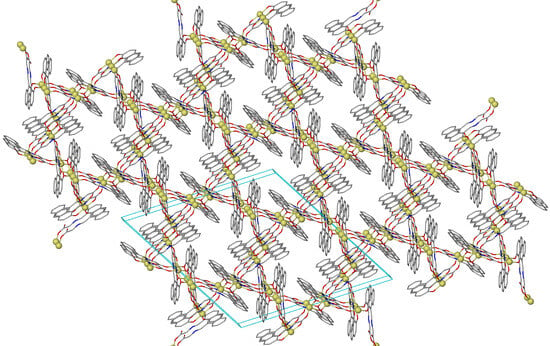
The overall packing of the 1 describes a 2D layered-based network (Figure 3), in which neighboring 2D layers are stacked at a distance of ~14.5 Å (closest ZnZn distance), stabilized via inter-layer C-H … π interactions, at ~3.73 Å.
Complex 2 crystallizes in the trigonal space group R-3, forming a 3D framework (Figure 4). Its ASU contains one crystallographically independent zinc centre bound to one dianionic L22− ligand found in a η2: η1: η1: η1: μ3 coordination mode, forming a six-member chelate ring around each metal centre, while bridging to two neighboring Zn centres via the alkoxide group and one Ocarboxylate atom, as observed for L12− in 1. However, the carboxylate unit of the amino acid part of the L12− ligands now adopts a anti, anti- η1: η1: μ coordination mode vs. the syn, anti- η1: η1: μ mode found in 1, which we believe is the main reason for the different dimensionality between 1 and 2. Again, as in 1, the zinc ions are five-coordinate, but now are found in a severely distorted (τ = 0.46) square-pyramidal {O4N} environment. In the extended structure of 2, cylindrical channels are running along the c axis, formed by the aromatic rings of L22−, resulting in a solvent-accessible volume of ~531 Å3 per unit cell (~10.1% of the unit cell volume).
As in 1, {Zn2(L22−)2} dimers are present in the structure serving as building units, in which the metal atoms are linked via the μ-Oalkoxide groups of the L22− ligands. The Zn ⋯ Zn distance within each dimer is ~3.15 Å, while neighboring {Zn2} are located at ~5.64 Å linked via the anti, anti- η1: η1: μ carboxylate groups of the L22− ligands. However, a comparison between the {Zn2} building units in 1 and 2 reveals that, although the {Zn2(OR)2} metallic core of each dimer is planar in both cases, the relative orientation of the planar aromatic rings with the respect to the {Zn2(OR)2} plane differs; more specifically, in 1 the angle between the naphthalene aromatic ring and the {Zn2(OR)2} plane is 21.7°, while in 2 the angle was found ~40.6°.

3.2. Optical Properties

Figure 5 shows the UV-Vis spectra recorded for methanolic solutions of complexes 1 and 2, between 200 and 425 nm. The purity of the samples was verified by means of pXRD comparison to their theoretical patterns (Figure S2).
Three main bands (metal-to-ligand charge transfer, MLCT) are observed for both complexes, with peak maxima at 238 (b1), 317 (b2), and 380 (b3) nm, among which the band at 238 nm was found to be the strongest. In addition, vibrational structure was observed in all the absorption bands, that was identical in shape and wavelength positioning for both complexes. Furthermore, the similar features of the UV-Vis absorption spectra reveal the presence of the same chromophore in the species, as expected from their crystal structures. The only significant difference between the UV-Vis absorption spectra of complexes 1 and 2 lies on the reversed trend in absorbance relative intensity between bands b2 and b3, with b2 being stronger for complex 1 and weaker for complex 2. The UV-Vis absorption spectra recorded for the two pretreated solutions of the complexes were utilized to measure the fluorescence spectra of the complexes. In particular, the fluorescence spectra of solutions 1 and 2 were measured at excitation wavelengths, for which the absorbance values were equal for the two complexes, i.e., 229, 273, and 325 nm. Note that the solution fluorescence spectra for the two complexes were also recorded at the b2 maximum absorbance, 317 nm, as well as their excitation spectra using λem = 441 nm (Figure S3). In Figure 6 the solution steady-state fluorescence spectra of 1 and 2 are displayed, using as excitation energy the three wavelengths at which complexes 1 and 2 absorb equally.
Although shape-wise the fluorescence spectra of the two complexes are very similar, a slight, but systematic red-shift of the fluorescence maximum intensity was observed, in the case of complex 2, with λ1,max and λ2,max to be at 442 and 445 nm, respectively. Further, the ~15% lower fluorescence intensity observed for complex 2, at the three different excitation wavelengths where the absorbance was equal, indicates that for complex 1 the fluorescence probability is higher, in excellent agreement with its smaller fluorescence lifetime (vide infra). Figure 7 shows the solid-state fluorescence spectra recorded at excitation wavelength λexc = 317 nm, for 1 and 2; again, the two fluorescence spectra are very similar, and present great similarity to those in solution. However, in the solid-state the intensity maxima occur at shorter wavelengths, ~435 nm, as anticipated since the effect of solvent relaxation, i.e., solvent molecules reorientation around the excited-state dipole, effectively lowers the energy and shifts the fluorescence to longer wavelengths.
The solid-state fluorescence of 1-OH-2-naphthaldehyde was also recorded for comparison purposes (Figure S4) using 317 nm as excitation wavelength, and it can be observed that its maximum fluorescence signal, λmax = 451 nm, is substantially blue-shifted, ~16 nm, in 1 and 2. Finally, solid-state fluorescence lifetimes for 1 and 2 were also measured using the output of a pulsed picosecond diode laser (375 ± 10 nm) as excitation wavelength and are presented in Figure 8. The fluorescence decay was fitted to a double-exponential kinetic expression, yielding two decay times for each complex, in consistence with multiple conformational states for each complex [23].
The fluorescence temporal profiles were fitted to a double exponential decay presented in Equation (1):
F(t) = Y0 + a1 × exp[−(tt0)/τ1] + a2 × exp[−(tt0)/τ2]
where F(t) is the temporal fluorescence signal, τ1 and τ2 are the two decay times, t0 is the zero-time where decay begins, and a1 and a2 are the normalized weighted contributions of the two decay times. For complex 1, τ1 and τ2 were determined to be 0.36 and 3.59 ns, respectively, with an average time τav = 2.0 ns. The relative contribution of the fast decay to the fluorescence decay was 90%, while the contribution of the slow decay amounted for 10%. For complex 2, the fast decay was substantially slower compared to complex 1, τ1 = 0.62 ns, while the slow decay was similar τ2 = 3.42 ns. The latter led to a larger average lifetime for 2, τav = 2.45 ns (Table 2). As far as the contribution of the two decays in 2 is concerned, significant differences compared to 1 were observed, with the fast and the slow decays contributing for 75 and 25%, respectively. The observed slower fluorescence and the larger contribution of the slow decay in complex 2 are both consistent with the increased dimensionality in the structure of 2 (3-D) compared to the structure of 1 (2-D).

4. Conclusions

In conclusion, in this work we present the synthesis, characterization, and fluorescence properties of two Zn coordination polymers, based upon the use of amino acid Schiff-base ligands. More specifically, we were able to isolate complex {[ZnL1]}2n (1), which describes a 2D coordination polymer, while upon replacing the metahylalanine amino acid ligand with glycine we were able to isolate complex {[ZnL2]·0.33MeOH}3n (2.0.33MeOH), which describes a 3D coordination polymer. Furthermore, both polymers strongly emit at ~435 nm (λexc = 317 nm) in solution and solid state, with complex 2 displaying a slightly larger lifetime of τav = 2.45 ns vs. τav = 2.02 ns for 1. Finally, we feel this work demonstrates an alternative and easy method for increasing the dimensionality of coordination polymers, upon suitable selection of Schiff-base amino acid ligands.

Supplementary Materials

The following supporting information can be downloaded at: https://www.mdpi.com/article/10.3390/chemistry5030121/s1, Figure S1. FTIR-ATR spectra of 1 and 2. Figure S2. PXRD patterns for 1 (top) and 2 (bottom). Figure S3. Solutions excitation (λem = 441 nm) and fluorescence (λex = 317 nm) spectra for complexes 1 and 2. Figure S4. Solid-state fluorescence spectrum of 1-OH-2-naphthaldehyde using 317 nm as excitation wavelength.

Author Contributions

Conceptualization, C.J.M.; methodology, C.J.M. and V.C.P.; formal analysis, C.J.M. and V.C.P.; experimental execution, R.K., P.A.T., and M.-A.I.S.; S.J.D. solved the single-crystal XRD data; writing—original draft preparation, review, and editing, C.J.M. and V.C.P.; supervision, C.J.M. and V.C.P. All authors have read and agreed to the published version of the manuscript.

Funding

This research received no external funding.

Data Availability Statement

Data are available upon request to corresponding authors.

Acknowledgments

This work was carried out in fulfillment of the requirements for the Master thesis of EKA according to the curriculum of the International Graduate Program in “Inorganic Biological Chemistry”, which operates at the University of Ioannina within the collaboration of the Departments of Chemistry of the Universities of Ioannina, Athens, Thessaloniki, Patras, and Crete, and the Department of Chemistry of the University of Cyprus.

Conflicts of Interest

The authors wish to declare no conflict of interest.

References

  1. Hoskins, B.F.; Robson, R. Infinite Polymeric Frameworks Consisting of Three Dimensionally Linked Rod-like Segments. J. Am. Chem. Soc. 1989, 111, 5962–5964. [Google Scholar] [CrossRef]
  2. Zhou, H.-C.; Long, J.R.; Yaghi, O.M. Introduction to Metal–Organic Frameworks. Chem. Rev. 2012, 112, 673–674. [Google Scholar] [CrossRef] [PubMed]
  3. Stock, N.; Biswas, S. Synthesis of Metal-Organic Frameworks (MOFs): Routes to Various MOF Topologies, Morphologies, and Composites. Chem. Rev. 2012, 112, 933–969. [Google Scholar] [CrossRef] [PubMed]
  4. Howarth, A.J.; Liu, Y.; Li, P.; Li, Z.; Wang, T.C.; Hupp, J.T.; Farha, O.K. Chemical, Thermal and Mechanical Stabilities of Metal–Organic Frameworks. Nat. Rev. Mater. 2016, 1, 15018. [Google Scholar] [CrossRef]
  5. Saeb, M.R.; Rabiee, N.; Mozafari, M.; Mostafavi, E. Metal-Organic Frameworks (MOFs)-Based Nanomaterials for Drug Delivery. Materials 2021, 14, 3652. [Google Scholar] [CrossRef]
  6. Zou, D.; Yu, L.; Sun, Q.; Hui, Y.; Tengjisi; Liu, Y.; Yang, G.; Wibowo, D.; Zhao, C.-X. A General Approach for Biomimetic Mineralization of MOF Particles Using Biomolecules. Colloids Surf. B Biointerfaces 2020, 193, 111108. [Google Scholar] [CrossRef]
  7. Li, H.; Li, L.; Lin, R.-B.; Zhou, W.; Zhang, Z.; Xiang, S.; Chen, B. Porous Metal-Organic Frameworks for Gas Storage and Separation: Status and Challenges. EnergyChem 2019, 1, 100006. [Google Scholar] [CrossRef]
  8. Dhakshinamoorthy, A.; Li, Z.; Garcia, H. Catalysis and Photocatalysis by Metal Organic Frameworks. Chem. Soc. Rev. 2018, 47, 8134–8172. [Google Scholar] [CrossRef]
  9. Sohrabi, H.; Ghasemzadeh, S.; Ghoreishi, Z.; Majidi, M.R.; Yoon, Y.; Dizge, N.; Khataee, A. Metal-Organic Frameworks (MOF)-Based Sensors for Detection of Toxic Gases: A Review of Current Status and Future Prospects. Mater. Chem. Phys. 2023, 299, 127512. [Google Scholar] [CrossRef]
  10. Thorarinsdottir, A.E.; Harris, T.D. Metal–Organic Framework Magnets. Chem. Rev. 2020, 120, 8716–8789. [Google Scholar] [CrossRef]
  11. Yang, J.; Yang, Y. Metal–Organic Frameworks for Biomedical Applications. Small 2020, 16, 1906846. [Google Scholar] [CrossRef]
  12. Torresi, S.; Famulari, A.; Martί-Rujas, J. Kinetically Controlled Fast Crystallization of M12L8 Poly-[n]-catenanes Using the 2,4,6-Tris(4-pyridyl)benzene Ligand and ZnCl2 in an Aromatic Environment. J. Am. Chem. Soc. 2020, 142, 9537–9543. [Google Scholar] [CrossRef]
  13. Martí-Rujas, J.; Elli, S.; Famulari, A. Kinetic trapping of 2,4,6-tris(4-pyridyl)benzene and ZnI2 into M12L8 poly-[n]-catenanes using solution and solid-state processes. Sci. Rep. 2023, 13, 5605. [Google Scholar] [CrossRef] [PubMed]
  14. Moghadam, P.Z.; Li, A.; Wiggin, S.B.; Tao, A.; Maloney, A.G.P.; Wood, P.A.; Ward, S.C.; Fairen-Jimenez, D. Development of a Cambridge Structural Database Subset: A Collection of Metal–Organic Frameworks for Past, Present, and Future. Chem. Mater. 2017, 29, 2618–2625. [Google Scholar] [CrossRef] [Green Version]
  15. Brugger, J. Zinc. In Encyclopedia of Geochemistry; White, W.M., Ed.; Springer International Publishing: Cham, Switzerland, 2016; pp. 1–4. ISBN 9783319391939. [Google Scholar]
  16. Dumur, F.; Beouch, L.; Tehfe, M.-A.; Contal, E.; Lepeltier, M.; Wantz, G.; Graff, B.; Goubard, F.; Mayer, C.R.; Lalevée, J.; et al. Low-Cost Zinc Complexes for White Organic Light-Emitting Devices. Thin Solid Film. 2014, 564, 351–360. [Google Scholar] [CrossRef]
  17. Safdar Ali, R.; Meng, H.; Li, Z. Zinc-Based Metal-Organic Frameworks in Drug Delivery, Cell Imaging, and Sensing. Molecules 2021, 27, 100. [Google Scholar] [CrossRef] [PubMed]
  18. Bahrani, S.; Hashemi, S.A.; Mousavi, S.M.; Azhdari, R. Zinc-Based Metal–Organic Frameworks as Nontoxic and Biodegradable Platforms for Biomedical Applications: Review Study. Drug Metab. Rev. 2019, 51, 356–377. [Google Scholar] [CrossRef]
  19. McKinlay, A.C.; Morris, R.E.; Horcajada, P.; Férey, G.; Gref, R.; Couvreur, P.; Serre, C. BioMOFs: Metal-Organic Frameworks for Biological and Medical Applications. Angew. Chem. Int. Ed. 2010, 49, 6260–6266. [Google Scholar] [CrossRef] [PubMed]
  20. Dou, Z.; Yu, J.; Cui, Y.; Yang, Y.; Wang, Z.; Yang, D.; Qian, G. Luminescent Metal–Organic Framework Films As Highly Sensitive and Fast-Response Oxygen Sensors. J. Am. Chem. Soc. 2014, 136, 5527–5530. [Google Scholar] [CrossRef]
  21. Martί-Rujas, J.; Elli, S.; Sacchetti, A.; Castiglione, F. Mechanochemical synthesis of mechanical bonds in M12L8 poly-[n]-catenanes. Dalton Trans. 2022, 51, 53–58. [Google Scholar] [CrossRef]
  22. Blackman, A.G.; Schenk, E.B.; Jelley, R.E.; Krenske, E.H.; Gahan, L.R. Five-Coordinate Transition Metal Complexes and the Value of τ5: Observations and Caveats. Dalton Trans. 2020, 49, 14798–14806. [Google Scholar] [CrossRef] [PubMed]
  23. Lakowicz, J.R. Principles of Fluorescence Spectroscopy, 2nd ed.; Kluwer Academic/Plenum Publishers: New York, NY, USA, 1999; ISBN 0-306-46093-9. [Google Scholar]
Figure 1. The H2L1 and H2L2 ligands reported in this work and their coordination modes in 1 and 2, respectively.
Chemistry 05 00121 g001
Figure 2. The crystal structure of 1 viewed on the bc plane, highlighting the formation of {Zn2} units. Colour code: ZnII = yellow; O = red; N = blue; and C = grey. H-atoms are omitted for clarity.
Chemistry 05 00121 g002
Figure 3. The crystal packing of 1 viewed on the ab plane. Different layers are depicted in different colours. H-atoms are omitted for clarity.
Chemistry 05 00121 g003
Figure 4. The crystal structure of 2 viewed down the c axis (top), its extended structure (middle) and the channels running down c axis (bottom). Colour code: the same as in Figure 2. H-atoms are omitted for clarity.
Chemistry 05 00121 g004
Figure 5. UV-Vis absorption spectra recorded for complexes 1 and 2.
Chemistry 05 00121 g005
Figure 6. Solution normalized fluorescence spectra of complexes 1 and 2, recorded with excitation wavelengths 229, 273, and 325 nm, at which UV-Vis absorbance values for the two species were measured to be equal.
Chemistry 05 00121 g006
Figure 7. Solid-state normalized fluorescence spectra of complexes 1 and 2 recorded with excitation wavelength, λexc = 317 nm.
Chemistry 05 00121 g007
Figure 8. Fluorescence decays of complex 1 (light grey solid circles) and 2 for solid-state (yellow solid triangles), stimulated by the output of a pulsed picosecond diode laser (375 nm). The solid lines represent fit of the data (see the text for details).
Chemistry 05 00121 g008
Table 1. Crystal data and structure refinement for complexes 1 and 2.
12
Empirical formulaC15H13NO3ZnC13.33H10.33NO3.33Zn
Formula weight320.63303.17
Temperature/K210210
Crystal systemMoniclinicTrigonal
Space groupP21/cR-3
a14.5062 (3)26.7293 (3)
b9.6958 (2)26.7293 (3)
c9.7987 (2)8.5248 (2)
α
β107.697 (1)
γ 120
Volume/Å31312.96 (5)5274.61 (17)
Z418
ρcalc g/cm31.6221.718
μ/mm−12.6602.962
F(000)656.02771.0
Crystal size/mm30.32 × 0.30 × 0.240.28 × 0.22 × 0.20
RadiationCuKα (λ = 1.54178)CuKα (λ = 1.54178)
2Θ range for data collection/°11.148 to 139.5946.614 to 139.834
Index ranges−17 ≤ h ≤ 17,−11 ≤ k ≤ 11,−11 ≤ l ≤ 11−31 ≤ h ≤ 32,−31 ≤ k ≤ 20,−10 ≤ l ≤ 10
Reflections collected14,3117111
Independent reflections24642214
Data/restraints/parameters2464/0/1832214/0/163
Goodness-of-fit on F21.0621.051
Final R indexes (I ≥ 2σ (I))R1 = 0.0219, wR2 = 0.0580R1 = 0.0243, wR2 = 0.0603
Final R indexes (all data)R1 = 0.0233, wR2 = 0.0592R1 = 0.0278, wR2 = 0.0626
Largest diff. peak/hole/e Å−30.31/−0.260.27/−0.30
Table 2. Decay times and weighted contributions for 1 and 2.
Complexesa1τ1 (ns)a2τ2 (ns)τav (ns)
10.9040.360.0963.592.02
20.7470.620.2533.422.45
Disclaimer/Publisher’s Note: The statements, opinions and data contained in all publications are solely those of the individual author(s) and contributor(s) and not of MDPI and/or the editor(s). MDPI and/or the editor(s) disclaim responsibility for any injury to people or property resulting from any ideas, methods, instructions or products referred to in the content.