Review

Cryptosporidiosis in Reptiles from Brazil: An Update for Veterinary Medicine

1
Department of Microbiology and Parasitology, Biomedical Institute, Fluminense Federal University, Rua Professor Hernani Mello, 101, São Domingos, Niterói 24210-130, Brazil
2
Laboratory of Clinical Research on Dermatozoonosis in Domestic Animals, Evandro Chagas Clinical Research Institute, Oswaldo Cruz Foundation, Av. Brasil, 4365, Rio de Janeiro 21040-360, Brazil
*
Author to whom correspondence should be addressed.
Parasitologia 2022, 2(3), 228-236; https://doi.org/10.3390/parasitologia2030019
Submission received: 1 July 2022 / Revised: 26 July 2022 / Accepted: 28 July 2022 / Published: 3 August 2022

Abstract

:
The concern about the protection of wildlife has been gathering attention from researchers worldwide. Zoos and aquariums have become widely recognized sites for the conservation of wildlife. However, the persistence of the illegal trade of wild animals, such as reptiles, and their use as pets can endanger not only the preservation of the species, but also allow the introduction of new pathogens and zoonotic diseases. It is important to highlight that preventive exams should be carried out prior to introducing these animals into a new facility to guarantee zoological management strategies. There are several reports of parasitism in reptiles, some of them with zoonotic potential, such as the genus Cryptosporidium spp. In Brazil, reports that explore the prevalence of cryptosporidiosis in reptiles are scarce, and very few have used molecular methods for the detection of Cryptosporidium spp., or the genotyping of its species and subtypes. This review aims to help professionals in the area and encourage them to increase their attention to this protozoan, which is usually neglected.

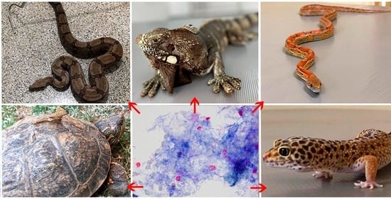
Graphical Abstract

1. Introduction

At present, the Reptilia class is divided into three subclasses and four orders. The subclass Anapsida includes the Order Testudines, represented by tortoises and turtles. The subclass Diapsida includes: The order Rhynchocephalia, which is represented by tuataras, the order Squamata, which encompasses the suborders Sauria (Lacertilia), and Amphisbaenia, which are represented by lizards, and the suborder Ophidia (Serpentes) is represented by snakes. The subclass Archosauria includes the order Crocodylia, which is represented by crocodiles, alligators, gharials, and caimans [1].
According to the Reptile Database, there are more than 11,050 species recognized in the world [1]. Australia leads the world in reptile species richness, and Mexico takes second place. An updated checklist of Brazilian reptile species has highlighted Brazil as being the third-richest in the world, regarding reptile fauna, with 795 species: 36 Testudines, 6 Crocodylia, and 753 Squamata (72 amphisbaenians, 276 “lizards”, and 405 snakes) with almost half (47%) of Brazilian reptiles endemic to the country [2].
In the survey conducted by Costa and Bérnils [2], the Northern region is the richest in species of reptiles (453), Squamata (423), snakes (243), lizards (152), chelonians (25), and alligators (5)—the latter group on an equal footing with the Midwest region. The Northeast region is the second-richest for these groups, except for alligators and snakes, and is the region with the most Amphisbaenia taxa (35), while the least wealth regarding the number of reptiles of all groups is found in the South.
In a fast-changing world with growing concerns about biodiversity loss, zoos and aquariums have become widely recognized sites for the ex situ conservation of wildlife. These sites are essential not only for the protection of endangered species, but also to improve research regarding conservation strategies, captive care, environmental education, and the elucidation of diseases [3,4].
In general, captivity is often associated with frequent exposure to stressors, as the animals are restricted to a smaller common area, which may favor the transmission of diseases. Parasites are likely to become a major challenge for maintaining wildlife populations of endangered species in this preservation modality. Determining the presence of gastrointestinal parasites is critical for more appropriate decision making in the management of these populations, as its requires careful control to minimize loss diversity [5,6]
Currently, the popularity of exotic pets has been increasing, and they have been drawing heightened attention. Many of them are collected from the wild at the point of their origin, or are the offspring of wild-caught animals [7]. Inadequate capture techniques, and poor and/or improper shipping, are causes of death for many reptiles before they even reach the pet stores.
Unfortunately, reptiles are among the most inhumanely treated animals in the pet trade. Rataj et al. [8] highlights that about 90% of wild-caught reptiles die in the first year of captivity, mainly because of physical trauma or because, for many species, the basic requirements for housing are unknown and their owners lack knowledge concerning their nutritional management, making them highly susceptible to metabolic diseases.
Additionally, the introduction of exotic species into the country, probably derived from the illegal pet trade which occurs freely in shopping portals and on social networks on the internet, increases the risk of establishing those animals in a new natural area. After the successful establishment of those specimens, control and management costs will become higher, and total eradication may be impossible, in most cases [9].
Nevertheless, it is important to highlight that the practice of keeping exotic or wild animals as pets may pose a risk to human health, as those animals can carry diseases, with possibly serious effects on the increase in invasive pathogens, such as viruses (e.g., West Nile virus), bacteria (e.g., Salmonella spp., Leptospira sp., Mycobacterium sp.), fungus (e.g., Candida sp., Trichosporon sp.), and protozoans (e.g., Cryptosporidium sp.) [8,10] outside of their native distributions. Moreover, captive environments can be stressful to these animals, as they usually live in high densities and limited spaces, compromising their immune systems, which favor the presence of parasitic diseases [11]. On the other hand, the anthropic influence on the environment has been constantly cited as a potential risk factor, as well as and cross-transmission of pathogens between wild animals, domestic animals, and humans [12].
All reptiles should be examined for specific pathogens (endo and ectoparasites, Salmonella spp., Leptospira spp., etc.) before introducing them into a new facility. It is essential to perform preventive exams to better understand which parasites can be found in captive animals to guarantee zoological management strategies [11].

2. Cryptosporidium spp.

There are several reports of parasitism in reptiles. The most common protozoa that may have public health implications belong to the genus Cryptosporidium [13,14,15,16,17].
Cryptosporidiosis is a disease caused by a protozoan parasite of the genus Cryptosporidium that infects epithelial cells in the microvillus border of the gastrointestinal tract of a broad range of vertebrates worldwide, including amphibians, reptiles, mammals, and birds [18,19,20].
Until recently, this apicomplexan parasite was grouped as a coccidian. However, Cavalier-Smith [21] reclassified Cryptosporidium from class Coccidiomorphea, subclass Coccidia, to class Gregarinomorphea, within a new subclass, Cryptogregaria, and a new order, Cryptogregarida, within the Family Cryptosporidiidae. According to Ryan et al. [22] similarities with gregarines rather than other coccidia include completing host-free life cycles, exhibiting sizable extracellular gamonts, syzygy, and a changing cell architecture to adapt to diverse environments, e.g., biofilms, coelom, intestines, soil, and water, and even the lack of an apicoplast, with the ability to complete its life cycle in the absence of a host cell in vitro.
Cryptosporidium spp. is an intracellular, but extra-cytoplasmic, parasite. Even though some scientific publications describe Cryptosporidium spp. as an epicellular parasite, this term does not best define them. The life cycle is monoxenous, requiring a single host. Cosmopolite transmission occurs by the ingestion of oocysts, which are highly resistant to environmental conditions [23].
More than 38 species of Cryptosporidium have been identified, and there are more than 40 additional genotypes of unknown status yet to be formally named. Nevertheless, only four species of Cryptosporidium are known to infect reptiles, and only two have been shown to cause disease in snakes [24,25,26].

3. Cryptosporidium spp. in Reptiles

Cryptosporidium infections are common in reptiles, and may affect many different species [19,20,27]. Reptilian Cryptosporidium species can also be distinguished by their predilection sites, e.g., gastric or intestinal. Most of them affect the gastrointestinal tract. Cryptosporidium serpentis and Cryptosporidium testudines are gastrointestinal parasites, whereas Cryptosporidium varanii (Cryptosporidium saurophilum) and Cryptosporidium ducismarci are intestinal parasites species. Additionally, some species differ in morphology, as C. serpentis oocysts are bigger than those of C. varanii [24,27,28].
Other species of Cryptosporidium that have been isolated from reptilian feces include Cryptosporidium baileyi, Cryptosporidium muris, Cryptosporidium parvum mouse genotype, and C. parvum bovine genotype. Although animals can present oocysts that are detected in the feces, it is important to recognize that an infected prey can be a source of oocysts that are ingested by the reptilian, undergo a passive oocyst transfer through the gastrointestinal tract, and do not cause subsequent infection [29]. Prior to recent studies, no infections in humans had been linked with reptilian Cryptosporidium species [30,31].
In wild animals, infection occurs predominantly asymptomatically, but apparently, there are some animal groups that are more sensitive than others. Cryptosporidiosis is a disease with a generally chronic course, and it can manifest in two ways: clinical, causing gastritis, enteritis, and gastroenteritis, or subclinical, in which the infected animal plays an important role as a carrier of oocysts to the environment [32]. Unlike in other animals, in which Cryptosporidium infection is usually self-limiting in immunocompetent individuals, in reptiles, it is frequently chronic and sometimes lethal, especially in snakes [19,27,33].
In lizards, protozoan infections have been associated with acute enteritis and bacterial gastritis, with clinical signs including diarrhea, anorexia, lethargy, and weight loss which may even be a reason for euthanasia in these animals [32,34]. There are reports of polyps forming in an iguana (Iguana iguana) ear canal, and the parasite has also been described as causing prolapse and cystitis, associated with severe lesions, in the gastrointestinal tract of these animals, in hosts of the same species, which demonstrates the versatility of the breeding site [35,36].

4. Cryptosporidium spp. in Reptiles from Brazil

In Brazil, few reports have investigated the prevalence of cryptosporidiosis in reptiles. Meireles [37] summarized the occurrence of Cryptosporidium spp. in several animal species. Despite the ten years difference in publication, the only other information regarding Cryptosporidium spp. in reptiles was presented by Karasawa et al. [29]. Studies aiming to classify the species of this protozoan and some aspects of cryptosporidiosis in reptiles developed in Brazil remain scarce, albeit they are growing in number (Table 1).
Karasawa et al. [29] investigate the prevalence of Cryptosporidium oocysts in Crotalus durissus terrificus using the centrifuge sedimentation technique and fecal smears stained using a modified acid-fast and auramine–rhodamine method. The authors believed that the infection rate could be underestimated due to the techniques employed, when compared with studies that used the detection of anti-Cryptosporidium antibodies in serum, as well as antigens in the feces of snakes [43]. However, these techniques can yield false-positive results which do not represent a real infection [33]. Anyone engaged in screening for oocysts of Cryptosporidium spp. in fecal samples understands that it is far from an easy task, due to the characteristics of the oocysts, namely their intermittent elimination and reduced size (usually 4–6 µm), the reality that infections are subclinical, normally producing fewer oocysts in the stool, and the fact that animals could eliminate oocysts at the time of feeding and may not actually be infected by Cryptosporidium spp. The authors also recommend that the fecal smear technique should be used exclusively for the determination of Cryptosporidium-positive snakes, and not for the diagnosis of negativity. Even multiple, subsequent negative smears cannot be used as the basis for any conclusion regarding infection with characteristics of the excreted oocysts that are insufficient for species identification. Although some minimal differences have been reported in the size of Cryptosporidium spp. oocysts of snakes [27], morphology alone cannot be used to differentiate Cryptosporidium species [44].
Due to the occurrence of deaths in the snake hatcheries at the Zoological Park Foundation in São Paulo, Brazil encouraged da Paixão Sevá et al. [38] to research the possible causes. After detection of Cryptosporidium spp. oocysts in some snake stool, a nested polymerase chain reaction (PCR) was accomplished. Molecular analyses confirmed all as C. serpentis. The authors agreed that since resistant forms of this parasite are only intermittently eliminated, individuals with negative tests may be false-negatives, and these animals could be asymptomatic hosts. In the subclinical stage of this infection, the number of oocysts eliminated may be low, which thus may produce a misleading diagnosis.
In the same year, Ruggiero et al. [39] evaluated the prevalence of Cryptosporidium serpentis in a gastric aspirate from captive snakes from the serpentarium of the Butantan Institute in São Paulo. The authors found a high prevalence of cryptosporidiosis with subclinical status in the animals kept in captivity and highlighted that the gastric lavages may be a powerful tool for the diagnosis of subclinical cryptosporidiosis in snakes.
Paiva et al. [40] sought to standardize an indirect enzyme-linked immunosorbent assay (ELISA) to detect antibodies against C. serpentis and to evaluate the clinical, parasitological, and humoral immune response in snakes naturally infected with C. serpentis. The snakes developed a humoral immune response against C. serpentis, although in some animals, a fluctuation in antibody titer and, in some cases, a lack of humoral response, was found. The authors recommend the collection of at least five to seven fecal samples for the screening of Cryptosporidium infection, and microscopy is the most commonly recommended method for diagnosing cryptosporidiosis in snakes.
The research of Souza et al. [41] involved 56 snakes that were kept in captivity at the Vital Brazil Institute. To investigate the evolutionary forms of parasites, such as helminths and protozoa, sedimentation and flotation techniques were employed, and ELISA was carried out to detect antigens of Cryptosporidium spp. Furthermore, discordance has been found between the results from microscopic 5/56 (8.9%) and immunological techniques for detecting antigens of Cryptosporidium spp. 34/56 (60.7%). The ELISA kit used was only able to detect the genus; differentiation between the pathogenic and pseudo parasitic species was not possible.
Da Silva et al. [42] explored the use of real-time PCR for diagnosing cryptosporidiosis in reptiles in the serpentarium of the Butantan Institute in São Paulo, Brazil. Nested PCR has the disadvantage of being a two-step process (involving both PCR and nested PCR). Moreover, both techniques require electrophoresis, the purification of amplified fragments, and sequencing for the identification of Cryptosporidium species. The real-time PCR approach presented in this work, given its characteristics of high sensitivity and specificity, represents a rapid and specific alternative for diagnosing C. serpentis infection from fecal samples. However, if the objective is to detect Cryptosporidium species that infect reptiles, PCR followed by sequencing is still the method of choice.

5. Transmission

Studies have suggested that the captive environment is conducive to cross-transmission of the parasite between different host species [25], with ex situ infection being the most prevalent [32].
The physical characteristics of the facilities, sanitary management, artificial conditions of ambient temperature, and high humidity are some of the factors that can maintain and prolong the viability of oocysts in these locations. Furthermore, the density of the animals in the enclosures, the proximity between enclosures, and the potential stress caused by captivity are factors that induce immunodepression, which contributes to the maintenance and transmission of enteropathogens [45,46]. It is also known that environmental pollution, including human and animal fecal material, is recognized as a potential dissemination route, for other animal species as well [46]
A work carried out in a zoo in Barcelona, Spain, attributed the dissemination of Cryptosporidium to the physical structure of the enclosures, suggesting that the primary transmission of protozoa in captivity occurs as a result of individuals with chronic infection, who keep the protozoa in the environment and cause successive re-infections in other animals [46].

6. Diagnosis

Different approaches can be carried out to diagnose Cryptosporidium, such as microscopy, immunofluorescent antibody (IFA), enzyme-linked immunosorbent assay (ELISA), and DNA-based detection methods [24,47]. Microscopy is regarded as the gold standard in the diagnosis of cryptosporidiosis [48]. Morphologic identification of oocysts through examination of stool smears is still commonly used, and is relatively simple and cost-efficient [31]. However, oocyst morphology is not a reliable way to speciate Cryptosporidium, and all positive results require further testing to rule out pseudo-parasitism [44].
The development of IFA, ELISA, and immunochromatographic assays (dipsticks) has enhanced Cryptosporidium identification as an alternative screening test, allowing for an evaluation of a larger number of samples in less time, ease of performance, and the ability to perform testing without the aid of a trained microscopist. Although many of these tests are designed especially for the diagnosis of Cryptosporidum parvum and C. hominis in humans, it is possible to identify C. serpentis in snakes with more sensitivity than by using fecal smear stains [33]. Despite the fact that these techniques may present greater sensitivity than conventional microscopy, a potential problem with false-positives may occur, due to the cross-reaction with other species of Cryptosporidium, including C. parvum and C. muris, so the results need to be interpreted and evaluated with caution [33].
The polymerase chain reaction (PCR) techniques have enabled specific sensitive detection of oocysts in clinical samples [47]. DNA sequencing of 18S has been required to reliably detect Cryptosporidium at the species levels [19,27,31,47,49,50]. The most commonly used genetic locus for subtyping Cryptosporidium spp. is the 60 kDa glycoprotein gene (gp60) [51]. Eventually, it is expected that molecular methods will replace microscopy altogether [48].
An overview of the advantages and disadvantages associated with the various diagnostic methods discussed herein, which are the main techniques applied in reptiles, was presented by [48].

7. Treatment and Control

Current treatment options for cryptosporidiosis are limited. Medication in wild animals is still carried out on an experimental basis [32]. It is known that protozoa of the genus Cryptosporidium present an atypical response to traditional anticoccidials, besides presenting biological and physiological characteristics that can hinder the action of the drugs. For these reasons, some authors only value support therapy, others recommend treatment with specific drugs, some indicate the use of immunostimulants, and there are those who suggest that the best practice would be the environmental monitoring of the agent and the adoption of prophylactic measures [52].
The control of resistant and environmental forms is the most important measure for the prophylaxis of the disease, but the inactivation of oocysts by disinfectants is not very efficient. Therefore, appropriate management and monitoring measures of the agent represent the most significant interventions in this context [32,53]. In the captive environment, preventive and biosecurity measures must be taken, as well as the adoption of good sanitary hygiene practices and the use of personal protection equipment by workers, who are important carriers of oocysts between enclosures [53]. Synanthropic animals (insects, rodents, birds, and others) are also considered carriers; it is therefore necessary to control the access of these creatures to the enclosures [53].
The adoption of measures involving the isolation and treatment of infected animals, parasitological monitoring of the herd, correct management of waste, keeping the density of individuals low in the enclosures, restricting contact between visitors and animals on display (in the case of institutions which allow public access), and avoiding stressful events for the animals is important for the prevention of the disease [53].
The elimination of environmental or nutritional problems and other diseases seems to be more effective than the use of anti-Cryptosporidium drugs to reduce the infection [29].
Prevention of C. serpentis infection is difficult and is mainly accomplished by the adoption of strict hygiene measures, quarantine, and screening for C. serpentis to prevent the introduction of positive snakes into negative collections [42].
Captive animals must be handled very carefully. Furthermore, diagnostic tests should be periodically carried out, even among clinically healthy animals, as a preventive measure. Periodic testing is essential. The early diagnosis of subclinical C. serpentis infection makes it possible to use emergency prevention and control actions, such as the isolation of snakes in captive populations, and the careful handling of the animals to reduce the incidence of pathogens, thereby promoting hatchery enhancement and preventing economic losses [38].
The practice of euthanizing Cryptosporidium-infected reptiles, such as snakes, as a control measure which would prevent the spread of infection to other animals is not recommended. It is important to emphasize that it is difficult to differentiate pathogenic oocysts from those which merely pass through the gastrointestinal tract. This control strategy can lead to the killing of uninfected animals [54].

8. Conclusions

In Brazil, despite the growing scientific studies regarding Cryptosporidium spp. infections in reptiles, there is a paucity of data on reptile infections, and almost all of the data are focused on snakes. The use of different diagnosis techniques, such as an integrative approach with PCR, microscopy, and immunological tests, would result in a more precise diagnosis of cryptosporidiosis. Therefore, future research is necessary to determine the susceptibility of different groups of reptile hosts to Cryptosporidium spp. in Brazil, which could also be useful for understanding the epidemiological scenario regarding the prognosis and severity of infections in these animals.

Author Contributions

Conceptualization, B.B.; methodology, B.B.; writing—original draft preparation, E.B.; M.S. and L.K.; writing—review and editing, B.B.; visualization, B.B. and L.K.; supervision, B.B.; project administration, B.B. All authors have read and agreed to the published version of the manuscript.

Funding

This research received no external funding.

Institutional Review Board Statement

Not applicable.

Informed Consent Statement

Not applicable.

Conflicts of Interest

The authors declare no conflict of interest.

References

  1. Uetz, P.; Freed, P.; Hošek, J. (Eds.) The Reptile Database. 2019. Available online: http://www.reptile-database.org/ (accessed on 1 April 2020).
  2. Costa and Bernils. Sociedade Brasileira De Herpetologia. Lista de Anfíbios e Répteis do Brasil. 2019. Available online: http://www.sbherpetologia.org.br/ (accessed on 15 July 2020).
  3. Barbosa, A.S.; Pinheiro, J.L.; dos Santos, C.R.; de Lima, C.S.C.C.; Dib, L.V.; Echarte, G.V.; Augusto, A.M.; Bastos, A.C.M.P.; Antunes Uchôa, C.M.; Bastos, O.M.P.; et al. Gastrointestinal Parasites in Captive Animals at the Rio de Janeiro Zoo. Acta Parasitol. 2020, 65, 237–249. [Google Scholar] [CrossRef] [PubMed]
  4. Reis, F.C.; Minuzzi-Souza, T.T.C.; Neiva, M.; Timbó, R.V.; Morais, I.O.B.; Lima, T.M.; Hecht, M.; Nitz, N.; Gurgel-Gonçalves, R. Trypanosomatid infections in captive wild mammals and potential vectors at the Brasilia Zoo, Federal District, Brazil. Vet. Med. Sci. 2019, 6, 248–256. [Google Scholar] [CrossRef] [PubMed] [Green Version]
  5. Brener, B.; Rique, A.A.; Diniz, A.L.; Millar, P.R. Parasitismo em serpentes. Med. Veterinária 2020, 14, 141–150. [Google Scholar] [CrossRef]
  6. Villar Echarte, G.; Da Silva Barbosa, A.; Pinheiro, J.L.; Segon, A.M.R.; Augusto, A.M.; Chenard, M.G.; Amendoeira, M.R.R. Toxoplasma gondii and other parasites studied in feline feces in zoos in Cuba and Brazil. Rev. De Patol. Trop./J. Trop. Pathol. 2020, 48, 211–221. [Google Scholar] [CrossRef]
  7. Ras-Norynska, M.; Sokól, R. Internal parasites of reptiles. Ann. Parasitol. 2015, 61, 115–117. [Google Scholar]
  8. Rataj, A.; Lindtner-Knific, R.; Vlahović, K.; Mavri, U.; Dovč, A. Parasites in pet reptiles. Acta Vet. Scand. 2011, 53, 33. [Google Scholar] [CrossRef] [PubMed] [Green Version]
  9. Fonseca, É.; Solé, M.; Rödder, D.; de Marco, P. Pet snakes illegally marketed in Brazil: Climatic viability and establishment risk. PLoS ONE 2017, 12, e0183143. [Google Scholar] [CrossRef] [Green Version]
  10. Mani, I.; Maguire, J.H. Small animal zoonoses and immuncompromised pet owners. Top. Companion Anim. Med. 2009, 24, 164–174. [Google Scholar] [CrossRef]
  11. Chagas, C.R.F.; Gonzalez, I.H.L.; Salgado, P.A.B.; Rodrigues, B.; Ramos, P.L. Giardia spp.; ten years of parasitological data in the biggest zoo of Latin America. Ann. Parasitol. 2019, 65, 35–51. [Google Scholar] [PubMed]
  12. Graczyk, T.K.; Aguirre, A.A.; Ostfeld, R.S.; Tabor, G.M.; House, C.; Pearl, M.C. (Eds.) Zoonotic infections and conservation. In Conservation Medicine: Ecological Health in Practice; Oxford University Press: Oxford, UK, 2002; pp. 40–61. [Google Scholar]
  13. Grego, K.F.; Gardiner, C.H.; Catao-Dias, J.L. Comparative pathology of parasitic infections in free-ranging and captive pit vipers (Bothrops jararac). Vet. Rec. 2004, 154, 559–562. [Google Scholar] [CrossRef] [PubMed]
  14. Lallo, M.A.; Pereira, A.; Araújo, R.; Favorito, S.E.; Bertolla, P.; Bondan, E.F. Ocorrência de Giardia, Cryptosporidium e microsporídios em animais silvestres em área de desmatamento no Estado de São Paulo, Brasil. Ciência Rural. 2009, 39, 1465–1470. [Google Scholar] [CrossRef] [Green Version]
  15. Marenzoni, M.; Zicavo, A.; Veronesi, F. Microbiological and parasitological investigation on chelonians reared in Italian facilities. Vet. Ital. 2015, 51, 173–178. [Google Scholar] [CrossRef] [PubMed]
  16. Papini, R.; Manetti, C.; Mancianti, F. Coprological survey in pet reptiles in Italy. Vet. Rec. 2011, 169, 207. [Google Scholar] [CrossRef]
  17. Rinaldi, L.; Capasso, M.; Mihalca, A.D.; Cirillo, R.; Cringoli, G.; Cacciò, S. Prevalence and molecular identification of Cryptosporidium isolates from pet lizards and snakes in Italy. Parasite 2012, 19, 437–440. [Google Scholar] [CrossRef] [Green Version]
  18. Ryan, U. Cryptosporidium in birds, fish and amphibians. Exp. Parasitol. 2010, 124, 113–120. [Google Scholar] [CrossRef] [PubMed] [Green Version]
  19. Ryan, U.; Xiao, L. Taxonomy and Molecular Taxonomy. In Cryptosporidium: Parasite and Disease; Cacciò, S.M., Widmer, G., Eds.; Springer: Vienna, Austria, 2014; pp. 3–41. [Google Scholar] [CrossRef]
  20. Santín, M. Clinical and subclinical infections with Cryptosporidium in animals. N. Z. Vet. J. 2013, 61, 1–10. [Google Scholar] [CrossRef]
  21. Cavalier-Smith, T. Gregarine site-heterogeneous 18S rDNA trees, revision of gregarine higher classification, and the evolutionary diversification of Sporozoa. Eur. J. Protistol. 2014, 50, 472–495. [Google Scholar] [CrossRef]
  22. Ryan, U.; Paparini, A.; Monis, P.; Hijjawi, N. It’s official—Cryptosporidium is a gregarine: What are the implications for the water industry? Water Res. 2016, 105, 305–313. [Google Scholar] [CrossRef] [Green Version]
  23. Bones, A.J.; Jossé, L.; More, C.; Miller, C.N.; Michaelis, M.; Tsaousis, A.D. Past and future trends of Cryptosporidium in vitro research. Exp. Parasitol. 2019, 196, 28–37. [Google Scholar] [CrossRef] [PubMed]
  24. Bogan, J.E. Gastric Cryptosporidiosis in Snakes, a Review. J. Herpetol. Med. Surg. 2019, 29, 71. [Google Scholar] [CrossRef]
  25. Feng, Y.; Xiao, L. Zoonotic Potential and Molecular Epidemiology of Giardia Species and Giardiasis. Clin. Microbiol. Rev. 2011, 24, 110–140. [Google Scholar] [CrossRef] [PubMed] [Green Version]
  26. Thompson, R.C.A.; Ash, A. Molecular epidemiology of Giardia and Cryptosporidium infections—What’s new? Infect. Genet. Evol. 2019, 75, 103951. [Google Scholar] [CrossRef]
  27. Xiao, L.; Ryan, U.M.; Graczyk, T.K.; Limor, J.; Li, L.; Kombert, M.; Junge, R.; Sulaiman, I.M.; Zhou, L.; Arrowood, M.J.; et al. Genetic Diversity of Cryptosporidium spp. in Captive Reptiles. Appl. Environ. Microbiol. 2004, 70, 891–899. [Google Scholar] [CrossRef] [PubMed] [Green Version]
  28. Šlapeta, J. DNA barcoding of Cryptosporidium. Parasitology 2018, 145, 574–584. [Google Scholar] [CrossRef]
  29. Karasawa, A.S.M.; da Silva, R.J.; Mascarini, L.M.; Barrella, T.H.; de Magalhães Lopes, C.A. Occurrence of Cryptosporidium (Apicomplexa, Cryptosporidiidae) in Crotalus durissus terrificus (Serpentes, Viperidae) in Brazil. Memórias Inst. Oswaldo Cruz 2002, 97, 779–781. [Google Scholar] [CrossRef] [PubMed] [Green Version]
  30. Zahedi, A.; Paparini, A.; Jian, F.; Robertson, I.; Ryan, U. Public health significance of zoonotic Cryptosporidium species in wildlife: Critical insights into better drinking water management. Int. J. Parasitol. Parasites Wildl. 2016, 5, 88–109. [Google Scholar] [CrossRef] [PubMed] [Green Version]
  31. Zahedi, A.; Ryan, U. Cryptosporidium–an update with an emphasis on foodborne and waterborne transmission. Res. Vet. Sci. 2020, 132, 500–512. [Google Scholar] [CrossRef]
  32. Cubas, Z.S.; Silva, J.C.R.; Catão Dias, J.L. Tratado de Animais Selvagens: Medicina Veterinária; Roca: Sao Paulo, Brazil, 2014; p. 207. [Google Scholar]
  33. Graczyk, T.K.; Cranfield, M.R.; Fayer, R. A comparative assessment of direct fluorescence antibody, modified acid-fast stain, and sucrose flotation techniques for detection of Cryptosporidium serpentis oocysts in snake fecal specimens. J. Zoo Wildl. Med. 1995, 26, 396–402. [Google Scholar]
  34. Gałęcki, R.; Sokół, R. Treatment of cryptosporidiosis in captive green iguanas (Iguana iguana). Vet. Parasitol. 2018, 252, 17–21. [Google Scholar] [CrossRef] [PubMed]
  35. Greiner, E.C.; Mader, D.R. Parasitology. In Reptile Medicine and Surgery, 2nd ed.; Mader, D.R., Ed.; Elsevier: St. Louis, MO, USA, 2006; p. 343364. [Google Scholar]
  36. Kik, M.J.; van Asten, A.J.; Lenstra, J.A.; Kirpensteijn, J. Cloaca prolapse and cystitis in green iguana (Iguana iguana) caused by a novel Cryptosporidium species. Vet. Parasitol. 2011, 175, 165–167. [Google Scholar] [CrossRef] [PubMed]
  37. Meireles, M.V. Cryptosporidium infection in Brazil: Implications for veterinary medicine and public health. Rev. Bras. Parasitol. Veterinária 2010, 19, 197–204. [Google Scholar] [CrossRef] [PubMed] [Green Version]
  38. da Paixão Sevá, A.; Sercundes, M.K.; Martins, J.; de Souza, S.O.; da Cruz, J.B.; Lisboa, C.S.; Correa, S.H.R.; Soares, R.M. Occurrence and Molecular Diagnosis of Cryptosporidium serpentis in Captive Snakes in São Paulo, Brazil. J. Zoo Wildl. Med. 2011, 42, 326–329. [Google Scholar] [CrossRef] [PubMed]
  39. Ruggiero, P.C.; Zacariotti, R.L.; Bondan, E.F.; Lallo, M.A. Prevalence of Cryptosporidium serpentis in captive snakes. Ciência Rural. 2011, 41, 1975–1978. [Google Scholar] [CrossRef] [Green Version]
  40. Paiva, P.R.S.O.; Grego, K.F.; Lima, V.M.F.; Nakamura, A.A.; da Silva, D.C.; Meireles, M.V. Clinical, serological, and parasitological analysis of snakes naturally infected with Cryptosporidium serpentis. Vet. Parasitol. 2013, 198, 54–61. [Google Scholar] [CrossRef] [PubMed] [Green Version]
  41. de Souza, J.L.; da Silva Barbosa, A.; Vazon, A.P.; Uchôa, C.M.A.; Nunes, B.C.; Cortez, M.B.V.; da Silva, V.L.; Más, L.B.; Melgarejo, A.R.; Bastos, O.M.P. Parasitological and immunological diagnoses from feces of captive-bred snakes at Vital Brazil Institute. Rev. Bras. Parasitol. Veterinária 2014, 23, 123–128. [Google Scholar] [CrossRef]
  42. da Silva, D.C.; Paiva, P.R.S.O.; Nakamura, A.A.; Homem, C.G.; de Souza, M.S.; Grego, K.F.; Meireles, M.V. The detection of Cryptosporidium serpentis in snake fecal samples by real-time PCR. Vet. Parasitol. 2014, 204, 134–138. [Google Scholar] [CrossRef]
  43. Graczyk, T.K.; Cranfield, M.R. Detection of Cryptosporidium-specific serum immunoglobulins in captive snakes by a polyclonal antibody in the indirect ELISA. Vet. Res. 1997, 28, 131–142. [Google Scholar]
  44. Fall, A.; Thompson, R.C.A.; Hobbs, R.P.; Morgan-Ryan, U. Morphology is not a reliable tool for delineating species within Cryptosporidium. J. Parasitol. 2003, 89, 399–402. [Google Scholar] [CrossRef]
  45. Appelbee, A.J.; Thompson, R.A.; Olson, M.E. Giardia and Cryptosporidium in mammalian wildlife—Current status and future needs. Trends Parasitol. 2005, 21, 370–376. [Google Scholar] [CrossRef]
  46. Gracenea, M.; Gomez, M.S.; Torres, J.; Carne, E.; Fernández-Morán, J. Transmission dynamics of Cryptosporidium in primates and herbivores at the Barcelona zoo: A long-term study. Vet. Parasitol. 2002, 104, 19–26. [Google Scholar] [CrossRef]
  47. Santos, H.L.C.; Rebello, K.M.; Bomfim, T.C.B. State of the Art and Future Directions of Cryptosporidium spp. In Parasitology and Microbiology Research; IntechOpen: London, UK, 2019. [Google Scholar]
  48. O’Leary, J.K.; Sleator, R.D.; Lucey, B. Cryptosporidium spp. diagnosis and research in the 21st century. Food Waterborne Parasitol. 2021, 24, e00131. [Google Scholar] [CrossRef] [PubMed]
  49. Pavlasek, I.; Ryan, U. Cryptosporidium varanii takes precedence over C. saurophilum. Exp. Parasitol. 2008, 118, 434–437. [Google Scholar] [CrossRef]
  50. Pedraza-Díaz, S.; Ortega-Mora, L.M.; Carrión, B.A.; Navarro, V.; Gómez-Bautista, M. Molecular characterisation of Cryptosporidium isolates from pet reptiles. Vet. Parasitol. 2009, 160, 204–210. [Google Scholar] [CrossRef] [PubMed]
  51. Feng, Y.; Ryan, U.M.; Xiao, L. Genetic Diversity and Population Structure of Cryptosporidium. Trends Parasitol. 2018, 34, 997–1011. [Google Scholar] [CrossRef] [PubMed]
  52. Xiao, L.; Fayer, R. Molecular characterisation of species and genotypes of Cryptosporidium and Giardia and assessment of zoonotic transmission. Int. J. Parasitol. 2008, 38, 1239–1255. [Google Scholar] [CrossRef]
  53. Samuel, W.M.; Pybus, M.J.; Kocan, A.A. Parasitic Diseases of Wild Mammals, 2nd ed.; Iowa State University Press: Ames, IA, USA, 2001. [Google Scholar]
  54. Plutzer, J.; Karanis, P. Molecular identification of a Cryptosporidium saurophilum from corn snake (Elaphe guttata guttata). Parasitol. Res. 2007, 101, 1141–1145. [Google Scholar] [CrossRef] [PubMed]
Table 1. Cryptosporidium spp. found in reptiles from Brazil using different diagnostic techniques.
HostLocalityDiagnostic TechniqueSpeciesGene TargetReference
SnakesSão PauloMECryptosporidium spp.NEKarasawa et al. [29]
SnakesSão PauloME,
PCR
C. serpentis18SDa Paixão Sevá et al. [38]
SnakesSão PauloME,
PCR
C. serpentis18SRuggiero et. al. [39]
SnakesSão PauloME,
PCR
C. serpentis18SPaiva et. al. [40]
SnakesRio de JaneiroME,
ELISA
Cryptosporidium spp.NESouza et. al. [41]
SnakesSão PauloPCR,
Real-time PCR
C. serpentis18S,
HSP70
da Silva et. al. [42]
ME: microscopic examination; NE: not evaluated; PCR: polymerase chain reaction; ELISA: enzyme-linked immunosorbent assay.
Publisher’s Note: MDPI stays neutral with regard to jurisdictional claims in published maps and institutional affiliations.