Article

17O-Dynamic NMR and DFT Investigation of Bis(acyloxy)iodoarenes

1
Laboratoire de RMN Haute Résolution CP 160/08, Université Libre de Bruxelles, Av. F.-D. Roosevelt 50, 1050 Brussels, Belgium
2
Dipartimento di Scienze Chimiche e Geologiche, Università di Cagliari, Complesso Universitario, S.S. 554, Bivio per Sestu, I-09042 Monserrato (CA), Italy
*
Authors to whom correspondence should be addressed.
Molecules 2012, 17(11), 12718-12733; https://doi.org/10.3390/molecules171112718
Submission received: 11 September 2012 / Revised: 10 October 2012 / Accepted: 19 October 2012 / Published: 26 October 2012
(This article belongs to the Special Issue Organic Iodine Chemistry 2012)

Abstract

:
Bis(acetoxy)iodobenzene and related acyloxy derivatives of hypervalent I(III) were studied by variable temperature solution-state 17O-NMR and DFT calculations. The 17O-NMR spectra reveal a dynamic process that interchanges the oxygen atoms of the acyloxy groups. For the first time, coalescence events could be detected for such compounds, allowing the determination of activation free energy data which are found to range between 44 and 47 kJ/mol. The analysis of the 17O linewidth measured for bis(acetoxy)iodobenzene indicates that the activation entropy is negligible. DFT calculations show that the oxygen atom exchange arises as a consequence of the [1,3]-sigmatropic shift of iodine. The calculated activation barriers are in excellent agreement with the experimental results. Both the 17O-NMR and DFT studies show that the solvent and chemical alterations, such as modification of the acyl groups or para- substitution of the benzene ring, hardly affect the energetics of the dynamic process. The low I-O Wiberg bond index (0.41–0.42) indicates a possible explanation of the invariance of both the energy barrier and the 17O chemical shift with para-substitution.

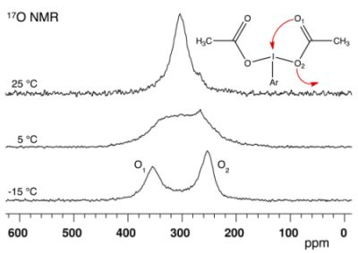
Graphical Abstract

1. Introduction

Hypervalent iodine chemistry is a field of great interest as shown by the large body of literature dealing with its various aspects [1,2,3,4,5,6]. For some years our group has been interested in studying the I-O bond and the dynamical behavior of some important I(III) and I(V) organic derivatives, such as bis(acyloxy)iodoarenes, benziodoxolones and the Dess-Martin periodinane (Figure 1), by combining solution-state 17O-NMR and Density Functional Theory (DFT) calculations [7,8,9].
Figure 1. Structure of (A) bis(acetoxy)iodobenzene, (B) (acetoxy)benziodoxolone and (C) the Dess-Martin periodinane.
Molecules 17 12718 g001
At room temperature, the 17O-NMR spectrum of all the studied bis(acyloxy)iodoarenes showed a single signal for both the carbonylic (O1) and ester-like (O2) oxygen atoms of the carboxylic groups. Similarly, a single signal was observed for the lateral acetoxy groups of the Dess-Martin reagent and, on the other hand, for the central acetoxy group of this λ5 iodane. With benziodoxolones, in contrast, distinctive 17O-NMR signals were observed at 25 °C for the O1 and O2 atoms of the carboxylic group not involved in the iodoxolone ring. However, at 45 °C, these two signals could not be detected. DFT calculations revealed that, among the various possible explanations, the [1,3]-sigmatropic shift of iodine between the two oxygen atoms of the carboxylic residue(s) (see Figure 2) is the actual cause of the experimental observations. The corresponding activation barriers were calculated and found to be in agreement with the 17O-NMR results. Summing up, our previous studies showed that the I-O bond of either I(III) or I(V) acyloxy derivatives is fluxional as a consequence of a [1,3]-sigmatropic shift mechanism. The activation barrier is much higher in the studied λ3 iodane than in the Dess-Martin λ5 iodane. Furthermore, in both the I(III) and I(V) acetoxy derivatives, the barrier is higher if the I-O bond is coplanar with the aromatic ring. The energetics and, consequently, the kinetics of the degenerate [1,3]-sigmatropic shift of iodine are thus affected by the environment of the carboxylic group. On the other hand, in a series of para-substituted bis(acetoxy)iodobenzenes, we observed a remarkable invariance of the 13C and 17O chemical shifts of the acetoxy groups. This is not yet understood. Indeed, the hypothesis of tight ion pairs that was first considered [7] is invalidated by our subsequent studies summarized above [8,9].
Dynamic NMR (DNMR) of low-sensitivity and fast-relaxing nuclei, among which is 17O, is definitely not routine in the study of organic molecules. Indeed, near the coalescence temperature, baseline distortion and poor signal-to-noise ratio may prevent the acquisition of NMR spectra that are suitable for line shape analysis while, in the intermediate-slow or -fast exchange regime, the natural linewitdh is not negligible and, moreover, may show a strong temperature dependence. Actually, coalescence could not be observed in any of our previous 17O-NMR studies of hypervalent iodine compounds. The acyloxy signal(s) of the I(III) derivatives, for which the coalescence temperature is accessible to solution-state NMR, broadened beyond detection as a consequence of the chemical exchange process itself or, at low temperature, as a consequence of quadrupole relaxation. Only rough estimations of the activation free energy (ΔG#) could therefore be provided. Recently, using acetoxysilanes as model compounds, we showed that currently available NMR instrumentations and pulse sequences allow for quantitative 17O-DNMR studies to be carried out without enrichment being needed [10]. This opened the perspective to precisely determine ΔG# values for the [1,3]-sigmatropic shift of iodine in I(III) acyloxy derivatives and, also, to determine the activation enthalpy (ΔH#) and the activation entropy (ΔS#) of this dynamic process. It is noteworthy that, in acetoxysilanes of the general formula (CH3COO)nSi(CH3)4-n with n = 1–4, the [1,3]-sigmatropic shift of the silicon atom is characterized by a significant activation entropy [10].
Figure 2. Schematic representation of the [1,3]-sigmatropic shift of iodine in bis(acetoxy)iodoarenes.
Molecules 17 12718 g002
The present work reports on several bis(acyloxy)iodoarenes: bis(acetoxy)iodobenzene (1), bis(trifluoroacetoxy)iodobenzene (2), para-methoxy-bis(acetoxy)iodobenzene (3) and para-nitro-bis(acetoxy)iodobenzene (4) (Figure 3). These λ3 iodanes were studied by 17O-DNMR with the primary goal of investigating the effect of electron-withdrawing and electron-donating groups on the energetics of the [1,3]-sigmatropic shift of iodine. Possible solvent effects were also investigated. The experimental data are compared to the results of DFT calculations.
Figure 3. Studied compounds with atom labeling and the angle I-Cac-O1 in bold.
Molecules 17 12718 g003

2. Results and Discussion

2.1. Bis(acetoxy)iodobenzene (1)

17O-NMR spectra of compound 1 dissolved in chloroform-d1 (CDCl3) were recorded at 14.1 T and various temperatures ranging between −30 and 110 °C (Figure 4). Two signals of similar integrated intensity are observed at low temperature (see Supporting Information). On the basis of chemical shift data reported for similar compounds [8,9,10,11,12], the signal at about 350 ppm is assigned to the carbonylic O1 oxygen atoms and, conversely, the signal at about 250 ppm is assigned to the ester-like O2 oxygen atoms. At −30 °C, the chemical shift (δ) and the full linewidth at half height (LW), as determined by fitting Lorentzian lines, are respectively 349 ppm and 3.28 kHz for the O1 signal and 246 ppm and 1.49 kHz for the O2 signal. Both these signals broaden for increasing temperature and coalescence occurs at about 5 °C. At 10 °C, a single signal is observed at δ ≈ 300 ppm, i.e., at the average of the O1 and O2 chemical shift values measured at low temperature. The LW of this signal decreases for increasing temperatures while the chemical shift is essentially constant.
Figure 4. 17O-NMR spectra recorded at 14.1 T and various temperatures for compound 1 dissolved in CDCl3. No apodization of the FID was used before Fourier transform.
Molecules 17 12718 g004
These observations are consistent with the evolution expected for an equally populated two-site exchange. At the coalescence temperature (Tc), the first order rate constant characterizing such an exchange process is directly related to the difference in resonance frequency in each site (Δν = ν1 ‑ ν2); the activation free energy at Tc can then be determined via the Eyring equation (see Supporting Information). For a chemical shift difference Δδ = (δ1 − δ2) = 103 ppm (Δν = 8.30 kHz at 14.1 T) and Tc = 5 °C, ΔG# is estimated to be (45.2 ± 0.5) kJ/mol. The error on ΔG# was estimated considering an absolute error on Tc of ± 3 °C. It is noteworthy that an error of ±5 ppm on Δδ affects ΔG# by only ±0.1 kJ mol−1.
Further information can be gained from the temperature dependence of the LW, which is shown in Figure 5 and was analyzed according to Equations (1–4), as in our previous 17O-DNMR investigation [10].
Molecules 17 12718 i001
Molecules 17 12718 i002
Molecules 17 12718 i003
Molecules 17 12718 i004
Molecules 17 12718 i005
In Equation (1), LWq is the natural linewidth due to quadrupole relaxation and LWex is the contribution due to chemical exchange. The temperature dependence of LWq is accounted for by an Arrhenius-type expression (Equation (2)) where the pre-exponential factor is the natural linewidth at 298 K and Eq is the activation energy. Identical activation energy was assumed for the quadrupole relaxation of both the O1 and O2 oxygen nuclei. Above the coalescence temperature, LWq is given by the arithmetic average of the natural linewidth values predicted for O1 and O2. The broadening due to chemical exchange between two equally populated sites is given by Equations (3a) or (3b), depending on whether the kinetics is slow or fast on the NMR spectral time scale. In Equation (3b), ν1− ν2 is the frequency difference between the O1 and O2 signals, k is the first-order rate constant, the temperature dependence of which is given by the Eyring equation (Equation (4)). ΔS# and ΔH# are the activation entropy and the activation enthalpy, respectively, which are assumed to be temperature independent, and the other symbols have their usual meaning. The best-fit of Equations (1)–(4) to the experimental LW data, using a least-squares technique based on the relative residuals [13], yields ΔG# = (45.5 ± 0.1) kJ/mol at 25 °C, which agrees with the value estimated at the coalescence (vide supra), ΔH# = (45 ± 1) kJ/mol and ΔS# = (−1 ± 5) J mol−1 K−1 [14]. Interestingly, the LW analysis reveals that the entropic contribution is not significant.
In order to investigate possible solvent effects, 17O-NMR spectra were recorded at 14.1 T and several temperatures for compound 1 dissolved in dichloromethane-d2 (CD2Cl2) and in acetonitrile-d3 (CD3CN). The spectra recorded in these more polar solvents are highly similar to the spectra recorded at the same temperature in CDCl3 (Figure 6). Tc is found to be about 10 °C in CD2Cl2 and is somewhat higher, ~15 °C, in CD3CN. Similar chemical shift values are observed, leading to similar ΔG# values (Table 1).
Figure 5. Full linewidth at half height (LW) measured at 14.1 T for the 17O-NMR signal(s) of compound 1 dissolved in CDCl3. The LW data were determined by fitting two Lorentzian lines of equal integrated intensity to the experimental spectra recorded at T ≤ −10 °C and one single Lorentzian line at T ≥ 20 °C. The solid lines are the results of the best fit analysis according to Equations (1)–(4). The natural linewidth contributions (shown as dashed lines) are characterized by the following parameters: Eq = (12.6 ± 0.2) kJ/mol, LWq298 = (0.94 ± 0.02) kHz and (0.40 ± 0.02) kHz for O1 and O2, respectively [14].
Molecules 17 12718 g005
Figure 6. 17O-NMR spectra recorded at 14.1 T and various temperatures for compound 1 dissolved in (A) CDCl3, (B) CD2Cl2 and (C) CD3CN. The asterisk indicates the peak of acetic acid. The spectra recorded at low temperature in CD3CN exhibit a low signal-to-noise ratio as a consequence of poor solubility of 1.
Molecules 17 12718 g006

2.2. Bis(trifluoroacetoxy)iodobenzene (2)

17O-NMR spectra of compound 2 dissolved in CDCl3 were recorded at 14.1 T and various temperatures ranging between −20 and 90 °C (Figure 7A). At low temperature, the signal of trifluoroacetic acid, which is present as an impurity in the sample, is the most intense signal because 2 is not entirely soluble and also because the 17O-NMR signals of 2, which could be detected at about 350 ppm (O1) and 220 ppm (O2), are highly broadened. At 0 °C in CD3CN (Figure 7B), the O1 and O2 signals are observed at 341 ppm and 218 ppm, respectively. Similarly to the spectra of compound 1, the spectra of 2 reveal the occurrence of a dynamic process which interchanges O1 and O2. The coalescence is observed at about 20 °C in CDCl3 and ΔG# is estimated to be (47.3 ± 0.5) kJ/mol.
Figure 7. 17O-NMR spectra recorded at 14.1 T for compound 2 dissolved in (A) CDCl3 and (B) CD3CN. An exponential multiplication of the FID with a line broadening factor of 100 Hz was used before Fourier transform. The dotted lines are guides for the eyes. The asterisk indicates the peak of trifluoroacetic acid.
Molecules 17 12718 g007

2.3. para-Methoxy-Bis(acetoxy)iodobenzene (3) and para-Nitro-Bis(acetoxy)iodobenzene (4)

17O-NMR spectra of compound 3 dissolved in CDCl3 were recorded at 14.1 T and various temperatures ranging between −25 and 12.5 °C (Figure 8A). The 17O-NMR signal of the methoxy group was detected at 62 ppm, i.e., in the expected range of chemical shift [15] (see Supporting Information). At −25 °C, the O1 and O2 signals are observed at 349 ppm and 254 ppm, respectively. The coalescence occurs somewhat below 0 °C; using Tc = −5 °C, ΔG# is estimated to be 43.7 kJ/mol.
For solubility reasons, compound 4 was dissolved in a 98/2 (v/v) mixture of CDCl3 and DMSO-d6 rather than in pure CDCl3. The 17O-NMR spectra were recorded at 14.1 T and various temperatures ranging between −25 and 37.5 °C (Figure 8B). The signal of the NO2 group was observed at 573 ppm, i.e., in the expected range of chemical shift [15] (see Supporting Information). At −25 °C, the O1 signal is observed at 359 ppm. The solubility of 4 is rather low and, consequently, acetic acid gives rise to an intense signal which masks the O2 signal at about 255 ppm. Coalescence is observed between 0 and 12.5 °C; using Tc = 5 °C, ΔG# is estimated to be 45.2 kJ/mol.
Figure 8. 17O-NMR spectra recorded at 14.1 T and various temperatures (A) for compound 3 dissolved in CDCl3 and (B) for compound 4 dissolved in CDCl3/DMSO-d6 98/2. An exponential multiplication of the FID with a line broadening factor of 100 Hz was used before Fourier transform. The dotted lines are guides for the eyes. The asterisk indicates the peak of acetic acid.
Molecules 17 12718 g008
Table 1. Activation barriers calculated at the PBE0/LANL2DZDP level and summary of the experimental results for compounds 14. Unless otherwise stated, the activation barrier (ΔE) was computed in-vacuo, the chemical shift of the O1 and O2 signals (δ1 and δ2) were determined by fitting Lorentzian lines to the spectrum recorded at the lowest investigated temperature, the activation free energy (ΔG#) was determined at the coalescence temperature (Tc) and the error on ΔG# is estimated to be ±0.5 kJ mol−1. This error is due to the error on Tc, which is estimated to be ± 3 °C. n.d.: not determined.
ΔESolventδ1δ2ΔδTcΔG#
kJ mol1(ppm)(ppm)(ppm)(°C)kJ mol1
145.2
48.3 a
CDCl3349246103545.5 ± 0.1 b
CD2Cl23552511041046.0
CD3CN3582511071546.8
248.4CDCl3345 c221 c1242047.3
CD3CN341218123n.d.n.d.
344.4CDCl334925495−543.7
445.2CDCl3/DMSO-d6d359~255 e~104545.2
a Computed in CHCl3 described as a polarizable continuum. b Value at 25 °C determined by the analysis of the linewidth data. The value estimated at the coalescence temperature is 45.2 kJ mol−1. c Estimated on the spectrum recorded at 0 °C. d 98/2 (v/v). e Estimated on the spectrum without fitting.

2.4. Comparisons with DFT Calculations and Discussion

Table 1 shows that the chemical shift of O1 is barely affected by the trifluoromethyl group but the chemical shift of O2 is reduced by about 30 ppm with respect to the value measured for bis(acetoxy)iodobenzene 1. In contrast, the 17O chemical shift data are quite insensitive to the para-substitution of the aromatic ring. This confirms the previous observations made for the average signal detected in the fast exchange regime [7]. In the present work, the O1 and O2 signals are, for the first time, distinctively observed and it can now be stressed that the previous observations are not the consequence of opposite chemical shift variations.
The 17O-DNMR results obtained for compound 1 indicate that, in CDCl3, the activation entropy characterizing the oxygen atom exchange is negligible and suggest that the activation free energy, about 46 kJ/mol, is rather insensitive to the dielectric constant of the solvent. The 17O-DNMR results also indicate that ΔG# is hardly affected by the chemical alteration present in compounds 24. Indeed, the ΔG# data measured for 4 and 1 are not significantly different. The replacement of the acetoxy groups of 1 by trifluoroacetoxy groups increases ΔG# by less than 2 kJ/mol while a methoxy group in para-position has the opposite effect.
As mentioned in the Introduction, the oxygen atom exchange in 1 was ascribed to a [1,3]-sigmatropic shift of iodine [8]. The activation energy barrier (ΔE) of this process can be calculated as the total energy difference between the transition state (TS) and the most stable rotamer. Our previous DFT study of compound 1 yielded ΔE = 45.2 kJ/mol in vacuo [8], a value which is in excellent agreement with the experimental results (Table 1). In our previous study on 1 and related compounds, we observed that the calculated energy profiles do not vary substantially when including the solvent, which was described in the framework of the polarizable continuum model (PCM) [16,17,18,19]. The DFT calculations yielded ΔE = 48.3 kJ/mol for 1 in chloroform. No important solvent effect on the activation energy barrier was observed and this also agrees with the experimental results of this work. On the base of these considerations, we believe that in vacuo calculations provide reliable results for the systems under investigations. In principle, it is the calculated activation free energy, and not ΔE, which should be compared to the experimental ΔG# values. However, various comparisons of DFT and experimental results have shown that the best agreement is obtained when no thermodynamical correction is applied to the calculated total energy; this point is discussed in the recent review of Casarini et al. [20]. Reasonably, the comparison between calculated energy barriers and experimental activation free energies is meaningful if the activation entropy is negligible or small, as it is the case for compound 1. In the following, it is assumed that there is no significant entropic contribution to the activation free energy measured for compounds 24.
DFT calculations were completed for compounds 24 with the main objective of verifying if the interchange of O1 and O2 observed by NMR for these λ3 iodanes can also be explained by the [1,3]-sigmatropic shift of iodine. The minimum energy structures of 24 show strong resemblance with those of 1 (see Figure 9 for 2), although some differences in the relative energies can be noted (Table 2). The stable rotamers of 2 exhibit the smallest variations in total energy with essentially the same value for the conformations A and B.
Figure 9. Optimized Pbe0/LANL2DZDP stable conformations of compound 2. Two representations are given for each conformer; they are rotated by approximately 90° around the I-O bonds. Energy of each conformer is given in Table 2 using the same label (A, B, C or D) as in the figure. Color code: cyan = carbon, white = hydrogen, red = oxygen, violet = fluorine, pink = iodine.
Molecules 17 12718 g009
Table 2. Relative total PBE0/LANL2DZDP energy (kJ/mol) of the stable rotamers of compounds 1-4. Labels indicating the rotamers as in Figure 9.
Conformer1234
A0.00.40.00.0
B1.70.00.83.7
C5.71.68.48.1
D9.33.27.67.7
Compound 2 is the one showing the largest experimental differences with compound 1. To verify whether the differences might be related to differences in the exchange mechanism, the latter was studied as previously done for compound 1 in reference [8]. The calculated energy profile is reported in Table 3 together with relevant structural data. It is similar to the profile found for 1, in having the maximum for a I-Cac-O1 angle comprised between 55° and 60° and in involving a rotation of 90° of the carboxylate plane during the shift (in this way going from conformation A to B).
It is reasonable to assume that the same exchange mechanism occurs for compound 3 and 4. In all cases, the calculated TS (see Supporting Information) are similar to that of 1 and the normal mode corresponding to the imaginary frequency clearly involves the oxygen atoms exchange. The energy barriers calculated in vacuo for compounds 24 are given in Table 1. The agreement with the corresponding experimental activation free energy is excellent, indicating that all of the studied bis(acyloxy)iodobenzenes are fluxional as a consequence of the [1,3]-sigmatropic shift of iodine and corroborating that this process is indeed quite insensitive to chemical alterations such as para- substitution of the arene moiety or modification of the acyl groups.
Table 3. Selected dihedrals and PBE0/LANL2DZDP total energy differences, calculated in vacuo with respect to the most stable rotamer, at variable I-Cac-O1 angle for compound 2. Atom labeling as in Figure 3.
I-Cac-O1 (°)ΔE kJ/molDihedral angles (°)
Ci-I-O1-CacCo-Ci-I-O2a
800.6 0.258.5
753.41.362.3
7010.20.090.0
6522.00.090.0
6038.30.090.0
5526.3−78.2 90.0
5013.5−78.590.1
453.3−77.992.1
400.0−76.796.3
TS (58.3)48.4−41.976.5
a O2 belongs to the unconstrained moiety. For a direct comparison among the dihedrals of the considered atomic configurations of 1 reported in a previous paper [8], the conformational enantiomers with the smallest Co-Ci-I-O2 angle between the unconstrained acetoxy unit and the clockwise Co were considered (view along the I-Ci bond).
As mentioned in the Introduction, para-substitution of bis(acetoxy)iodobenzenes does not significantly affect the 13C nor 17O chemical shift of the acetoxy groups [7]. This contrasts with the observations on relatively similar systems, such as para-substituted benzyl alcohols [21] and phenylacetic acids [22]. We are not able, for the moment, to give a clear-cut explanation of this difference but the lack of electronic effect transmission between iodine and oxygen might be related to the bond order. X-ray measurements [23,24] on compound 1 showed that the I-O bond length is 216 pm, a figure slightly greater than the sum (201.9 pm) of the iodine (134.5 pm) and oxygen (67.4 pm) covalent radii [25], and smaller than the sum (348 pm) of the iodine (196 pm) and oxygen (152 pm) van der Waals radii [26]. Similar results are observed for the I-S distance, as reported by Varvoglis on some carbamoyl(diaryl)iodanes [27,28]. X-ray studies showed this distance to be 305.3 pm, a value comprised between the sum of the iodine and sulphur covalent radii (238.7 pm) and the sum of their van der Waals radii (400 pm). From these data, as well as other significant arguments, the I-S bond order was estimated to be 0.5 [27]. Good estimate of bond orders can be obtained by computation. In fact, Kiprof [29] has shown for a series of hypervalent iodine derivatives that there is a quite good correlation between the experimental I-O bond length and the I-O bond order as estimated by the Wiberg bond index. Kiprof's value of the I-O bond order of 1 is 0.41, in full agreement with the values computed in this work for the minimum energy conformation (Table 4). We note that the I-O bond order, which has been shown [29] to be affected by variation of the substituent at the iodine atom, is actually insensitive to change of the acyl groups or to para-substitution of the aromatic ring. It thus seems reasonable that the rather low energy barrier of the [1,3]-sigmatropic shift of iodine in compounds 14 is related to the low I(III)-O bond order in the stable conformations. Finally, it is noteworthy that the I-O bond order is somehow conserved in the transition state.
Table 4. Wiberg bond indices for compounds 14 calculated at the PBE0/LANL2DZDP level.
CompoundGlobal MinimumTransition State
I-O1I-O2I-O1I-O2
10.060.420.280.16
20.04 (0.02) a0.42 (0.42) a0.250.15
30.070.410.270.16
40.080.420.290.15
a Differently from the other compounds the global minimum of 2 is the conformation B with two non equivalent moieties (see Figure 9). In parenthesis are reported the data for the carboxylate group with the plane perpendicular to the I-Ci bond.

3. Experimental

Compounds 1 and 2 were purchased from Sigma-Aldrich (Bornem, Belgium) and used as received. Compounds 3 and 4 were synthesized according to a procedure described in literature [30,31]. CDCl3, CD2Cl2, and DMSO-d6 were purchased from Sigma-Aldrich, CD3CN was purchased from Euriso-top (Paris, France). The samples used for the 17O-NMR measurements were saturated solution prepared at room temperature in 5 mm J. Young valve NMR tubes.

3.1. NMR Measurements

The NMR spectra were recorded on a Varian VNMRS spectrometer operating at 14.1 T (599.9 MHz for 1H and 81.33 MHz for 17O) equipped with a 5 mm autoX dual broadband probe and temperature regulation. The sample was left to reach equilibrium at the desired temperature within the magnet for at least 15 min before the NMR measurements. The 17O-NMR spectra were recorded with the improved RIDE (ring down elimination) pulse sequence of Kozminsky et al. [32] using a 100 μs cawurst adiabatic inversion pulse and the following acquisition parameters: spectral width of about 850 ppm (~69 kHz) centered at 290 ppm, 5 ms relaxation delay, 10 μs (90°) excitation pulse, 10 μs (rof2) preacquisition delay (alfa = 0), 5 ms acquisition time and a number of transients ranging between 5 × 10⁴ and 5 × 10⁶. The receiver ddrtc parameter was optimized to obtain spectra without first-order phase error. The spectra were recorded lock-on without sample spinning. Unless otherwise stated, the processing comprised correction of the first three points of the Free Induction Decay (FID) by backward linear prediction, zero filling, Fourier transform, zero-order phase correction and baseline correction. The chemical shift scale was calibrated at 25 °C with respect to a sample of pure H2O used as an external chemical shift reference (0 ppm).

3.2. Computational Details

Structure optimizations of all compounds were performed using the same theory level used in our previous investigations of hypervalent iodine compounds [8,9], which was shown to perform well on these classes of compounds. More specifically the calculations have been carried out at the DFT level employing the PBE0 functional [33], a parameter-free hybrid variant of the Perdew, Burke, and Ernzerhof (PBE) generalized gradient functional [34], as implemented in the commercially available Gaussian 03 suite of program [35]. The effective core-potential valence basis set LANL2DZ (i.e., D95V [36] basis set for the first row elements and the Los Alamos ECP plus DZ on iodine) [37] extended with polarization (d) and diffuse (p) functions [38,39] was employed for all atoms. Numerical integration was performed using a pruned grid having 99 radial shells and 509 angular points per shell. Vibrational analysis was carried out at the same level of theory to check the character of all stationary points. The starting geometries of the minima and TS states of compounds 24 were constructed by modifying (with appropriate substitutions) the minima and the TS states of compound 1, as obtained in a previous investigation [8]. The freely available program Molden was used for this purpose [40]. Cartesian coordinates and energies of the optimized geometries of minima and TS are reported in the Supporting Information. NBO version 3 [41] as implemented in Gaussian03 was employed for the analysis of the Wiberg bond index [42]. Graphics of molecular models were generated using the freely available VMD software [43].

4. Conclusions

The fluxional behaviour of bis(acetoxy)benzene and some of its derivatives has been studied by 17O dynamic NMR and DFT calculations. The activation free energy characterizing the [1,3]-sigmatropic shift of iodine between the oxygen atoms of the carboxylic group is shown to be rather insensitive to the solvent (CDCl3, CD2Cl2, and CD3CN), to the substitution of the acetate groups by trifluoroacetate groups and to the presence of electron-donor or electron-acceptor para-substituents.
The energy barriers calculated by DFT are in excellent agreement with the activation free energy data determined by NMR. The small or negligible variations with para-substitution is in agreement with the invariance of the I-O bond order, which has a rather small value of about 0.4 in all the studied compound. In the transition states, the partial breaking of the I-O bond is accompanied, and thus compensated, by the simultaneous formation of another I-O bond with the carbonylic oxygen atom, and the sum of their bond orders is in all cases between 0.4 and 0.5.
Importantly, this study confirms the recently demonstrated feasibility of 17O-Dynamic NMR to obtain quantitative kinetic data on organic molecules without the need of isotopic enrichment.

Supplementary Materials

Supplementary materials can be accessed at: https://www.mdpi.com/1420-3049/17/11/12718/s1.

Acknowledgements

L.F. and M.L. thank the “Fonds de la Recherche Scientifique” (F.R.S.-FNRS) and the Université Libre de Bruxelles (ULB) for financial support. F.M. and G.C. acknowledge Cagliari University for funding (ex 60% fund).

References and Notes

  1. Merritt, E.A.; Olofsson, B. Diaryliodonium salts: A journey from obscurity to fame. Angew. Chem. Int. Ed. 2009, 48, 9052–9070. [Google Scholar] [CrossRef]
  2. Ladziata, U.; Zhdankin, V.V. Hypervalent iodine(V) reagents in organic synthesis. ARKIVOC 2006, 9, 26–58. [Google Scholar]
  3. Zhu, C.; Sun, C.; Wei, Y. Direct oxidative conversion of alcohols, aldehydes and amines into nitriles using hypervalent iodine(III) reagent. Synthesis 2010, 24, 4235–4241. [Google Scholar]
  4. Yusubov, M.S.; Zhdankin, V.V. Hypervalent iodine reagents and green chemistry. Curr. Org. Synth. 2012, 9, 247–272. [Google Scholar]
  5. Dohi, T.; Maruyama, A.; Minamitsuji, Y.; Takenaga, N.; Kita, Y. First hypervalent iodine(III)-catalyzed C-N bond forming reaction: Catalytic spirocyclization of amides to N-fused spirolactams. Chem. Commun. 2007, 12, 1224–1226. [Google Scholar]
  6. Zagulyaeva, A.; Yusubov, M.S.; Zhdankin, V.V. A general and convenient preparation of [Bis(trifluoroacetoxy)iodo]perfluoroalkanes and [Bis(trifluoroacetoxy)iodo]arenes by oxidation of organic iodides using oxone and trifluoroacetic acid. J. Org. Chem. 2010, 75, 2119–2122. [Google Scholar]
  7. Cerioni, G.; Uccheddu, G. Solution structure of bis(acetoxy)iodoarenes as observed by O-17 NMR spectroscopy. Tetrahedron Lett. 2004, 45, 505–507. [Google Scholar] [CrossRef]
  8. Mocci, F.; Uccheddu, G.; Frongia, A.; Cerioni, G. Solution structure of some lambda(3) iodanes: An O-17 NMR and DFT study. J. Org. Chem. 2007, 72, 4163–4168. [Google Scholar]
  9. Fusaro, L.; Luhmer, M.; Cerioni, G.; Mocci, F. On the fluxional behavior of Dess-Martin periodinane: A DFT and O-17 NMR Study. J. Org. Chem. 2009, 74, 8818–8821. [Google Scholar] [CrossRef]
  10. Fusaro, L.; Mameli, G.; Mocci, F.; Luhmer, M.; Cerioni, G. Dynamic NMR of low-sensitivity fast-relaxing nuclei: 17O NMR and DFT study of acetoxysilanes. Magn. Reson. Chem. 2012, 50, 152–158. [Google Scholar]
  11. Lycka, A.; Holecek, J.; Handlir, K.; Pola, J.; Chvalovsky, V. 17O, 13C, and 29Si NMR spectra of some acyloxy- and diacetoxysilanes and acetoxygermanes. Collect. Czech. Chem. Commun. 1986, 51, 2582–2589. [Google Scholar]
  12. Boykin, D.W.; Baumstark, A.L. 17O NMR spectroscopic data for carbonyl compounds. In 17O NMR Spectroscopy in Organic Chemistry; CRC Press: Boca Raton, FL, USA, 1991; pp. 205–231. [Google Scholar]
  13. Saez, P.; Rittmann, B. Model-parameter estimation using least-squares. Water Res. 1992, 26, 789–796. [Google Scholar] [CrossRef]
  14. The errors are standard deviations provided by the analysis of a series of 1000 pseudo-experimental data sets obtained by adding random errors to the experimental LW data as well as to the temperature. These errors were generated from a Gaussian distribution considering a relative standard error on LW of 5% and an absolute standard error on the temperature of ±1 K.
  15. Gerothanassis, I.P. Oxygen-17 NMR spectroscopy: Basic principles and applications (Part I). Prog. Nucl. Magn. Reson. Spectrosc. 2010, 56, 95–197. [Google Scholar]
  16. Mennucci, B.; Tomasi, J. Continuum solvation models: A new approach to the problem of solute’s charge distribution and cavity boundaries. J. Chem. Phys. 1997, 106, 5151–5158. [Google Scholar]
  17. Cances, E.; Mennucci, B.; Tomasi, J. A new integral equation formalism for the polarizable continuum model: Theoretical background and applications to isotropic and anisotropic dielectrics. J. Chem. Phys. 1997, 107, 3032–3041. [Google Scholar] [CrossRef]
  18. Cossi, M.; Barone, V.; Mennucci, B.; Tomasi, J. Ab initio study of ionic solutions by a polarizable continuum dielectric model. Chem. Phys. Lett. 1998, 286, 253–260. [Google Scholar] [CrossRef]
  19. Cossi, M.; Scalmani, G.; Rega, N.; Barone, V. New developments in the polarizable continuum model for quantum mechanical and classical calculations on molecules in solution. J. Chem. Phys. 2002, 117, 43–54. [Google Scholar] [CrossRef]
  20. Casarini, D.; Mazzanti, A.; Lunazzi, L. Recent advances in stereodynamics and conformational analysis by dynamic nmr and theoretical calculations. Eur. J. Org. Chem. 2010, 2035–2056. [Google Scholar]
  21. Balakrishnan, P.; Baumstark, A.L.; Boykin, D.W. 17O NMR spectroscopy: Unusual substituent effects in para-substituted benzyl alcohols and acetates. Tetrahedron Lett. 1984, 25, 169–172. [Google Scholar] [CrossRef]
  22. Monti, D.; Orsini, F.; Ricca, G.S. Oxygen-17 NMR Spectroscopy: Effect of Substituents on Chemical Shifts for o− m− p− Substituted Benzoic Acids, Phenylacetic and Methyl Benzoates. Spectroscopy Lett. 1986, 19, 91–99. [Google Scholar] [CrossRef]
  23. Alcock, N.W.; Countryman, R.M.; Esperas, S.; Sawyer, J.F. Secondary bonding. Part 5. The crystal and molecular structures of phenyliodine(III) diacetate and bis(dichloroacetate). J. Chem. Soc. Dalton Trans. 1979, 5, 854–860. [Google Scholar]
  24. Kokkou, S.C.; Cheer, C.J. Structure of diacetato(m-tolyl)iodine(III). Acta Cryst. 1986, C42, 1159–1161. [Google Scholar]
  25. Pyykko, P. Refitted tetrahedral covalent radii for solids. Phys. Rev. B 2012, 024115. [Google Scholar]
  26. Bondi, A. Van der waals volumes and radii. J. Phys. Chem. 1964, 68, 441–451. [Google Scholar]
  27. Kotali, E.; Varvoglis, A.; Bozopoulos, A.; Rentzeperis, P. A stable dibenzoiodolyl pyrrolidinedithiocarbamate. J. Chem. Soc. Chem. Commun. 1985, 24, 1819–1820. [Google Scholar]
  28. Kotali, E.; Varvoglis, A. Dialkylcarbamoyl(diaryl)iodanes. J. Chem. Soc. Perkin Trans. I 1987, 12, 2759–2763. [Google Scholar] [CrossRef]
  29. Kiprof, P. The nature of iodine oxygen bonds in hypervalent 10-I-3 iodine compounds. ARKIVOC 2005, 4, 19–25. [Google Scholar]
  30. Kazmierczak, P.; Skulski, L. A simple, two-step conversion of various iodoarenes to (Diacetoxyiodo)arenes with Chromium(VI) oxide as the oxidant. Synthesis 1998, 1721–1723. [Google Scholar]
  31. Merkushev, E.B.; Novikov, A.N.; Makarchenko, S.S.; Moskalchuk, A.S.; Glushova, V.V.; Kogai, T.Y.; Polyakova, L.G. Organic compounds of polyvalent iodine. VIII. Simple synthesis of phenyliodosocarboxylates. Zh. Org. Khim. 1975, 11, 1259–1262. [Google Scholar]
  32. Kozminski, W.; Jackowski, K. Application of adiabatic inversion pulses for elimination of baseline distortions in Fourier transform NMR. A natural abundance 17O NMR spectrum for gaseous acetone. Magn. Reson. Chem. 2000, 38, 459–462. [Google Scholar]
  33. Adamo, C.; Barone, V. Toward reliable density functional methods without adjustable parameters: The PBE0 model. J. Chem. Phys. 1999, 110, 6158–6179. [Google Scholar]
  34. Perdew, J.P.; Burke, K.; Ernzerhof, M. Generalized gradient approximation made simple. Phys. Rev. Lett. 1996, 77, 3865–3868. [Google Scholar] [CrossRef]
  35. Frisch, M.J.; Trucks, G.W.; Schlegel, H.B.; Scuseria, G.E.; Robb, M.A.; Cheeseman, J.R.; Montgomery, J.A., Jr.; Vreven, T.; Kudin, K.N.; Burant, J.C.; et al. Gaussian 03, Revision C.02; Gaussian, Inc.: Wallingford, CT, USA, 2004. [Google Scholar]
  36. Dunning, T.H., Jr.; Hay, P.J. Modern Theoretical Chemistry: Methods of Electronic Structure Theory; Schaefer, H.F., III, Ed.; Plenum: New York, NY, USA, 1976; Volume 3, pp. 1–28. [Google Scholar]
  37. Wadt, W.R.; Hay, P.J. Ab initio effective core potentials for molecular calculations. Potentials for main group elements Na to Bi. J. Chem. Phys. 1985, 82, 284–298. [Google Scholar] [CrossRef]
  38. Check, C.E.; Faust, T.O.; Bailey, J.M.; Wright, B.J.; Gilbert, T.M.; Sunderlin, L.S. Addition of polarization and diffuse functions to the LANL2DZ basis set for P-Block elements. J. Phys. Chem. A 2001, 105, 8111–8116. [Google Scholar]
  39. Basis sets were obtained from the Extensible Computational Chemistry Environment Basis Set Database, Version 02/25/04, as developed and distributed by the Molecular Science Computing Facility, Environmental and Molecular Sciences Laboratory, which is part of the Pacific Northwest Laboratory, P.O. Box 999, Richland, Washington 99352, and funded by the U.S. Department of Energy.
  40. Schaftenaar, G.; Noordik, J.H. The effect of isodensity surface sampling on ESP derived charges and the effect of adding bond-centers on DMA derived charges. J. Comput. Aided Mol. Des. 2000, 14, 123–134. [Google Scholar] [CrossRef]
  41. Weinhold, F. Natural bond orbital methods. In Encyclopedia of Computational Chemistry; Schleyer, P.V.R., Allinger, N.L., Clark, T., Gasteiger, J., Kollman, P.A., Schaefer, H.F., III, Schreiner, P.R., Eds.; John Wiley & Sons: Chichester, UK, 1998; Volume 3, pp. 1792–1811. [Google Scholar]
  42. Wiberg, K.B. Application of the pople-santry-segal CNDO method to the cyclopropylcarbinyl and cyclobutyl cation and to bicyclobutane. Tetrahedron 1968, 24, 1083–1096. [Google Scholar] [CrossRef]
  43. Humphrey, W.; Dalke, A.; Schulten, K. VMD: Visual molecular dynamics. J. Mol. Graph. 1996, 14, 33–38. [Google Scholar] [CrossRef]
  • Sample Availability: Not available.