Article

Mechanisms Underlying Vasorelaxation Induced in Rat Aorta by Galetin 3,6-Dimethyl Ether, a Flavonoid from Piptadenia stipulacea (Benth.) Ducke

by
Cibério L. Macêdo
1,*,†,
Luiz H. C. Vasconcelos
1,*,†,
Ana C. De C. Correia
1,
Italo R. R. Martins
1,
Daysianne P. De Lira
2,
Bárbara V. De O. Santos
1,3,
Fabiana De A. Cavalcante
1,4 and
Bagnólia A. Da Silva
1,3
1
Programa de Pós-graduação em Produtos Naturais e Sintéticos Bioativos, Centro de Ciências da Saúde, Universidade Federal da Paraíba, João Pessoa, PB 58051-900, Brazil
2
Departamento de Farmácia, Faculdade Santa Maria (FSM), Cajazeiras, PB 58900-000, Brazil
3
Departamento de Ciências Farmacêuticas, Centro de Ciências da Saúde, Universidade Federal da Paraíba, João Pessoa, PB 58051-970, Brazil
4
Departamento de Fisiologia e Patologia, Centro de Ciências da Saúde, Universidade Federal da Paraíba, João Pessoa, PB 58051-970, Brazil
*
Authors to whom correspondence should be addressed.
These authors contributed equally to this work.
Molecules 2014, 19(12), 19678-19695; https://doi.org/10.3390/molecules191219678
Submission received: 10 September 2014 / Revised: 17 November 2014 / Accepted: 17 November 2014 / Published: 27 November 2014
(This article belongs to the Collection Bioactive Compounds)

Abstract

:
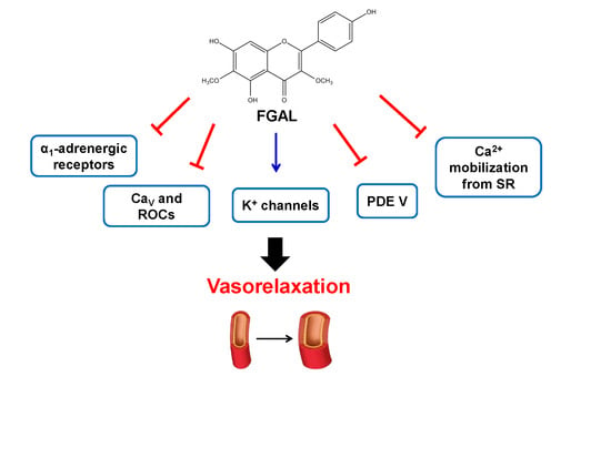
In this study, we investigated the relaxant action of galetin 3,6-dimethyl ether (FGAL) on rat aorta. The flavonoid relaxed both PMA‑ and phenylephrine (Phe)-induced contractions (pD2 = 5.36 ± 0.11 and 4.17 ± 0.10, respectively), suggesting the involvement of PKC and Phe pathways or α1 adrenergic receptor blockade. FGAL inhibited and rightward shifted Phe-induced cumulative concentration‑response curves, indicating a noncompetitive antagonism of α1 adrenergic receptors. The flavonoid was more potent in relaxing 30 mM KCl- than 80 mM KCl-induced contractions (pD2 = 5.50 ± 0.22 and 4.37 ± 0.12). The vasorelaxant potency of FGAL on Phe-induced contraction was reduced in the presence of 10 mM TEA+. Furthermore, in the presence of apamin, glibenclamide, BaCl2 or 4-AP, FGAL-induced relaxation was attenuated, indicating the participation of small conductance calcium-activated K+ channels (SKCa), ATP-sensitive K+ channels (KATP), inward rectifier K+ channels (Kir) and voltage-dependent K+ channels (KV), respectively. FGAL inhibited and rightward shifted CaCl2-induced cumulative concentration-response curves in both depolarizing medium (high K+) and in the presence of verapamil and phenylephrine, suggesting inhibition of Ca2+ influx through voltage-gated calcium channels (CaV) and receptor operated channels (ROCs), respectively. Likewise, FGAL inhibited Phe-induced contractions in Ca2+-free medium, indicating inhibition of Ca2+ release from the sarcoplasmic reticulum (SR). FGAL potentiated the relaxant effect of aminophylline and sildenafil but not milrinone, suggesting the involvement of phosphodiesterase V (PDE V). Thus, the FGAL vasorelaxant mechanism involves noncompetitive antagonism of α1 adrenergic receptors, the non-selective opening of K+ channels, inhibition of Ca2+ influx through CaV or ROCs and the inhibition of intracellular Ca2+ release. Additionally, there is the involvement of cyclic nucleotide pathway, particularly through PDE V inhibition.

Graphical Abstract

1. Introduction

Flavonoids are a large class of polyphenolic substances found in plants [1] known for their interesting activities in vascular diseases [2,3,4]. Several pharmacological effects have been described for this class of secondary metabolites, such as inhibition of enzymes involved in the synthesis of reactive oxygen species, such as xanthine oxidase, NADPH oxidase and lipoxygenase [5], increase in nitric oxide (NO) production in vascular smooth muscle [6] and spasmolytic activity in various models of smooth muscle [7,8]. Furthermore, some flavonoids are known for their actions on vascular tone, such as quercetin, kaempferol, luteolin, apigenin, catechin and epicatechin [1].
Galetin 3,6-dimethyl ether (FGAL) (Figure 1), a flavonoid isolated from the plant Piptadenia stipulacea (Benth.) Ducke, has exhibited some pharmacological activities, such as antiviral [9], antinociceptive and anti-inflammatory activities in mice [2], as well as non-selective spasmolytic activity in smooth muscles (e.g., guinea-pig ileum and trachea and rat uterus and aorta). Moreover, this flavonoid has shown the highest relaxant potency in rat aorta, and this effect is independent of endothelium-derived relaxant factors (EDRF) [10].
Regarding the pharmacological effects described for flavonoids, these secondary metabolites appear to be candidates for the treatment of various diseases caused by disorders of smooth muscle, especially those affecting the cardiovascular system, such as hypertension, atherosclerosis and ischemic infarction [1]. Therefore, the aim of this work was to characterize the mechanisms involved in vasorelaxation induced by the flavonoid FGAL in rat aorta.
Figure 1. Chemical structure of galetin 3,6-dimethyl ether (FGAL).
Molecules 19 19678 g001

2. Results and Discussion

In blood vessels, the endothelium plays an important role in regulating vascular smooth muscle tone by releasing endothelium-derived relaxing factors (EDRF) [11], including endothelium‑derived hyperpolarizing factor (EDHF), nitric oxide (NO), prostacyclins and epoxyeicosatrienoic acids [12]. Despite this, it was previously demonstrated that the vasorelaxation induced by FGAL is independent of EDRF, since it relaxed aorta in both the absence and presence of endothelium in an equipotent manner [10]. On the other hand, several mechanisms are involved in endothelium-independent vasorelaxation, such as Ca2+ channel blockade, K+ channel opening, protein kinase C (PKC) inhibition, attenuation of Ca2+ release from the sarcoplasmic reticulum (SR) and phosphodiesterase (PDE) pathway inhibition [13]. Moreover, it has been demonstrated that flavonoids can produce vasorelaxation by different mechanisms, such as NO release from endothelium [14], PKC and PDE inhibition [15,16], blockade of Ca2+ influx through voltage‑sensitive Ca2+ channels (CaV) [17] and K+ channel activation (IKCa and BKCa) [18,19].
PKC is a key protein to vascular smooth muscle contraction [20]. This protein kinase can both activate CaV and inhibit K+ channels, leading to Ca2+ influx and contributing to the contractile process. Phorbol esters, such as phorbol 12‑myristate 13-acetate (PMA), which are described as PKC stimulators, are used as exogenous activators of this protein kinase. Also, they have been known to induce sustained contraction in several arterial tissues [21,22]. FGAL (10−8 to 10−3 M) relaxed aorta pre‑contracted with 3 × 10⁻⁷ M phenylephrine (Phe) (pD2 = 5.36 ± 0.11) or 10−6 M PMA (pD2 = 4.17 ± 0.10). According to the pD2 values, FGAL was about 16-fold more potent in relaxing aorta pre‑contracted with Phe than with PMA. The vehicle did not show a significant relaxant effect in rat aorta pre‑contracted with either contractile agent (Figure 2).
The fact that PMA and Phe elicit contraction by different pathways suggests that the relaxant effect of FGAL in rat aorta involves both mechanisms. Therefore, the flavonoid can act on PKC (PMA exclusive target) as well as CaV, K+ channels and Ca2+ mobilization from SR, which are involved in Phe signaling but are not directly affected by PMA [23]. Figure 2 shows that the inhibition of PKC is possibly involved, but that other targets (e.g., CaV, K+ channels or others) are involved in a major manner. Moreover, we do not discard a possible direct antagonism of α1 adrenergic receptors by FGAL.
To assess the blockade of α1 adrenergic receptors by FGAL, the effect of the flavonoid on cumulative concentration‑response curves to phenylephrine (10−11 to 3 × 10⁻⁵ M) was investigated. FGAL (3 × 10⁻⁶ to 3 × 10⁻⁵ M) rightward shifted these curves in a non‑parallel manner with reduction in Emax and pD2 values of phenylephrine (Table 1, Figure 3). The FGAL pD2 value in inhibiting the effect of phenylephrine was 4.94 ± 0.04. In presence of a noncompetitive antagonist, the agonist has its maximum effect suppressed and abolished at higher concentrations of the antagonist [24]. Thus, according to the data obtained, FGAL showed the profile of a noncompetitive antagonist acting on α1 adrenergic receptors, which can be associated with a blockade of downstream pathways, such as CaV, K+ channels and SR Ca2+ mobilization.
Figure 2. Effect of FGAL or vehicle on tonic contractions induced by 3 × 10⁻⁷ M Phe (FGAL: ▲, n = 5; vehicle: ♦, n = 3) or 10−6 M PMA (FGAL: Δ, n = 5; vehicle: ◊, n = 3) in rat aorta. Symbols and vertical bars represent the mean ± S.E.M., respectively. Student’s t-test, *** p < 0.001 (Phe vs. PMA).
Molecules 19 19678 g002
Figure 3. Cumulative concentration-response curves to phenylephrine in both the absence (■, control, n = 5) and presence of 3 × 10⁻⁶ (□, n = 3), 10−5 (●, n = 3) and 3 × 10⁻⁵ M (○, n = 3) of FGAL. Symbols and vertical bars represent the mean and S.E.M., respectively.
Molecules 19 19678 g003
K+ channel activation and CaV blockade are two important mechanisms to promote vasorelaxation and are signaling pathway targets of α1 adrenergic receptor agonists, such as phenylephrine. Thus, we investigated whether these channels would be involved in the relaxant effect of FGAL. Accordingly, we evaluated the relaxation induced by FGAL in aorta pre-contracted with high (30 and 80 mM KCl) extracellular K+ concentration ([K+]o) [25]. By altering [K+]o, it is possible to determine if a drug has activity as a K+ channel opener or a CaV blocker, since K+ channel openers are more potent in relaxing a muscle pre-contracted with 30 mM than with 80 mM KCl, because this increase in [K+]o to 80 mM prevents ion efflux, even in the case of the opening of K+ channels in the plasma membrane [26].
Table 1. Emax and pD2 values of phenylephrine in both the absence (control) and presence of FGAL (3 × 10⁻⁶ to 3 × 10⁻⁵ M) in rat aorta. Data are expressed as the mean ± S.E.M. (n = 3). One-way ANOVA followed by Bonferroni’s post-test: *** p < 0.001 (control vs. FGAL), ## p < 0.01 (3 × 10⁻⁶ vs. 10−5 M FGAL), ¥¥¥ p < 0.001 (10−5 vs. 3 × 10⁻⁵ M FGAL). Nd = not determined.
[FGAL] MEmax (%)pD2
Control100.0 ± 0.08.13 ± 0.18
3 × 10−694.9 ± 2.77.75 ± 0.10
10−560.9 ± 8.2 *** ##7.49 ± 0.16
3 × 10−53.5 ± 2.3 *** ¥¥¥Nd
FGAL (10−8 to 10−3 M) relaxed aorta pre-contracted with both 30 mM KCl (pD2 = 5.50 ± 0.22) and 80 mM (pD2 = 4.37 ± 0.12). According to the pD2 values, the flavonoid showed about 10-fold greater relaxant potency in rat aorta pre-contracted with 30 mM than with 80 mM KCl (Figure 4). This result indicates that FGAL activates K+ channels in relaxing rat aorta.
Figure 4. Effect of FGAL on tonic contractions induced by 30 mM KCl (●) or 80 mM KCl (○) in rat aorta. Symbols and vertical bars represent mean ± S.E.M., respectively. Student’s t-test, ** p < 0.01 (30 mM vs. 80 mM KCl).
Molecules 19 19678 g004
To confirm the activation of K+ channels by FGAL, the non‑selective blocker of these channels TEA+ (10 mM) was employed as a pharmacological tool. The relaxant potency of FGAL was reduced in the presence of the blocker (about 4-fold), confirming the participation of K+ channels in relaxation induced by FGAL (Figure 5, Table 2). The vascular smooth muscle expresses multiple K+ channel subtypes, where KATP, Kir, KV, BKCa and SKCa are the most expressed subtypes [27]. Hence, the involvement of these subtypes of K+ channels was evaluated by employing their selective blockers. The relaxation induced by FGAL was not changed in the presence of 1 mM TEA+, excluding the participation of BKCa. Conversely, the relaxant potency of the flavonoid was reduced in the presence of glibenclamide, BaCl2, 4-AP and apamin, indicating the activation of KATP, Kir, KV and SKCa, respectively, by FGAL to induce vasorelaxation in rat aorta (Table 2).
Figure 5. Representative records of relaxant effect of FGAL in rat aorta pre-contracted with 3 × 10⁷ M Phe in both absence (A) and presence (B) of 10 mM TEA+. The arrows represent the addition of substances.
Molecules 19 19678 g005
Table 2. Emax (%) and pD2 values of FGAL in both the absence and presence of K+ channel blockers in rat aorta. Data are expressed as the mean ± S.E.M. (n = 5). One-way ANOVA followed by Dunnett’s post-test. * p < 0.05, ** p < 0.01 (FGAL vs. FGAL + blockers).
CompoundsEmax (%)pD2
FGAL100.0 ± 0.05.35 ± 0.11
10 mM TEA+ + FGAL98.7 ± 1.34.71 ± 0.06 **
1 mM TEA+ + FGAL92.8 ± 3.45.57 ± 0.17
10−5 M glibenclamide + FGAL98.8 ± 1.24.79 ± 0.05 *
5 × 10−8 M apamin + FGAL98.3 ± 1.14.75 ± 0.11 *
10−4 M BaCl2 + FGAL89.1 ± 4.6 *4.62 ± 0.17 **
10−3 M 4-AP + FGAL86.2 ± 3.7 **4.82 ± 0.11 *
Similar results are described in the literature for other flavonoids that promote relaxation in rat aorta through the activation of K+ channels; for example, pinocembrin activates KATP, KCa and KV [28], (‒)-epigallocatechin-3-gallate activates KATP, SKCa, IKCa and BKCa [29], and quercetin acts on BKCa [30].
Cytosolic calcium concentration ([Ca2+]i) increase in vascular smooth muscle cell is essential for its contraction and can occur by ion influx from the extracellular medium [31] or by release from intracellular stores [32]. The Ca2+ influx in vascular smooth muscle cells involves the opening of CaV and ROCs [33]. Pharmacological assays showed that the tonic contraction induced by high [K+]o is mainly due to the depolarization of smooth muscle cells and consequent Ca2+ influx through CaV, while the contraction induced by Phe is caused by Ca2+ influx through both CaV and ROCs [34,35,36]. Given this and since FGAL caused more potent relaxation (about 6-fold) in aorta pre-contracted with Phe (pD2 = 5.36 ± 0.11) than that with 80 mM KCl (pD2 = 4.37 ± 0.12), it is suggested that FGAL may reduce the Ca2+ influx through both CaV and ROCs.
To assess this hypothesis, we evaluated the effect of FGAL on CaCl2‑induced contractions in depolarizing medium nominally without Ca2+ to determine the inhibition of CaV by the flavonoid, and its effect on CaCl2‑induced cumulative concentration-response curves (10−7 to 10−1 M) in the presence of verapamil (CaV blocker) and Phe to determine the inhibition of ROCs. FGAL (10−5 to 3 × 10⁻⁴ M) inhibited these cumulative concentration‑response curvesand this effect was concentration dependent. These curves were rightward shifted in a non‑parallel manner with reduction in Emax and pD2 values of CaCl2 (Table 3, Figure 6). The pD2 value of FGAL in inhibiting the effect of CaCl2 was 4.44 ± 0.05.
Table 3. Emax and pD2 values of CaCl2 in both the absence (control) and presence of FGAL (10−5 to 3 × 10⁻⁴ M) in rat aorta. Data are expressed as the mean ± S.E.M. (n = 5). One‑way ANOVA followed by Bonferroni’s post-test: ** p < 0.01 and *** p < 0.001 (control vs. FGAL), ### p < 0.001 (10−5 vs. 3 × 10⁻⁵ M FGAL), ¥¥¥ p < 0.001 (3 × 10⁵ vs. 104 M FGAL). Nd = not determined.
[FGAL] MEmax (%)pD2
Control100.0 ± 0.02.52 ± 0.10
10−594.3 ± 2.71.95 ± 0.12 **
3 × 10−561.7 ± 8.1 *** ###2.69 ± 0.08
10−49.7 ± 1.9 *** ¥¥¥Nd
3 × 10−41.1 ± 0.8 ***Nd
Figure 6. Cumulative concentration-response curves to CaCl2 in depolarizing medium (high K+) nominally without Ca2+ in both the absence (■, control, n = 5) and presence of 10−5 (□, n = 5), 3 × 10⁻⁵ (●, n = 5), 10−4 (○, n = 5) and 3 × 10⁻⁴ M (▲, n = 5) of FGAL. Symbols and vertical bars represent the mean and S.E.M., respectively.
Molecules 19 19678 g006
In a similar way, FGAL (3 × 10⁻⁶ to 3 × 10⁻⁵ M) inhibited CaCl2-induced cumulative contractions (10−5 to 3 × 10⁻¹ M) in the presence of verapamil and Phe (Table 4, Figure 7). The FGAL pD2 value in inhibiting the effect of CaCl2 was 5.07 ± 0.06. Together, these results confirm that FGAL inhibits Ca2+ influx by blocking both CaV and ROCs to relax vascular smooth muscle. However, comparing the inhibitory effect of FGAL in Figure 6 and Figure 7, the potency of the flavonoid was higher in the presence of verapamil and Phe than in their absence. This therefore indicates that FGAL acts mainly by inhibiting ROCs.
Table 4. Emax and pD2 values of CaCl2 in the presence of verapamil (10−6 M) and Phe (10−6 M), in both the absence (control) and presence of FGAL (3 × 10⁻⁶ to 3 × 10⁻⁵ M) in rat aorta. Data are expressed as the mean ± S.E.M. (n = 5). One-way ANOVA followed by Bonferroni’s post-test: * p < 0.05, ** p < 0.01 and *** p < 0.001 (control vs. FGAL), ### p < 0.001 (3 × 10⁻⁶ vs. 10−5 M FGAL), p ¥¥¥ < 0.001 (10−5 vs. 3 × 10⁻⁵ M FGAL). Nd = not determined.
[FGAL] MEmax (%)pD2
Control100.0 ± 0.02.97 ± 0.09
3 × 10−681.3 ± 4.6 **2.28 ± 0.18 *
10−547.6 ± 4.6 *** ###2.43 ± 0.20
3 × 10−54.1 ± 1.9 *** ¥¥¥Nd
Figure 7. Cumulative concentration-response curves to CaCl2 in the presence of verapamil (10−6 M) and Phe (10−6 M), in both the absence (■, control, n = 5) and presence of 3 × 10⁻⁶ (□, n = 5), 10−5 (●, n = 5) and 3 × 10⁻⁵ M (○, n = 5) of FGAL. Symbols and vertical bars represent the mean and S.E.M., respectively.
Molecules 19 19678 g007
Increase in [Ca2+]i may also occur due to Ca2+ release from intracellular stores, especially SR [37]; hence, in Ca2+-free medium, Phe-induced contraction occurs mainly due to Ca2+ release from SR. As can be seen in Figure 8, FGAL inhibited Phe-induced contractions in Ca2+-free medium in a concentration-dependent manner (Emax = 96.6% ± 1.5%; IC50 = 1.0 ± 0.1 × 10⁻⁵ M), supporting our hypothesis that the relaxant effect of FGAL involves the inhibition of Ca2+ release from SR.
Figure 8. Effect of FGAL on contractions induced by 10−6 M Phe in Ca2+-free medium in rat aorta. One-way ANOVA followed by Bonferroni’s post-test, *** p < 0.001 (control vs. FGAL), ### p < 0.001 (3 × 10⁻⁶ vs. 10−5 M FGAL), p ¥¥ < 0.01 (10−5 vs. 3 × 10⁻⁵ M FGAL), §§§ p < 0.001 (3 × 10⁻⁵ vs. 10−4 M FGAL), n = 5.
Molecules 19 19678 g008
PDEs are widely distributed in mammalian tissues and hydrolyze cAMP and cGMP, resulting in their inactive products 5'-AMP and 5'-GMP, which do not activate PKA and PKG, respectively, thus stopping the cell signaling mechanism dependent on increased cyclic nucleotides [38]. Substances able to raise intracellular levels of cAMP or cGMP show a strong relaxant effect, which can be due to PDE inhibition [39]. Some flavonoids from different plant species inhibit PDEs [40], for instance (‒)-epigallocatechina-3-gallate in rat aorta [29].
Therefore, to assess the possible involvement of cyclic nucleotide-PDE pathway, we determined the relaxant effect of aminophylline, a non-selective PDE inhibitor, in both the absence and presence of FGAL. The relaxant effect induced by aminophylline (10−10 to 10−3 M, positive control) (pD2 = 4.36 ± 0.09) was potentiated about 4-fold in the presence of FGAL (pD2 = 5.13 ± 0.24), confirming that FGAL inhibits cyclic nucleotide-PDE pathways to relax rat aorta. Additionally, in vascular smooth muscle, PDE III and V are the most expressed PDE subtypes [38], and interestingly, the relaxation curve induced by sildenafil (10−8 to 10−4 M) (pD2 = 6.84 ± 0.25), a PDE V inhibitor, was potentiated about 9-fold in the presence of FGAL (pD2 = 7.62 ± 0.22); however, the relaxation induced by milrinone (10−8 to 3 × 10⁻⁴ M) (pD2 = 7.23 ± 0.23), a PDE III inhibitor, was not changed in the presence of the flavonoid (pD2 = 7.28 ± 0.09) (Figure 9).
Thus, these results indicate that PDE V but not PDE III pathway is involved in relaxation induced by FGAL in rat aorta.
Figure 9. Effect of aminophylline (A, ●) sildenafil (B, ) and milrinone (C, ) on tonic contractions induced by 3 × 10⁻⁷ M Phe in both the absence and presence of 3 × 10⁻⁶ M FGAL (○, ∆ and □, respectively). Symbols and vertical bars represent the mean and S.E.M., respectively. Student’s t-test, * p < 0.05 (aminophylline/sildenafil vs. FGAL + aminophylline/sildenafil), n = 5.
Molecules 19 19678 g009

3. Experimental

3.1. Chemicals

The flavonoid FGAL was obtained according to the reported method [2]. Aminophylline, apamin, barium chloride (BaCl2), Cremophor EL®, glibenclamide, phenylephrine (Phe), phorbol 12-myristate-13-acetate (PMA), tetraethylammonium chloride (TEA+), verapamil and 4-aminopyridine (4-AP) were obtained from Sigma-Aldrich (Duque de Caxias, RJ, Brazil). Milrinone was obtained from Sanofi-Aventis (São Paulo, SP, Brazil) and sildenafil was obtained from Nutrifarm (São Paulo, SP, Brazil). All substances were dissolved in distilled water, except glibenclamide, which was dissolved in ethanol and diluted in distilled water, and FGAL, which was dissolved in 3% Cremophor EL® and diluted in distilled water to 10−2 M and further diluted according to concentration required for each experimental protocol. The final concentration of Cremophor EL® in the organ bath never exceeded 0.3% (v/v), which was demonstrated to be devoid of significant observable effects on vascular muscle tone (Figure 2).

3.2. Animals

Male Wistar rats (Rattus norvegicus) weighing 250–350 g from Bioterium Professor Thomas George of Centro de Biotecnologia (CBiotec/UFPB) were used. The animals were maintained in a 12 h light/dark cycle (lights on: 06:00–18:00 h), under controlled temperature (21 ± 1 °C), with free access to food and water. Animal welfare and experimental procedures were followed in accordance with the Ethical Principles for Care and Use of Laboratory Animals of the Brazilian Society for Laboratory Animal Science, approved by the Animal Research Ethics Committee (CEPA) of “Laboratório de Tecnologia Farmacêutica” (LTF/UFPB, protocol 0105/10).

3.3. Preparation of Rat Aortic Rings

The animals were euthanized by cervical dislocation followed by sectioning of cervical vessels. Rat aorta was immediately removed, immersed in Krebs solutions and bubbled with carbogen mixture (95% O2 and 5% CO2). The Krebs solution composition was (mM): NaCl (118.0), KCl (4.55), MgSO4 (5.7), KH2PO4 (1.1), CaCl2 (2.52), NaHCO3 (25.0) and glucose (11.0), with pH adjusted to 7.4. The Krebs depolarizing solution nominally without Ca2+ was made with 80 mM KCl in equimolar exchange for NaCl, and the Ca2+‑free Krebs solution with the addition of 3 mM EDTA, both with omission of CaCl2. Aortic rings (2–3 mm) were immersed in organ baths with 5 mL of Krebs solution, at 37 °C and bubbled with carbogen mixture.
To record isometric contractions, aortic segments were suspended with steel rods and connected to force transducers (FORT-10) attached to an amplifier (TMB4M), both from World Precision Instruments (Sarasota, FL, USA) and connected to an A/D converter into a PC running Biomed® software (BioData, Ribeirão Preto, SP, Brazil). The resting time of aorta was 60 min in a preload tension of 1 g (baseline). During the organ resting phase, the solution was changed every 15 min to avoid metabolite accumulation. In the experiments, the integrity of endothelium was not verified, since the relaxant effect exhibited by FGAL was similar either in its absence and presence [10].

3.4. Experimental Protocols

3.4.1. Effect of FGAL on Rat Aorta Pre-Contracted with Phe or PMA

After the initial procedures, a contraction was evoked with 3 × 10⁻⁷ M Phe or 10−6 M phorbol 12-myristate 13-acetate (PMA), a protein kinase C (PKC) activator [41]. During the sustained phase of the contraction, FGAL (10−8 to 10−3 M) or the vehicle (distilled water + Cremophor®) was cumulatively added to obtain a concentration‑response curve [29]. The relaxation induced by FGAL was expressed as the reverse percentage of the initial contraction induced with both contractile agents, and the pD2 values of FGAL were calculated and compared.

3.4.2. Effect of FGAL on Phenylephrine-Induced Cumulative Contractions

After the stabilization period, two consecutive and similar cumulative concentration-response curves for Phe (10−10 to 3 × 10⁻⁵ M) were obtained in the absence of FGAL (control). FGAL (3 × 10⁻⁶ to 10−5 and 3 × 10⁻⁵ M) was then added at different concentrations and preparations for 15 min, and a third cumulative concentration-response curve with Phe was obtained. Each preparation was exposed to only one FGAL concentration. The maximum amplitude of concentration-response curves for Phe was considered as 100% (control), and all contractions in the presence of FGAL were assessed referring to it. The pD2 value of Phe was calculated on the basis of the Emax reached in the presence of different concentrations of FGAL [42].

3.4.3. Effect of FGAL on Rat Aorta Pre-Contracted with KCl (30 or 80 mM)

After the resting period, a contraction was evoked with 3 × 10⁻⁷ M Phe to test the organ responsiveness and maximum tension. Thirty minutes later, a second contraction was evoked with 30 or 80 mM KCl. During the sustained phase of the contraction, FGAL (10−8 to 10−3 M) was cumulatively added to obtain a relaxation curve. The relaxation induced by FGAL was expressed as the reverse percentage of the initial contraction elicited with 30 or 80 mM KCl, and the pD2 values of FGAL were calculated and compared [26].

3.4.4. Effect of FGAL on Rat Aorta Pre-Contracted with Phe in Both Absence and Presence of K+ Channel Blockers

After the initial procedures, a contraction was evoked with 3 × 10⁻⁷ M Phe in both the absence (control) and presence of 10 mM TEA+, a non-selective K+ channel blocker [43]; 1 mM TEA+, a BKCa blocker [43]; 10−5 M glibenclamide, a KATP blocker [44]; 10−4 M BaCl2, a Kir blocker [25]; 10−3 M 4-AP, a KV blocker [45] and 5 × 10⁻⁸ M apamin, a SKCa blocker [46], in independent experiments, which were added to the organ baths 20 min before the Phe-induced contraction. During the sustained phase of the contraction, FGAL (10−8 to 3 × 10⁻⁴ M) was cumulatively added to obtain a relaxation curve.
The relaxation induced by FGAL was expressed as the reverse percentage of the initial contraction induced with the agonist. The pD2 values of FGAL were calculated and compared.

3.4.5. Effect of FGAL on CaCl2-Induced Cumulative Contractions in Depolarizing Medium (80 mM KCl) Nominally Ca2+-free

After the stabilization period, the Krebs solution was replaced by a Krebs depolarizing solution nominally Ca2+‑free for 45 min. After this period, two consecutive and similar cumulative concentration‑response curves with CaCl2 were obtained in the absence of FGAL (control). Then, FGAL (10−5, 3 × 10⁻⁵, 10−4 and 3 × 10⁻⁴ M) was added, at different concentrations and in different preparations, for 15 min, and a third cumulative concentration-response curve with CaCl2 (10−7 to 10−1 M) was obtained [47]. The Emax obtained with the control was taken as 100%, and all concentration‑response curves in the presence of FGAL were assessed referring to it. The pD2 value of CaCl2 was calculed based on the Emax reached in the presence of different concentrations of FGAL.

3.4.6. Effect of FGAL on CaCl2-Induced Cumulative Contractions in the Presence of Verapamil and Phe

After the stabilization period, Krebs solution was replaced by a Krebs solution nominally Ca2+-free for 45 min. Next, 10−6 M verapamil was added to the organ bath for 10 min, followed by a contraction induced with 10−6 M Phe. Ten minutes later, two consecutive and similar cumulative concentration-response curves with CaCl2 (10−5 to 3 × 10⁻¹ M) were obtained in the absence of FGAL (control). The preparations were then washed with Krebs solution nominally Ca2+-free, 10−6 M verapamil was added to the organ bath for 10 min, FGAL (3 × 10⁻⁶, 10−5, and 3 × 10⁻⁵ M) was incubated at different concentrations and in different preparations, and a third cumulative concentration‑response curve with CaCl2 was obtained [48]. The Emax obtained with the control was taken as 100%, and all concentration‑response curves to CaCl2 in the presence of FGAL were assessed referring to it. The pD2 value of CaCl2 was determined on the basis of the Emax obtained in the presence of different concentrations of FGAL.

3.4.7. Effect of FGAL on Phe-Sensitive Ca2+ Mobilization from Sarcoplasmic Reticulum (SR)

After the stabilization period, Krebs solution was replaced by a Ca2+-free Krebs solution (with the addition of 3 mM EDTA) for 10 min, followed by phasic contractions with 10−6 M Phe until depleting the intracellular Ca2+ stores in SR. The bath solution was replaced by Krebs solution for 15 min to promote the replenishment of Ca2+ stores. The Ca2+-free Krebs solution was replaced for 10 min, and then two similar phasic contractions with 10−6 M Phe (control) were induced. FGAL (3 × 10⁻⁶, 10−5, 3 × 10⁻⁵ and 10−4 M) was incubated for 15 min at different concentrations and in different preparations, and another phasic contraction was evoked with 10−6 M Phe [28]. The inhibition of the phasic contractions induced with Phe was calculated by comparing the contractile response in both the absence and presence of FGAL.

3.4.8. Effect of Phosphodiesterase (PDE) Inhibitors on Rat Aorta Pre-Contracted with Phe in Both Absence and Presence of FGAL

After the stabilization period, a contraction with 3 × 10⁻⁷ M Phe was evoked in both the absence (control) and presence of 3 × 10⁻⁶ M FGAL for 20 min [8]. During the sustained phase of the contraction, aminophylline (10−10 to 10−3 M), a non‑selective PDE inhibitor, milrinone (10−8 to 10−4 M), a PDE III inhibitor, and sildenafil (10−8 to 10−4 M), a PDE V inhibitor [38], were added to the organ bath, in different experiments, to obtain a relaxation curve. Their relaxation potency was evaluated by comparing their pD2 values in both the absence and presence of FGAL.

3.5. Statistical Analysis

Data were expressed as the mean and standard error of the mean (S.E.M.). The negative logarithm of FGAL, Phe or Ca2+ concentration that produced a half-maximal response (pD2, also denoted as pEC50) and the concentration of FGAL that inhibits 50% of maximal response to Phe or CaCl2 (IC50) were determined by non-linear regression [49,50]. Mean differences were statistically compared using Student’s t-test, for two independent groups, or one‑way ANOVA followed by Bonferroni’s or Dunnett’s post-test, for multiple comparisons. The null hypothesis was discarded when p < 0.05. All values were obtained using GraphPad Prism® 5.01 software (GraphPad Software Inc., La Jolla, CA, USA).

4. Conclusions

In this work, the mechanism underlying the vasorelaxant action of galetin 3,6-dimethyl ether in rat aorta was elucidated for the first time. This mechanism involves the noncompetitive antagonism of α1 adrenergic receptors and includes the non‑selective opening of K+ channels, the inhibition of Ca2+ influx through CaV or ROCs and the inhibition of intracellular Ca2+ release, which may be by direct or indirect mechanisms. Additionally, there is the involvement of cyclic nucleotide pathways, particularly through PDE V inhibition (Figure 10). Taken together, these data suggest that FGAL is a promising flavonoid to be used in the treatment of conditions associated with vascular smooth muscle disorders, such as hypertension or ischemia.
Figure 10. Overall study scheme representing the effect of FGAL on inducing rat aorta relaxation.
Molecules 19 19678 g010

Acknowledgments

The authors thank José Crispim Duarte for providing technical assistance. This work was supported by Coordenação de Aperfeiçoamento de Pessoal de Nível Superior (CAPES) and Conselho Nacional de Desenvolvimento Científico e Tecnológico (CNPq), Brazil.

Author Contributions

C.L.M. and L.H.C.V. are the authors who mainly contributed to this research, performing pharmacological experiments, analysis of the data and writing the manuscript. A.C.C.C. was involved in experimental work and analysis of the data. I.R.R.M. was involved in drafting the manuscript. D.P.L. and B.V.O.S. performed the chemical isolation of the compound studied. F.A.C. and B.A.S. analyzed the data and interpreted the results. All authors read and approved the final manuscript.

Conflicts of Interest

The authors declare no conflict of interest.

References

  1. Perez-Vizcaino, F.; Duarte, J. Flavonols and cardiovascular disease. Mol. Asp. Med. 2010, 31, 473–494. [Google Scholar] [CrossRef]
  2. Queiroz, A.C.; Lira, D.P.; Dias, T.L.M.F.; Souza, E.T.; Matta, C.B.B.; Aquino, A.B.; Cavalcante‑Silva, L.H.A.; Silva, D.J.C.; Mella, E.A.C.; Agra, M.F.; et al. The antinociceptive and anti‑inflammatory activities of Piptadenia stipulacea Benth. (Fabaceae). J. Ethnopharmacol. 2010, 128, 377–383. [Google Scholar] [CrossRef] [PubMed]
  3. Grassi, D.; Aggio, A.; Onori, L.; Croce, G.; Tiberti, S.; Ferri, C.; Ferri, L.; Desideri, G. Tea, flavonoids and nitric oxide-mediated vascular reactivity. J. Nutr. 2008, 138, 1554S–1560S. [Google Scholar] [PubMed]
  4. Grassi, D.; Desideri, G.; Croce, G.; Tiberti, S.; Aggio, A.; Ferri, C. Flavonoids, vascular function and cardiovascular protection. Curr. Pharm. Des. 2009, 15, 1072–1084. [Google Scholar] [CrossRef] [PubMed]
  5. Mladenka, P.; Zatloukalová, I.; Filipsky, T.; Hrdina, R. Cardiovascular effects of flavonoids are not caused only by direct antioxidant activity. Free Radic. Biol. Med. 2010, 49, 963–975. [Google Scholar] [CrossRef] [PubMed]
  6. Andriantsitohaina, R.; Auger, C.; Chataigneau, T.; Étienne-Selloum, N.; Li, H.; Martínez, M.C.; Schini-Kerth, V.B.; Laher, I. Molecular mechanisms of the cardiovascular protective effects of polyphenols. Br. J. Nutr. 2012, 108, 1532–1549. [Google Scholar] [CrossRef] [PubMed] [Green Version]
  7. Lima, J.T.; Almeida, J.R.G.S.; Barbosa-Filho, J.M.; Assis, T.S.; Silva, M.S.; Cunha, E.V.L.; Braz-Filho, R.; Silva, B.A. Spasmolytic action of diplotropin, a furanoflavon from Diplotropis ferruginea Benth., involves calcium blockade in guinea-pig ileum. Z. Naturforsch. B 2005, 60, 1093–1100. [Google Scholar]
  8. Sato, Y.; He, J.X.; Nagai, H.; Tani, T.; Akao, T. Isoliquiritigenin, one of the antispasmodic principles of Glycyrrhiza ularensis roots, acts in the lower part of intestine. Biol. Pharm. Bull. 2007, 30, 145–149. [Google Scholar] [CrossRef]
  9. Elsohly, H.N.; El-Feraly, F.S.; Joshi, A.S.; Walker, L.A. Antiviral flavonoids from Alkanna orientalis. Planta Med. 1997, 63, 384. [Google Scholar] [CrossRef] [PubMed]
  10. Macêdo, C.L.; Vasconcelos, L.H.C.; Correia, A.C.C.; Martins, I.R.R.; Lira, D.P.; Santos, B.V.O.; Silva, B.A. Spasmolytic effect of galetin 3,6-dimethyl ether, a flavonoid obtained from Piptadenia stipulacea (Benth) Ducke. J. Smooth Muscle Res. 2011, 47, 123–134. [Google Scholar] [CrossRef] [PubMed]
  11. Török, J. Histamine-induced relaxation in pulmonary artery of normotensive and hypertensive rats: Relative contribution of prostanoids, nitric oxide and hyperpolarization. Physiol. Res. 2000, 49, 107–114. [Google Scholar] [PubMed]
  12. Edwards, G.; Félétou, M.; Weston, A.H. Endothelium-derived hyperpolarizing factors and associated pathways: A synopsis. Pflug. Arch. 2010, 459, 863–879. [Google Scholar] [CrossRef]
  13. Chen, G.P.; Ye, Y.; Li, L.; Yang, Y.; Qian, A.B.; Hu, S.J. Endothelium-independent vasorelaxant effect of sodium ferulate on rat thoracic aorta. Life Sci. 2009, 84, 81–88. [Google Scholar] [CrossRef] [PubMed]
  14. Duarte, J.; Jimenez, R.; Villar, I.C.; Perez-Viscaino, F.; Jimenez, J.; Tamargo, J. Vasorelaxant effects of the bioflavonoid chrysin in isolated rat aorta. Planta Med. 2001, 67, 567–569. [Google Scholar] [CrossRef] [PubMed]
  15. Picq, M.; Dubois, M.; Prigent, A.F.; Nemoz, G.; Pacheco, H. Inhibition of the different cyclic nucleotide phosphodiesterase isoforms separated from rat brain by flavonoid compounds. Biochem. Int. 1989, 18, 47–57. [Google Scholar] [PubMed]
  16. Romero, M.; Jiménez, R.; Sánchez, M.; López-Sepúlveda, R.; Zarzuelo, M.J.; O’Valle, F.; Zarzuelo, A.; Pérez-Vizcaíno, F.; Duarte, J. Quercetin inhibits vascular superoxide production induced by endothelin-1: Role of NADPH oxidase, unclouped eNOS and PKC. Atherosclerosis 2009, 202, 58–67. [Google Scholar] [CrossRef] [PubMed]
  17. Scholz, E.P.; Zitron, E.; Katus, H.A.; Karle, C.A. Cardiovascular ion channels as a molecular target of flavonoids. Cardiovasc. Ther. 2010, 28, e46–e52. [Google Scholar] [CrossRef] [PubMed]
  18. Cogolludo, A.; Frazziano, G.; Briones, A.M.; Cobeno, L.; Moreno, L.; Lodi, F. The dietary flavonoid quercetin activates BKCa currents in coronary arteries via production of H2O2. Role in vasodilatation. Cardiovasc. Res. 2007, 73, 424–431. [Google Scholar] [CrossRef] [PubMed]
  19. Khoo, N.K.; White, C.R.; Pozzo-Miller, L.; Zhou, F.; Constance, C.; Inoue, T.; Patel, R.P.; Parks, D.A. Dietary flavonoid quercetin stimulates vasorelaxation in aortic vessels. Free Radic. Biol. Med. 2010, 49, 339–347. [Google Scholar] [CrossRef] [PubMed]
  20. Webb, R.C. Smooth muscle contraction and relaxation. Adv. Physiol. Educ. 2003, 27, 201–206. [Google Scholar] [PubMed]
  21. Baraban, J.M.; Gould, R.J.; Peroutka, S.J.; Snyder, S.H. Phorbol ester effects on neurotransmission: Interaction with neurotransmitters and calcium in smooth muscle. Proc. Natl. Acad. Sci. USA 1985, 82, 604–607. [Google Scholar] [CrossRef] [PubMed]
  22. Gleason, M.M.; Flaim, S.F. Phorbol ester contracts rabbit thoracic aorta by increasing intracellular calcium and by activating calcium influx. Biochem. Biophys. Res. Commun. 1986, 138, 1362–1369. [Google Scholar] [CrossRef] [PubMed]
  23. Alioua, A.; Mahajan, A.; Nishimaru, K.; Zarei, M.M.; Stefani, E.; Toro, L. Coupling of c-Src to large conductance voltage- and Ca2+-activated K+ channels as a new mechanism of agonist‑induced vasoconstriction. Proc. Natl. Acad. Sci. USA 2002, 99, 14560–14565. [Google Scholar] [CrossRef] [PubMed]
  24. May, L.T.; Leach, K.; Sexton, P.M.; Christopoulos, A. Allosteric modulation of G protein‑coupled receptors. Ann. Rev. Pharmacol. Toxicol. 2007, 47, 1–51. [Google Scholar] [CrossRef]
  25. Nelson, M.T.; Quayle, J.M. Physiological roles and properties of potassium channels in arterial smooth muscle. Am. J. Physiol. 1995, 268, C799–C822. [Google Scholar] [PubMed]
  26. Gurney, A.M. Mechanisms of drug-induced vasodilation. J. Pharm. Pharmacol. 1994, 46, 242–251. [Google Scholar] [CrossRef] [PubMed]
  27. Ko, E.A.; Han, J.; Jung, I.D.; Park, W.S. Physiological roles of K+ channels in vascular smooth muscle cells. J. Smooth Muscle Res. 2008, 44, 65–81. [Google Scholar] [CrossRef] [PubMed]
  28. Zhu, X.M.; Fang, L.H.; Li, Y.J.; Du, G.H. Endothelium-dependent and -independent relaxation induced by pinocembrin in rat aortic rings. Vasc. Pharmacol. 2007, 46, 160–165. [Google Scholar] [CrossRef]
  29. Álvarez, E.; Campos‑Toimil, M.; Justiniano‑Basaran, H.; Lugnier, C.; Orallo, F. Study of the mechanisms involved in the vasorelaxation induced by (‒)-epigallocatechin-3-gallate in rat aorta. Br. J. Pharmacol. 2006, 147, 269–280. [Google Scholar] [CrossRef] [PubMed]
  30. Iozzi, D.; Schubert, R.; Kalenchuk, V.U.; Neri, A.; Sgaragli, G.; Fusi, F.; Saponara, S. Quercetin relaxes rat tail main artery partly via a PKG-mediated stimulation of KCa1.1 channels. Acta Physiol. 2013, 208, 329–339. [Google Scholar] [CrossRef]
  31. Nelson, M.T.; Huang, Y.; Brayden, J.E.; Hescheler, J.; Standen, N.B. Arterial dilations in response to calcitonin gene-related peptide involve activation of K+ channels. Nature 1990, 344, 770–773. [Google Scholar] [CrossRef] [PubMed]
  32. Guibert, C.; Marthan, R.; Savineau, J.P. Angiotensin II-induced Ca2+-oscillations in vascular myocytes from the rat pulmonary artery. Am. J. Physiol. 1996, 270, L637–L642. [Google Scholar] [PubMed]
  33. Guibert, C.; Ducret, T.; Savineau, J.P. Voltage-independent calcium influx in smooth muscle. Prog. Biophys. Mol. Biol. 2008, 98, 10–23. [Google Scholar] [CrossRef] [PubMed]
  34. Horowitz, A.; Menice, C.B.; Laporte, R.; Morgan, K.G. Mechanisms of smooth muscle contraction. Physiol. Rev. 1996, 76, 967–1003. [Google Scholar] [PubMed]
  35. Noguera, M.A.; Ivorra, M.D.; Chuliá, S.; D’ocon, P. Capacitative Ca2+ entry associated with α1‑adrenoceptors in rat aorta. Naunyn Schmiedebergs Arch. Pharmacol. 1997, 356, 83–89. [Google Scholar] [CrossRef] [PubMed]
  36. Morello, S.; Vellecco, V.; Alfieri, A.; Mascolo, N.; Cicala, C. Vasorelaxant effect of the flavonoid galangin on isolated rat thoracic aorta. Life Sci. 2006, 78, 825–830. [Google Scholar] [CrossRef] [PubMed]
  37. Sammels, E.; Parys, J.B.; Missiaen, L.; Smedt, H.D.; Bultynck, G. Intracelular Ca2+ storage in health and disease: A dynamic equilibrium. Cell Calcium 2010, 47, 297–314. [Google Scholar] [CrossRef] [PubMed]
  38. Lugnier, C. Cyclic nucleotide phosphodiesterase (PDE) superfamily: A new target for the development of specific therapeutic agents. Pharmacol. Ther. 2006, 109, 366–398. [Google Scholar] [CrossRef] [PubMed]
  39. Bender, A.T.; Beavo, J.A. Cyclic nucleotide phosphodiesterases: Molecular regulation to clinical use. Pharmacol. Rev. 2006, 58, 488–520. [Google Scholar] [CrossRef] [PubMed]
  40. Rahimi, R.; Ghiasi, S.; Azimi, H.; Fakhari, S.; Abdollahi, M. A review of the herbal phosphodiesterase inhibitors: Future perspective of new drugs. Cytokine 2010, 49, 123–129. [Google Scholar] [CrossRef] [PubMed]
  41. Suenaga, H.; Kasuya, Y.; Kamata, K. Effects of calmodulin antagonist (W-7) on phorbol ester (PMA)-induced contractile response in isolated rat aorta. J. Smooth Muscle Res. 2001, 37, 1–7. [Google Scholar] [CrossRef] [PubMed]
  42. Dunne, A. Comparison of individual and cumulative dose-response curves [proceedings]. Br. J. Pharmacol. 1979, 67, 491P–492P. [Google Scholar] [PubMed]
  43. Niu, L.G.; Zhang, M.S.; Liu, Y.; Xue, W.X.; Liu, D.B.; Zhang, J.; Liang, Y.Q. Vasorelaxant effect of taurine is diminished by tetraethylammonium in rat isolated arteries. Eur. J. Pharmacol. 2008, 580, 169–174. [Google Scholar] [PubMed]
  44. Mishra, S.K.; Aaronson, P.I. A role for a glibenclamide-sensitive, relatively ATP-insensitive K+ current, in regulating membrane potential and current in rat aorta. Cardiovasc. Res. 1999, 44, 429–435. [Google Scholar] [CrossRef] [PubMed]
  45. Cole, W.C.; Clement-Chomienne, O.; Aiello, E.A. Regulation of 4‑aminopyridine-sensitive, delayed rectifier K+ channels in vascular smooth muscle by phosphorylation. Biochem. Cell Biol. 1996, 74, 439–447. [Google Scholar] [CrossRef] [PubMed]
  46. Van Der Staay, F.J.; Fanelli, R.J.; Blokland, A.; Schmidt, B.H. Behavioral effects of apamin, a selective inhibitor of the SKCa-channel, in mice and rats. Neurosci. Biobehav. Rev. 1999, 23, 1087–1110. [Google Scholar] [CrossRef] [PubMed]
  47. Fulton, D.J.R.; Hodgson, W.C.; Sikorski, B.W.; King, R.G. Attenuated responses to endothelin‑1, KCl and CaCl2, but not noradrenaline, of aortae from rats with streptozotocin‑induced diabetes mellitus. Br. J. Pharmacol. 1991, 104, 928–932. [Google Scholar] [CrossRef] [PubMed]
  48. Senejoux, F.; Girard, C.; Kerram, P.; Aisa, H.A.; Berthelot, A.; Bévalot, F.; Demougeot, C. Mechanisms of vasorelaxation induced by Ziziphora clinopodioides Lam. (Lamiaceae) extract in rat thoracic aorta. J. Ethnopharmacol. 2010, 132, 268–273. [Google Scholar] [CrossRef] [PubMed]
  49. Jenkinson, D.H.; Barnard, E.A.; Hoyer, D.; Humphrey, P.P.A.; Leff, P.; Shankley, N.P. International Union of Pharmacology Committee on receptor nomenclature and drug classification. IX. Recommendations on terms and symbols in quantitative pharmacology. Pharmacol. Rev. 1995, 47, 255–266. [Google Scholar] [PubMed]
  50. Neubig, R.R.; Spedding, M.; Kenakin, T.; Christopoulos, A. International Union of Pharmacology Committee on receptor nomenclature and drug classification. XXXVIII. Update on terms and symbols in quantitative pharmacology. Pharmacol. Rev. 2003, 55, 597–606. [Google Scholar] [CrossRef] [PubMed]
  • Sample Availability: Samples of galetin 3,6-dimethyl ether are available from the authors.