
Inhibition of Urinary Macromolecule Heparin on Aggregation of Nano-COM and Nano-COD Crystals

Abstract
:1. Introduction

2. Results and Discussion
2.1. HP Inhibits the Aggregation of Nano COM and Nano COD Crystallites in Aqueous Solution

- (1)
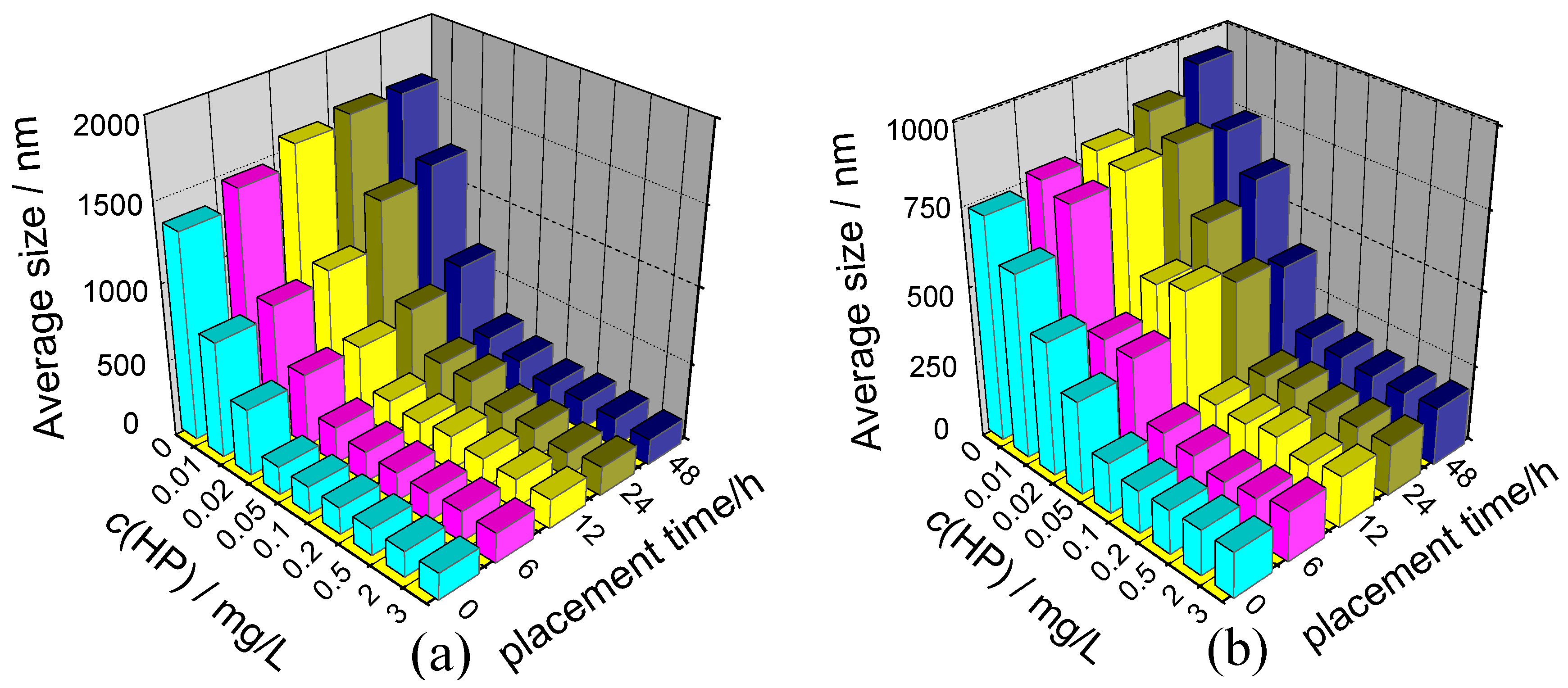
- At c(HP) = 0, wherein the crystals were dispersed in pure water, the mean diameters of nano COM and nano COD were 1343 and 734 nm, respectively, indicating serious aggregation of two nano crystals because the real diameters of nano COM and COD are about 50 nm.
- (2)
- The aggregation of nano COM or nano COD was inhibited after HP was added into the solution, leading to an obvious decrease in their mean diameters. When c(HP) was increased to 0.10 mg/L, the mean diameter of nano COM (Figure 2a) and nano COD (Figure 2b) crystals abruptly decreased to about 200 nm, respectively.
2.2. HP Makes the Zeta Potentials Became Negative on the Surface of Nano COM and COD Crystallites
- (1)
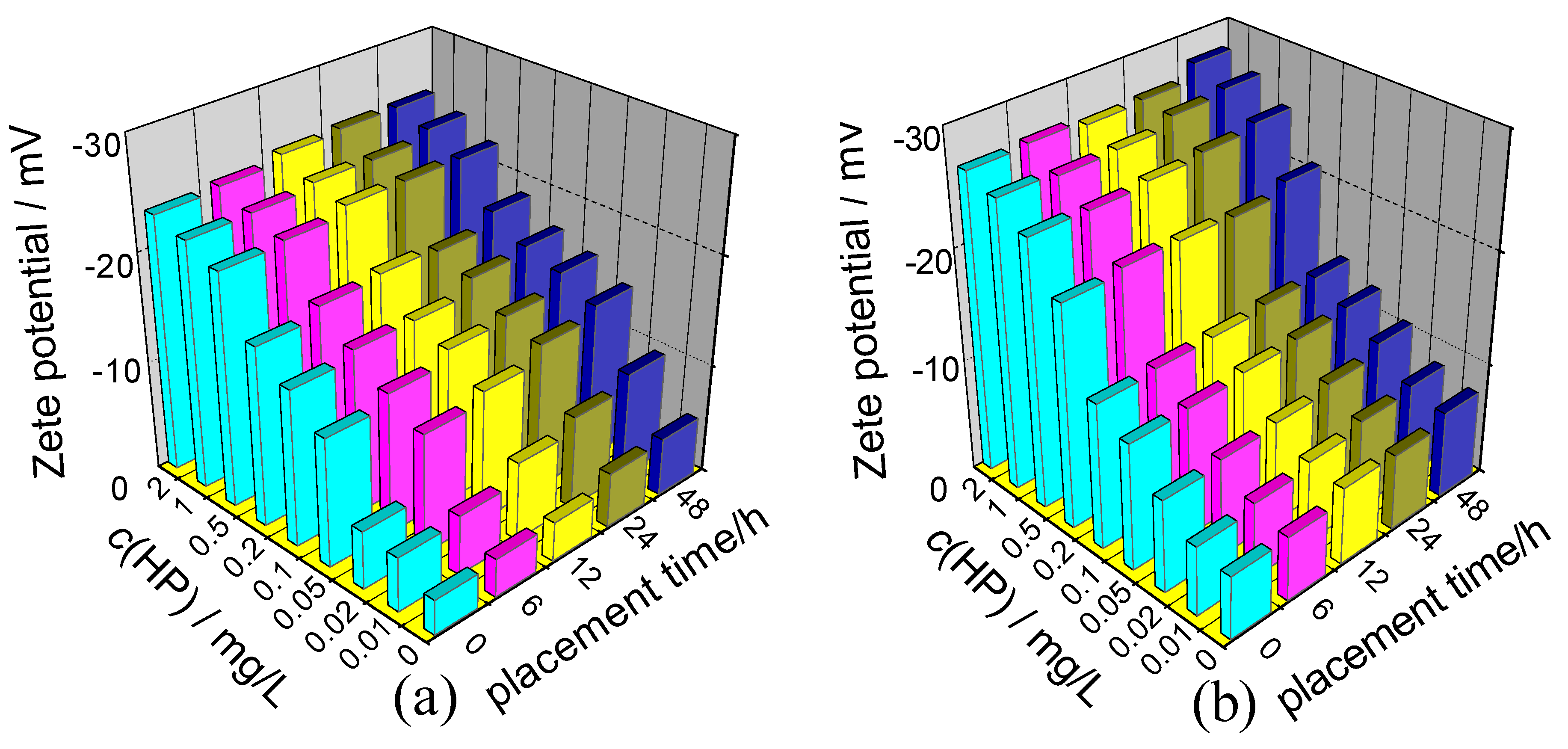
- All the Zeta potential values were negative. It did not matter how much the concentration of HP was (in the region from 0–2 mg/L), whether it was nano COM or nano COD, or whether HP was added or the solution was placed 48 h after HP was added. All the Zeta potential values were negative.
- (2)
- At low concentration (c(HP) = 0–0.10 mg/L), the absolute value of the Zeta potential on COM and COD crystals increased rapidly with the increase of c(HP). However, at higher concentration (c(HP) = 0.20–2.0 mg/L), this increase became slower because the adsorption of HP on the crystallite surface reached saturation at this concentration region.
- (3)
- The effect of placement time on Zeta potential was lesser. When the suspensions of nano COM and nano COD crystals were placed from 0 h to 48 h, the Zeta potential of the crystals became slightly negative because the adsorbed HP molecules on the crystallite surface became tighter and more orderly with the increase of placement time. This phenomenon led to an increase in the amount of the adsorbed HP molecules. The adsorbed amount of HP molecules increased finitely, so Zeta potential barely changed.

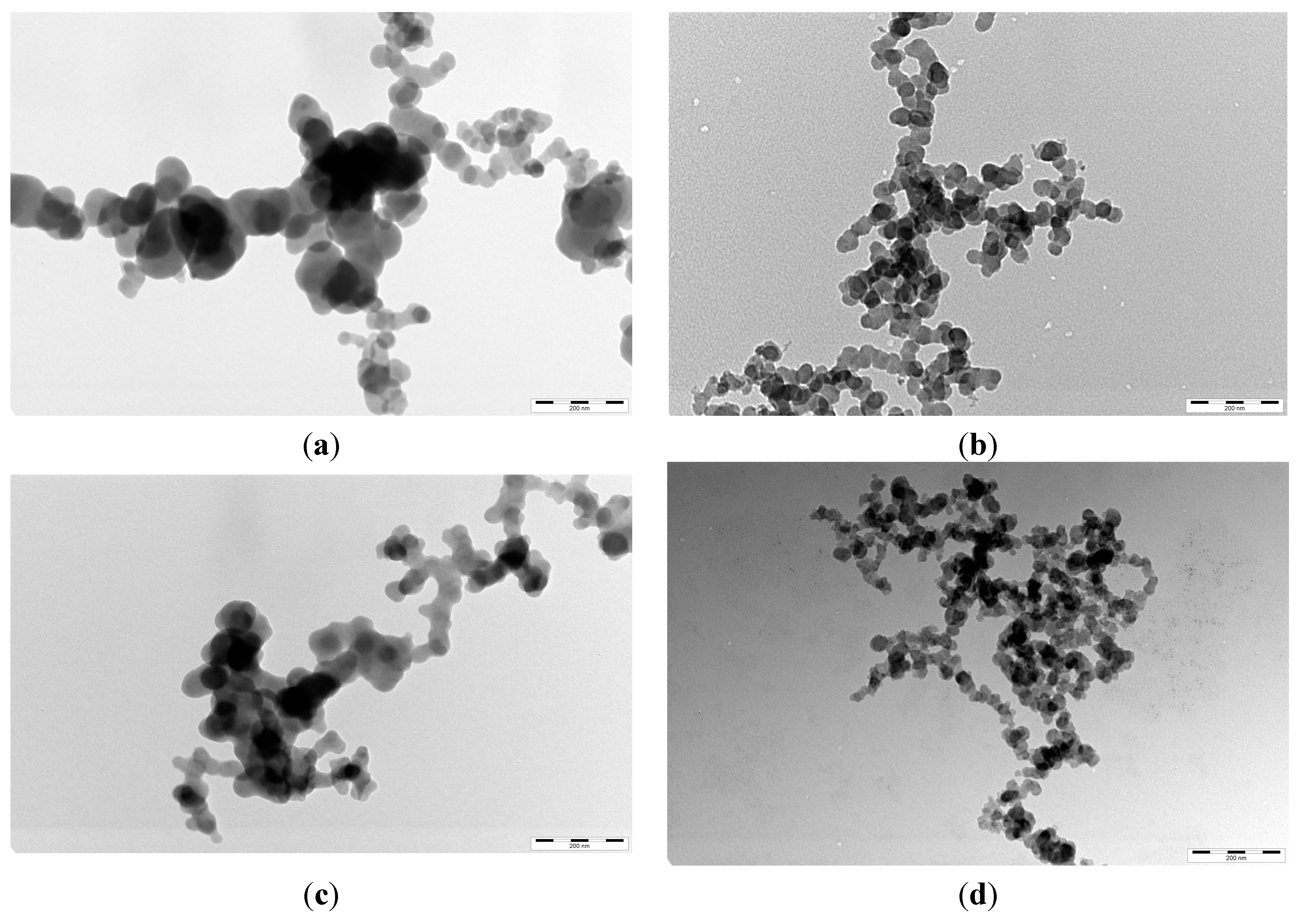
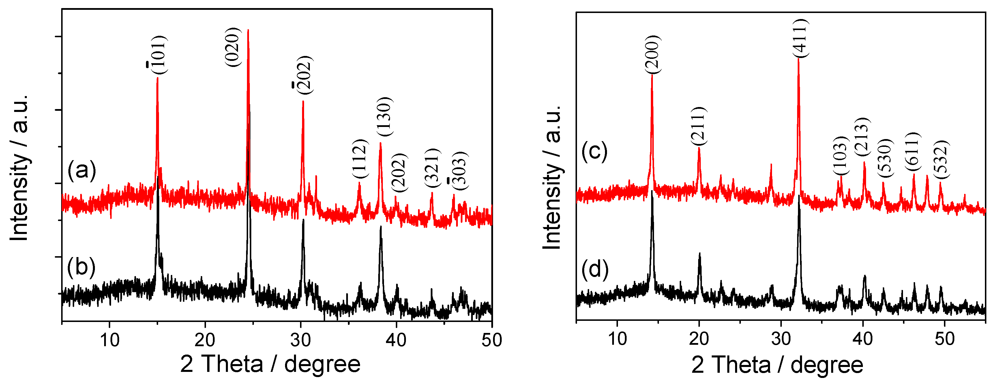
2.3. TEM Images and XRD Patterns of Nano COM and Nano COD Crystals in the Presence of HP


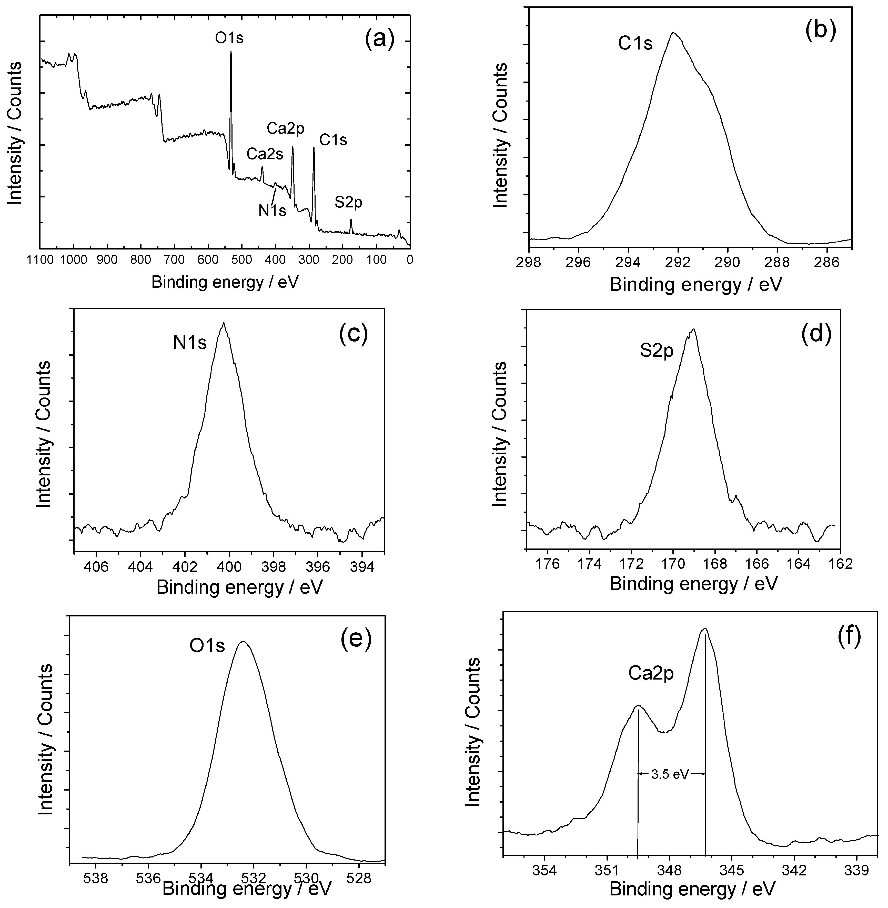
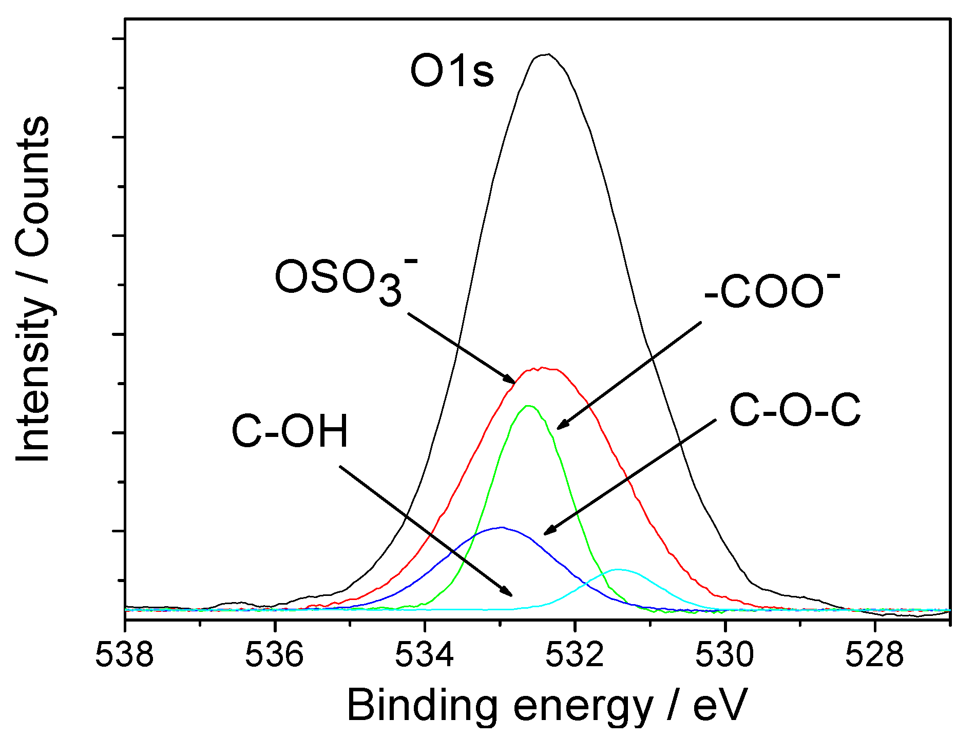
2.4. X-ray Photoelectron Spectroscopy (XPS) Spectra of the Interaction between HP and Crystals


| Sample | C1s | N1s | S2p | O1s | Ca2p1/2 | Ca2p3/2 |
|---|---|---|---|---|---|---|
| COM | 285.0 | 531.7 | 347.2 | 350.7 | ||
| COD | 285.0 | 531.9 | 347.4 | 350.9 | ||
| HP powder | 285.0 | 399.5 | 169.0 | 532.0 | ||
| COM-HP | 285.0 | 400.3 | 169.2 | 532.5 | 346.0 | 349.5 |
| COD-HP | 285.0 | 400.1 | 169.3 | 532.7 | 346.3 | 349.8 |

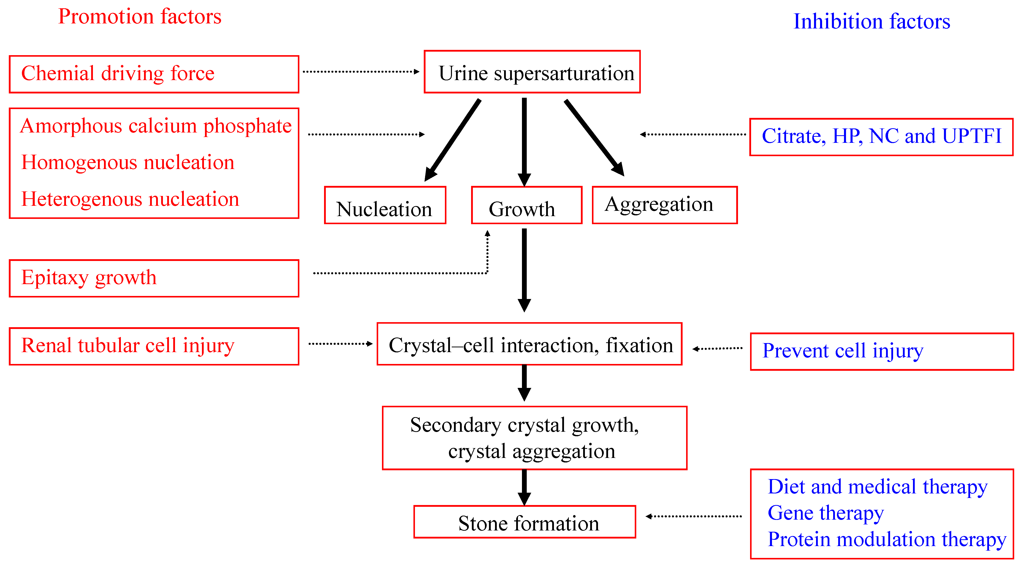
2.5. Mechanism of Calcium Oxalate Stone Formation

3. Experimental Section
3.1. Reagents and Apparatus
3.2. Preparation of Nano COM and COD Crystals
3.3. Effect of HP on Aggregation, Zeta Potential, and Phase Change of Nano COM and Nano COD Crystals
3.4. Measurement of Nano Crystals by High-Resolution Transmission Electron Microscopy (HRTEM)
3.5. X-ray Photoelectron Spectroscopy
4. Conclusions
Acknowledgments
Author Contributions
Conflicts of Interest
References
- Moe, O.W. Kidney stones: Pathophysiology and medical management. Lancet 2006, 367, 333–344. [Google Scholar] [CrossRef] [PubMed]
- Zhang, J.; Wang, G.Z.; Jiang, N.; Yang, J.W.; Gu, Y.; Yang, F. Analysis of urinary calculi composition by infrared spectroscopy: A prospective study of 625 patients in eastern China. Urol. Res. 2010, 38, 111–115. [Google Scholar] [CrossRef] [PubMed]
- Hesse, A.; Schneider, H.J.; Weitz, G.; Cruse, R.; Klee, W.E. Magnesium ammonium phosphate monohydrate-a hitherto undetected constituent of urinary calculi. Int. Urol. Nephrol. 1973, 5, 19–26. [Google Scholar] [CrossRef] [PubMed]
- Lee, T.; Lin, Y.C. Mimicking the initial development of calcium urolithiasis by screening calcium oxalate and calcium phosphate phases in various urinelike solutions, time points, and pH values at 37 °C. Cryst. Growth Des. 2011, 11, 2973–2992. [Google Scholar] [CrossRef]
- Marickar, Y.M.F.; Lekshmi, P.R.; Varma, L.; Koshy, P. EDAX versus FTIR in mixed stones. Urol. Res. 2009, 37, 271–276. [Google Scholar] [CrossRef] [PubMed]
- Gao, J.; Xue, J.-F.; Xu, M.; Gui, B.-S.; Wang, F.-X.; Ouyang, J.-M. Nanouric acid or nanocalcium phosphate as central nidus to induce calcium oxalate stone formation: A high resolution transmission electron microscopy study on urinary nanocrystallites. Int. J. Nanomed. 2014, 9, 4399–4409. [Google Scholar]
- Farmanesh, S.; Ramamoorthy, S.; Chung, J.; Asplin, J.R.; Karande, P.; Rimer, J.D. Specificity of growth inhibitors and their cooperative effects in calcium oxalate monohydrate crystallization. J. Am. Chem. Soc. 2013, 136, 367–376. [Google Scholar] [CrossRef] [PubMed]
- Kirejczyk, J.K.; Porowski, T.; Filonowicz, R.; Kazberuk, A.; Marta, S.; Wasilewska, A.; Debek, W. An association between kidney stone composition and urinary metabolic disturbances in children. J. Pediatr. Urol. 2014, 10, 130–135. [Google Scholar] [CrossRef] [PubMed]
- Hess, B.; Zipperle, L.; Jaeger, P. Citrate and calcium effects on Tamm-Horsfall glycoprotein as a modifier of calcium oxalate crystal aggregation. Am. J. Physiol. 1993, 265, F784–F791. [Google Scholar] [PubMed]
- Viswanathan, P.; Rimer, J.D.; Kolbach, A.M.; Ward, M.D.; Kleinman, J.G.; Wesson, J.A. Calcium oxalate monohydrate aggregation induced by aggregation of desialylated Tamm-Horsfall protein. Urol. Res. 2011, 39, 269–282. [Google Scholar] [CrossRef] [PubMed]
- Iida, S.; Takazono, I.; Suzuki, K.; Shimada, A.; Yahara, J.; Yoshii, S.; Matsuoka, K.; Noda, S. Analysis of matrix glycosaminoglycans (GAGs) in urinary stones by high-performance liquid chromatography. Scanning Microsc. 1999, 13, 173–181. [Google Scholar]
- Astrup, P. On the determination of heparin in blood plasma and urine. Acta Pharmacol. Toxicol. 1947, 3, 165–178. [Google Scholar] [CrossRef]
- Gohel, M.D.I.; Shum, D.K.; Tam, P.C. Electrophoretic separation and characterization of urinary glycosaminoglycans and their roles in urolithiasis. Carbohydr. Res. 2007, 342, 79–86. [Google Scholar] [CrossRef] [PubMed]
- Shirane, Y.; Kurokawa, Y.; Miyashita, S.; Komatsu, H.; Kagawa, S. Study of inhibition mechanisms of glycosaminoglycans on calcium oxalate monohydrate crystals by atomic force microscopy. Urol. Res. 1999, 27, 426–431. [Google Scholar] [CrossRef] [PubMed]
- Poon, N.W.; Gohel, M.D.I. Urinary glycosaminoglycans and glycoproteins in a calcium oxalate crystallization system. Carbohydr. Res. 2012, 347, 64–68. [Google Scholar] [CrossRef] [PubMed]
- Miyake, O.; Yoshimura, K.; Tsujihata, M.; Yoshioka, T.; Koide, T.; Takahara, S.; Okuyama, A. Possible causes for the low prevalence of pediatric urolithiasis. Urology 1999, 53, 1229–1234. [Google Scholar] [CrossRef] [PubMed]
- Ryall, R.L.; Harnett, R.M.; Marshall, V.R. The effect of urine, pyrophosphate, citrate, magnesium and glycosaminoglycans on the growth and aggregation of calcium oxalate crystals in vitro. Clin. Chim. Acta 1981, 112, 349–356. [Google Scholar] [CrossRef] [PubMed]
- Michelacci, Y.M.; Boim, M.A.; Bergamaschi, C.T.; Rovigatti, R.M.; Schor, N. Possible role for chondroitin sulfate in urolithiasis: In vivo studies in an experimental model. Clin. Chim. Acta 1992, 208, 1–8. [Google Scholar] [CrossRef] [PubMed]
- Guerrini, M.; Beccati, D.; Shriver, Z.; Naggi, A.; Viswanathan, K.; Bisio, A.; Capila, I.; Lansing, J.C.; Guglieri, S.; Fraser, B.; et al. Oversulfated chondroitin sulfate is a contaminant in heparin associated with adverse clinical events. Nat. Biotechnol. 2008, 26, 669–675. [Google Scholar] [CrossRef] [PubMed]
- Fischer, K.G. Essentials of anticoagulation in hemodialysis. Hemodial. Int. 2007, 11, 178–189. [Google Scholar] [CrossRef] [PubMed]
- Chevalier, F.; Angulo, J.; Nieto, P.M. The heparin-Ca2+ interaction: Structure of the Ca2+ binding site. Eur. J. Org. Chem. 2002, 14, 2367–2376. [Google Scholar] [CrossRef]
- Kavanagh, J.P. Methods for the study of calcium oxalate crystallisation and their application to urolithiasis research. Scanning Microsc. 1992, 6, 685–705. [Google Scholar] [PubMed]
- Kok, D.J.; Papapoulos, S.E.; Blomen, L.J.M.J.; Bijvoet, O.L.M. Modulation of calcium oxalate monohydrate crystallization kinetics in vitro. Kidney Int. 1988, 34, 346–350. [Google Scholar] [CrossRef] [PubMed]
- Xie, B.; Halter, T.J.; Borah, B.M.; Nancollas, G.H. Aggregation of calcium phosphate and oxalate phases in the formation of renal stones. Cryst. Growth Des. 2015, 15, 204–211. [Google Scholar] [CrossRef] [PubMed]
- Grases, F.; Costa-Bauza, A.; Conte, A. Studies on structure of calcium oxalate monohydrate renal papillary calculi. Mechanism of formation. Scanning Microsc. 1993, 7, 1067–1074. [Google Scholar] [PubMed]
- Khan, S.R.; Hackett, R.L. Role of organic matrix in urinary stone formation: An ultrastructural study of crystal matrix interface of calcium oxalate monohydrate stones. J. Urol. 1993, 150, 239–245. [Google Scholar] [PubMed]
- Shapur, N.K.; Uvarov, V.; Popov, I.; Katz, R.; Gofrit, O.N.; Landau, E.H.; Pode, D.; Duvdevani, M. Crystallite size—Is it a new predictor for renal stone burden? Urology 2012, 80, 980–985. [Google Scholar] [CrossRef] [PubMed]
- Duan, C.-Y.; Xia, Z.-Y.; Zhang, G.-N.; Gui, B.-S.; Xue, J.-F.; Ouyang, J.-M. Changes in urinary nanocrystallites in calcium oxalate stone formers before and after potassium citrate intake. Int. J. Nanomed. 2013, 8, 909–918. [Google Scholar]
- Iida, S.; Ishimatsu, M.; Chikama, S.; Inoue, M.; Matsuoka, K.; Akasu, T.; Noda, S.; Khan, S.R. Protective role of heparin/heparan sulfate on oxalate-induced changes in cell morphology and intracellular Ca2+. Urol. Res. 2003, 31, 198–206. [Google Scholar] [CrossRef] [PubMed]
- Gjaldbaek, J.C. Inhibition of chondroitin sulphate and heparin on the growth and agglomeration of calcium oxalate monohydrate crystals in vitro. Clin. Chim. Acta 1982, 120, 363–365. [Google Scholar] [CrossRef] [PubMed]
- Robertson, W.G.; Scurr, D.S. Modifiers of calcium oxalate crystallization found in urine. I. Studies with a continuous crystallizer using an artificial urine. J. Urol. 1986, 135, 1322–1326. [Google Scholar] [PubMed]
- Rej, R.N.; Holme, K.R.; Perlin, A.S. Observations on cation-binding characteristics and nuclear magnetic resonance spectra of the N-carboxymethyl and N-acetyl analogs of heparin. Can. J. Chem. 1990, 68, 1740–1745. [Google Scholar] [CrossRef]
- Chevalier, F.; Lucas, R.; Angulo, J. The heparin-Ca2+ interaction: The influence of the O-sulfation pattern on binding. Carbohydr. Res. 2004, 339, 975–983. [Google Scholar] [CrossRef] [PubMed]
- Rabenstein, D.L.; Robert, J.M.; Peng, J. Multinuclear magnetic resonance studies of the interaction of inorganic cations with heparin. Carbohydr. Res. 1995, 278, 239–256. [Google Scholar] [CrossRef] [PubMed]
- King, M.; McClure, W.F.; Andrews, L.C. (Eds.) Powder Diffraction File Alphabetic Index, Inorganic Phases/Organic Phases; International Center for Diffraction Data: Newtown Square, PA, USA, 1992.
- Safonov, V.A.; Lubnin, E.N.; Vykhodtseva, L.N.; Chusova, G.E.; Polukarov, Y.M. Surface layers on chromium in sulfuric acid solutions of organic substances: An X-ray photoelectron spectroscopy study. Russ. J. Electrochem. 2001, 37, 1244–1248. [Google Scholar] [CrossRef]
- Ouyang, J.-M.; Zheng, W.-J.; Huang, N.-X.; Tai, Z.-H. X-ray photoelectron spectroscopy study of Langmuir-Blodgett films N-octadecyl-8-hydroxy-2-quinoline carboxamide deposited from subphases containing metal ions. Thin Solid Films 1999, 340, 257–261. [Google Scholar] [CrossRef]
- Ouyang, J.-M.; Ling, W.-H.; Li, C.; Liu, H.-G.; Xue, Q.-B.; Xu, G.Y. Langmuir and Langmuir-Blodgett films of bilirubin. Mol. Cryst. Liquid Cryst. 2001, 363, 195–205. [Google Scholar] [CrossRef]
- Wagner, C.D.; Riggs, W.M.; Davis, J.E.; Moulder, J.F. Handbook of X-ray Photoelectron Spectroscopy; Muilenberg, G.E., Ed.; Physical Electronics Division, Perkin Elmer Corporation: Eden Prairie, MN, USA, 1979. [Google Scholar]
- Valdés, H.; Sánchez-Polo, M.; Rivera-Utrilla, J.; Zaror, C.A. Effect of ozone treatment on surface properties of activated carbon. Langmuir 2002, 18, 2111–2116. [Google Scholar] [CrossRef]
- Gardner, S.D.; Singamsetty, C.S.; Booth, G.L.; He, G.R.; Pittman, C.U. Surface characterization of carbon fibers using angle-resolved XPS and ISS. Carbon 1995, 33, 587–595. [Google Scholar] [CrossRef]
- Tsujihata, M. Mechanism of calcium oxalate renal stone formation and renal tubular cell injury. Int. J. Urol. 2008, 15, 115–120. [Google Scholar] [CrossRef] [PubMed]
- Moro, F.D.; Mancini, M.; Tavolini, I.M.; Marco, V.D.; Bassi, P. Cellular and molecular gateways to urolithiasis: A new insight. Urol. Int. 2005, 74, 193–197. [Google Scholar] [CrossRef] [PubMed]
- Canales, B.K.; Hatch, M. Kidney stone incidence and metabolic urinary changes after modern bariatric surgery: Review of clinical studies, experimental models, and prevention strategies. Surg. Obes. Relat. Dis. 2014, 10, 734–742. [Google Scholar] [CrossRef] [PubMed]
- Ma, M.-C.; Chen, Y.-S.; Huang, H.-S. Erythrocyte oxidative stress in patients with calcium oxalate stones correlates with stone size and renal tubular damage. Basic Transl. Sci. 2014, 510, 9–17. [Google Scholar]
- He, J.-Y.; Deng, S.-P.; Ouyang, J.-M. Morphology, size distribution, aggregation, and crystal phase of nanocrystallites in urines of healthy persons and lithogenic patients. IEEE Trans. Nanobiosci. 2010, 9, 156–163. [Google Scholar] [CrossRef]
- Tasian, G.E.; Lawrence, C. Evaluation and medical management of kidney stones in children. J. Urol. 2014, 192, 1329–1336. [Google Scholar] [CrossRef] [PubMed]
- Thomas, K.; Wong, K.; Withington, J.; Bultitude, M.; Doherty, A. Cystinuria—A urologist’s perspective. Nat. Rev. Urol. 2014, 11, 270–277. [Google Scholar] [CrossRef] [PubMed]
- Wang, B.; Wu, B.; Liu, J.; Yao, W.; Xia, D.; Li, L.; Chen, Z.; Ye, Z.; Yu, X. Analysis of altered microRNA expression profiles in proximal renal tubular cells in response to calcium oxalate monohydrate crystal adhesion: Implications for kidney stone disease. PLoS One 2014, 9, e101306. [Google Scholar] [CrossRef] [PubMed]
- Kulaksizoglu, S.; Sofikerim, M.; Cevik, C. Impact of various modifiers on calcium oxalate crystallization. Int. J. Urol. 2007, 14, 214–218. [Google Scholar] [CrossRef] [PubMed]
- Sample Availability: Samples of COM and COD are available from the authors.
© 2015 by the authors. Licensee MDPI, Basel, Switzerland. This article is an open access article distributed under the terms and conditions of the Creative Commons Attribution license ( http://creativecommons.org/licenses/by/4.0/).
Share and Cite
Ou, Y.; Xue, J.-F.; Tan, C.-Y.; Gui, B.-S.; Sun, X.-Y.; Ouyang, J.-M. Inhibition of Urinary Macromolecule Heparin on Aggregation of Nano-COM and Nano-COD Crystals. Molecules 2015, 20, 1626-1642. https://doi.org/10.3390/molecules20011626
Ou Y, Xue J-F, Tan C-Y, Gui B-S, Sun X-Y, Ouyang J-M. Inhibition of Urinary Macromolecule Heparin on Aggregation of Nano-COM and Nano-COD Crystals. Molecules. 2015; 20(1):1626-1642. https://doi.org/10.3390/molecules20011626
Chicago/Turabian StyleOu, Yan, Jun-Fa Xue, Cai-Yan Tan, Bao-Song Gui, Xin-Yuan Sun, and Jian-Ming Ouyang. 2015. "Inhibition of Urinary Macromolecule Heparin on Aggregation of Nano-COM and Nano-COD Crystals" Molecules 20, no. 1: 1626-1642. https://doi.org/10.3390/molecules20011626
APA StyleOu, Y., Xue, J.-F., Tan, C.-Y., Gui, B.-S., Sun, X.-Y., & Ouyang, J.-M. (2015). Inhibition of Urinary Macromolecule Heparin on Aggregation of Nano-COM and Nano-COD Crystals. Molecules, 20(1), 1626-1642. https://doi.org/10.3390/molecules20011626