
Biliary Atresia Animal Models: Is the Needle in a Haystack?


Abstract
:1. Introduction
2. Results

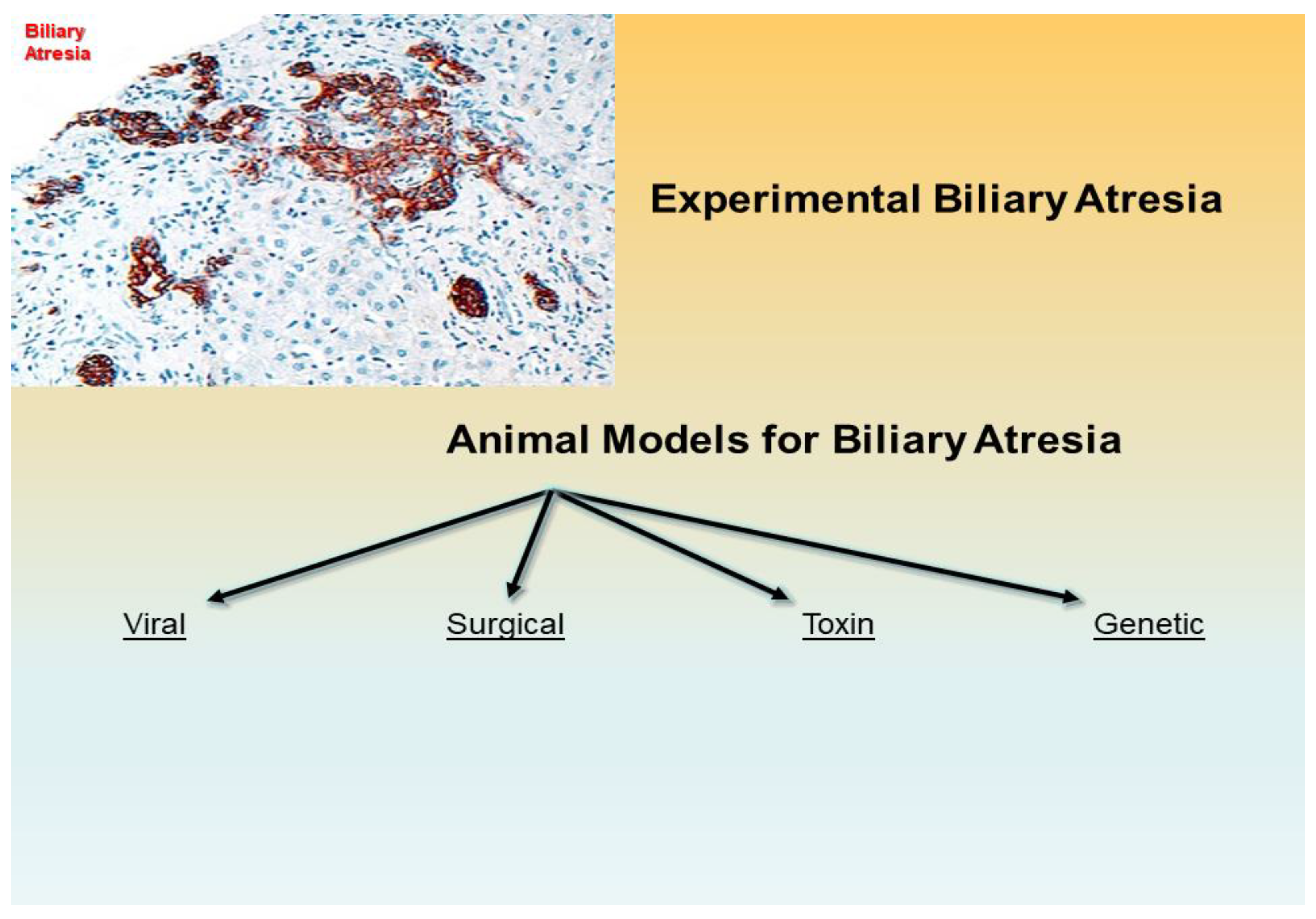
| Viral | Surgical | Toxin, Prenatal 1 | Toxin, Postnatal 2 | Genetic |
|---|---|---|---|---|
| gpCMV (Guinea pig): Infection-based => MNI, PC, Ch, F, BDP (Wang et al., 2011 [94]) | Post natal BDL (Rabbit, lamb, rat, pig, monkey): Surgery-based => MNI, PC, Ch, F, ~ BDP (temp.) (Cameron & Oakley 1932 [79]) (Holder & Ashcraft 1966 [80]) (Morgan et al., 1966) (Spitz 1980 [81]) | Phalloidin (Wistar rat): IP Administration => Canalicular Ch, ↑ Vol. peri-canalicular actin filaments (Hosoda et al., 1997 [101]) 1,4-phenylene-diisothio- cyanate (Wistar rat) Oral Administration => EHBDD, F, BDP (Ogawa et al., 1983 [112]) Monohydroxy bile acids (NZ white rabbit): IV Administration => BDO (in some offspring) (Jenner 1978 [102]) Biliatresone (BALB/c mouse): IP Administration => EHBD-A, MNI, F, BDP (Yang et al., 2020 [105]) | Phorbol myristate acetate (Golden hamsters): GB infusion => Peribiliary PMN, F (Schmeling et al., 1991 [103]) | inv mouse (OVE210 heterozygous invmutant mouse) IHBD-A (periportal), BDP, CBD patent (Shimadera et al., 2007 [106]) |
| RRV/HCR-3/WI-78 (BALB/c mouse): Infection -based => MNI, PC, Ch, ± F, -Atresia (Riepenhoff-Talty et al., 1993 [95]) | Obliterative micro-Sx (Wistar rat): Microsurgery -based => F, BDP (zones 1 & 2) (Aller et al., 2004 [113,114]) | Sox17 haploinsufficiency based mouse (C57BL/6 mouse) => Injury of the epithelial cells of the EHBDS, GB hypoplasia, BD stenosis/atresia (Uemura et al., 2013 [110]; Uemura et al., 2020 [54])) | ||
| Reo Virus 3 (mice): Infection-based => MNI, PC, Ch (Phillips et al., 1969 [115]) | Transplantation (C57BL/6 and B1O.A mice): Graft-based (Fetal/perinatal renal subcapsular allografts in adult congenic mice) => Fibrosclerosis (Schreiber et al., 1992 [89]) | Pkhd1-Nonobese diabetic (NOD) mouse (NOD.Abd3) => MNI, BDP (Huang et al., 2018 [111]) | ||
| Rotavirus Reassortant–Induced Model (RRRV: TR(VP2,VP4)) (Mouse): Infection-based => F, BDP (Mohanty et al., 2020 [98]) | Organ Culture (Embryonal liver culture) Cell culture-based (BD Induction in embryonic liver) (Petersen et al., 2001 [100]) |
3. Discussion
4. Materials and Methods
Author Contributions
Funding
Institutional Review Board Statement
Informed Consent Statement
Data Availability Statement
Acknowledgments
Conflicts of Interest
References
- Sergi, C.; Adam, S.; Kahl, P.; Otto, H.F. The remodeling of the primitive human biliary system. Early Hum. Dev. 2000, 58, 167–178. [Google Scholar] [CrossRef]
- Amella, C.; Cappello, F.; Kahl, P.; Fritsch, H.; Lozanoff, S.; Sergi, C. Spatial and temporal dynamics of innervation during the development of fetal human pancreas. Neuroscience 2008, 154, 1477–1487. [Google Scholar] [CrossRef] [PubMed]
- Sergi, C.M. The Paucity of Interlobular Bile Ducts. In Liver Diseases: A Multidisciplinary Textbook; Radu-Ionita, F., Pyrsopoulos, N.T., Jinga, M., Tintoiu, I.C., Sun, Z., Bontas, E., Eds.; Springer International Publishing: Cham, Switzerland, 2020; pp. 261–272. [Google Scholar] [CrossRef]
- Dorn, L.; Menezes, L.F.; Mikuz, G.; Otto, H.F.; Onuchic, L.F.; Sergi, C. Immunohistochemical detection of polyductin and co-localization with liver progenitor cell markers during normal and abnormal development of the intrahepatic biliary system and in adult hepatobiliary carcinomas. J. Cell. Mol. Med. 2009, 13, 1279–1290. [Google Scholar] [CrossRef] [Green Version]
- Sergi, C.; Kahl, P.; Otto, H.F. Contribution of apoptosis and apoptosis-related proteins to the malformation of the primitive intrahepatic biliary system in Meckel syndrome. Am. J. Pathol. 2000, 156, 1589–1598. [Google Scholar] [CrossRef] [Green Version]
- Sergi, C.; Adam, S.; Kahl, P.; Otto, H.F. Study of the malformation of ductal plate of the liver in Meckel syndrome and review of other syndromes presenting with this anomaly. Pediatr. Dev. Pathol. 2000, 3, 568–583. [Google Scholar] [CrossRef]
- Sergi, C.; Benstz, J.; Feist, D.; Nutzenadel, W.; Otto, H.F.; Hofmann, W.J. Bile duct to portal space ratio and ductal plate remnants in liver disease of infants aged less than 1 year. Pathology 2008, 40, 260–267. [Google Scholar] [CrossRef]
- Russo, P.; Magee, J.C.; Anders, R.A.; Bove, K.E.; Chung, C.; Cummings, O.W.; Finegold, M.J.; Finn, L.S.; Kim, G.E.; Lovell, M.A.; et al. Key Histopathologic Features of Liver Biopsies That Distinguish Biliary Atresia from Other Causes of Infantile Cholestasis and Their Correlation with Outcome: A Multicenter Study. Am. J. Surg. Pathol. 2016, 40, 1601–1615. [Google Scholar] [CrossRef] [Green Version]
- Sergi, C.M.; Gilmour, S. Biliary Atresia: A Complex Hepatobiliary Disease with Variable Gene Involvement, Diagnostic Procedures, and Prognosis. Diagnostics 2022, 12, 330. [Google Scholar] [CrossRef]
- Sergi, C.M. Genetics of Biliary Atresia: A Work in Progress for a Disease with an Unavoidable Sequela into Liver Cirrhosis following Failure of Hepatic Portoenterostomy. In Liver Cirrhosis-Debates and Current Challenges; Tsoulfas, G., Ed.; IntechOpen: London, UK, 2019. [Google Scholar] [CrossRef] [Green Version]
- Bove, K.E.; Thrasher, A.D.; Anders, R.; Chung, C.T.; Cummings, O.W.; Finegold, M.J.; Finn, L.; Ranganathan, S.; Kim, G.E.; Lovell, M.; et al. Inflammation, Active Fibroplasia, and End-stage Fibrosis in 172 Biliary Atresia Remnants Correlate Poorly with Age at Kasai Portoenterostomy, Visceral Heterotaxy, and Outcome. Am. J. Surg. Pathol. 2018, 42, 1625–1635. [Google Scholar] [CrossRef]
- Sergi, C.M.; Chiu, B. Targeting NLRP3 inflammasome in an animal model for Coronavirus Disease 2019 (COVID-19) caused by the Severe Acute Respiratory Syndrome Coronavirus 2 (SARS-CoV-2). J. Med. Virol. 2021, 93, 669–670. [Google Scholar] [CrossRef]
- Boaru, S.G.; Borkham-Kamphorst, E.; Tihaa, L.; Haas, U.; Weiskirchen, R. Expression analysis of inflammasomes in experimental models of inflammatory and fibrotic liver disease. J. Inflamm. 2012, 9, 49. [Google Scholar] [CrossRef] [PubMed] [Green Version]
- Longo, L.; Tonin Ferrari, J.; Rampelotto, P.H.; Hirata Dellavia, G.; Pasqualotto, A.C.P.O.; Thadeu Schmidt Cerski, C.; Reverbel da Silveira, T.; Uribe-Cruz, C.; Alvares-da-Silva, M.R. Gut Dysbiosis and Increased Intestinal Permeability Drive microRNAs, NLRP-3 Inflammasome and Liver Fibrosis in a Nutritional Model of Non-Alcoholic Steatohepatitis in Adult Male Sprague Dawley Rats. Clin. Exp. Gastroenterol. 2020, 13, 351–368. [Google Scholar] [CrossRef] [PubMed]
- Berauer, J.P.; Mezina, A.I.; Okou, D.T.; Sabo, A.; Muzny, D.M.; Gibbs, R.A.; Hegde, M.R.; Chopra, P.; Cutler, D.J.; Perlmutter, D.H.; et al. Identification of PKD1L1 Gene Variants in Children with the Biliary Atresia Splenic Malformation Syndrome. Hepatology 2019, 70, 899–910. [Google Scholar] [CrossRef] [PubMed]
- Nio, M.; Wada, M.; Sasaki, H.; Tanaka, H.; Watanabe, T. Long-term outcomes of biliary atresia with splenic malformation. J. Pediatr. Surg. 2015, 50, 2124–2127. [Google Scholar] [CrossRef]
- Goel, A.; Chaudhari, S.; Sutar, J.; Bhonde, G.; Bhatnagar, S.; Patel, V.; Bhor, V.; Shah, I. Detection of Cytomegalovirus in Liver Tissue by Polymerase Chain Reaction in Infants with Neonatal Cholestasis. Pediatr. Infect. Dis. J. 2018, 37, 632–636. [Google Scholar] [CrossRef]
- Zani, A.; Quaglia, A.; Hadzic, N.; Zuckerman, M.; Davenport, M. Cytomegalovirus-associated biliary atresia: An aetiological and prognostic subgroup. J. Pediatr. Surg. 2015, 50, 1739–1745. [Google Scholar] [CrossRef]
- Brindley, S.M.; Lanham, A.M.; Karrer, F.M.; Tucker, R.M.; Fontenot, A.P.; Mack, C.L. Cytomegalovirus-specific T-cell reactivity in biliary atresia at the time of diagnosis is associated with deficits in regulatory T cells. Hepatology 2012, 55, 1130–1138. [Google Scholar] [CrossRef] [Green Version]
- Domiati-Saad, R.; Dawson, D.B.; Margraf, L.R.; Finegold, M.J.; Weinberg, A.G.; Rogers, B.B. Cytomegalovirus and human herpesvirus 6, but not human papillomavirus, are present in neonatal giant cell hepatitis and extrahepatic biliary atresia. Pediatr. Dev. Pathol. 2000, 3, 367–373. [Google Scholar] [CrossRef]
- Saito, T.; Terui, K.; Mitsunaga, T.; Nakata, M.; Ono, S.; Mise, N.; Yoshida, H. Evidence for viral infection as a causative factor of human biliary atresia. J. Pediatr. Surg. 2015, 50, 1398–1404. [Google Scholar] [CrossRef]
- Saito, T.; Shinozaki, K.; Matsunaga, T.; Ogawa, T.; Etoh, T.; Muramatsu, T.; Kawamura, K.; Yoshida, H.; Ohnuma, N.; Shirasawa, H. Lack of evidence for reovirus infection in tissues from patients with biliary atresia and congenital dilatation of the bile duct. J. Hepatol. 2004, 40, 203–211. [Google Scholar] [CrossRef]
- Glaser, J.H.; Morecki, R. Reovirus type 3 and neonatal cholestasis. Semin. Liver Dis. 1987, 7, 100–107. [Google Scholar] [CrossRef] [PubMed]
- Hertel, P.M.; Estes, M.K. Rotavirus and biliary atresia: Can causation be proven? Curr. Opin. Gastroenterol. 2012, 28, 10–17. [Google Scholar] [CrossRef] [PubMed]
- Mahjoub, F.; Shahsiah, R.; Ardalan, F.A.; Iravanloo, G.; Sani, M.N.; Zarei, A.; Monajemzadeh, M.; Farahmand, F.; Mamishi, S. Detection of Epstein Barr virus by chromogenic in situ hybridization in cases of extra-hepatic biliary atresia. Diagn. Pathol. 2008, 3, 19. [Google Scholar] [CrossRef] [PubMed] [Green Version]
- Fjaer, R.B.; Bruu, A.L.; Nordbo, S.A. Extrahepatic bile duct atresia and viral involvement. Pediatr. Transplant. 2005, 9, 68–73. [Google Scholar] [CrossRef]
- Muraji, T.; Ohtani, H.; Ieiri, S. Unique manifestations of biliary atresia provide new immunological insight into its etiopathogenesis. Pediatr. Surg. Int. 2017, 33, 1249–1253. [Google Scholar] [CrossRef]
- Chen, G.; Xue, P.; Zheng, S.; Chen, L.; Ma, Y. A pathological scoring system in the diagnosis and judgment of prognosis of biliary atresia. J. Pediatr. Surg. 2015, 50, 2119–2123. [Google Scholar] [CrossRef]
- Safwan, M.; Ramachandran, P.; Vij, M.; Shanmugam, N.; Rela, M. Impact of ductal plate malformation on survival with native liver in children with biliary atresia. Pediatr. Surg. Int. 2015, 31, 837–843. [Google Scholar] [CrossRef]
- Czubkowski, P.; Cielecka-Kuszyk, J.; Rurarz, M.; Kaminska, D.; Markiewicz-Kijewska, M.; Pawlowska, J. The limited prognostic value of liver histology in children with biliary atresia. Ann. Hepatol. 2015, 14, 902–909. [Google Scholar] [CrossRef]
- Mukhopadhyay, S.G.; Roy, P.; Chatterjee, U.; Datta, C.; Banerjee, M.; Banerjee, S.; Basu, A.K.; Ganguli, M. A histopathological study of liver and biliary remnants in the long-term survivors (>10 years) of cases of biliary atresia. Indian J. Pathol. Microbiol. 2014, 57, 380–385. [Google Scholar] [CrossRef]
- Vukovic, J.; Grizelj, R.; Bojanic, K.; Coric, M.; Luetic, T.; Batinica, S.; Kujundzic-Tiljak, M.; Schroeder, D.R.; Sprung, J. Ductal plate malformation in patients with biliary atresia. Eur. J. Pediatr. 2012, 171, 1799–1804. [Google Scholar] [CrossRef]
- Yamaguti, D.C.; Patricio, F.R. Morphometrical and immunohistochemical study of intrahepatic bile ducts in biliary atresia. Eur. J. Gastroenterol. Hepatol. 2011, 23, 759–765. [Google Scholar] [CrossRef] [PubMed]
- Roy, P.; Chatterjee, U.; Ganguli, M.; Banerjee, S.; Chatterjee, S.K.; Basu, A.K. A histopathological study of liver and biliary remnants with clinical outcome in cases of extrahepatic biliary atresia. Indian J. Pathol. Microbiol. 2010, 53, 101–105. [Google Scholar] [CrossRef] [PubMed]
- Nakanuma, Y.; Harada, K.; Sato, Y.; Ikeda, H. Recent progress in the etiopathogenesis of pediatric biliary disease, particularly Caroli’s disease with congenital hepatic fibrosis and biliary atresia. Histol. Histopathol. 2010, 25, 223–235. [Google Scholar] [CrossRef]
- Campos-Sanchez, J.C.; Esteban, M.A. Review of inflammation in fish and value of the zebrafish model. J. Fish Dis. 2021, 44, 123–139. [Google Scholar] [CrossRef] [PubMed]
- Kent, M.L.; Sanders, J.L.; Spagnoli, S.; Al-Samarrie, C.E.; Murray, K.N. Review of diseases and health management in zebrafish Danio rerio (Hamilton 1822) in research facilities. J. Fish Dis. 2020, 43, 637–650. [Google Scholar] [CrossRef]
- Stevens, C.H.; Reed, B.T.; Hawkins, P. Enrichment for Laboratory Zebrafish-A Review of the Evidence and the Challenges. Animals 2021, 11, 698. [Google Scholar] [CrossRef]
- Lorent, K.; Yeo, S.Y.; Oda, T.; Chandrasekharappa, S.; Chitnis, A.; Matthews, R.P.; Pack, M. Inhibition of Jagged-mediated Notch signaling disrupts zebrafish biliary development and generates multi-organ defects compatible with an Alagille syndrome phenocopy. Development 2004, 131, 5753–5766. [Google Scholar] [CrossRef] [Green Version]
- Cui, S.; Eauclaire, S.F.; Matthews, R.P. Interferon-gamma directly mediates developmental biliary defects. Zebrafish 2013, 10, 177–183. [Google Scholar] [CrossRef]
- Cui, S.; Erlichman, J.; Russo, P.; Haber, B.A.; Matthews, R.P. Intrahepatic biliary anomalies in a patient with Mowat-Wilson syndrome uncover a role for the zinc finger homeobox gene zfhx1b in vertebrate biliary development. J. Pediatr. Gastroenterol. Nutr. 2011, 52, 339–344. [Google Scholar] [CrossRef]
- Cui, S.; Capecci, L.M.; Matthews, R.P. Disruption of planar cell polarity activity leads to developmental biliary defects. Dev. Biol. 2011, 351, 229–241. [Google Scholar] [CrossRef] [Green Version]
- Tian, L.; Ye, Z.; Kafka, K.; Stewart, D.; Anders, R.; Schwarz, K.B.; Jang, Y.Y. Biliary Atresia Relevant Human Induced Pluripotent Stem Cells Recapitulate Key Disease Features in a Dish. J. Pediatr. Gastroenterol. Nutr. 2019, 68, 56–63. [Google Scholar] [CrossRef]
- Tang, V.; Cofer, Z.C.; Cui, S.; Sapp, V.; Loomes, K.M.; Matthews, R.P. Loss of a Candidate Biliary Atresia Susceptibility Gene, add3a, Causes Biliary Developmental Defects in Zebrafish. J. Pediatr. Gastroenterol. Nutr. 2016, 63, 524–530. [Google Scholar] [CrossRef]
- Ke, J.; Zeng, S.; Mao, J.; Wang, J.; Lou, J.; Li, J.; Chen, X.; Liu, C.; Huang, L.M.; Wang, B.; et al. Common genetic variants of GPC1 gene reduce risk of biliary atresia in a Chinese population. J. Pediatr. Surg. 2016, 51, 1661–1664. [Google Scholar] [CrossRef]
- Ningappa, M.; Min, J.; Higgs, B.W.; Ashokkumar, C.; Ranganathan, S.; Sindhi, R. Genome-wide association studies in biliary atresia. Wiley Interdiscip. Rev. Syst. Biol. Med. 2015, 7, 267–273. [Google Scholar] [CrossRef]
- Smith, K. Biliary tract: GPC1 genetic risk further links Hedgehog signalling with pathogenesis of biliary atresia. Nat. Rev. Gastroenterol. Hepatol. 2013, 10, 127. [Google Scholar] [CrossRef]
- Cui, S.; Leyva-Vega, M.; Tsai, E.A.; EauClaire, S.F.; Glessner, J.T.; Hakonarson, H.; Devoto, M.; Haber, B.A.; Spinner, N.B.; Matthews, R.P. Evidence from human and zebrafish that GPC1 is a biliary atresia susceptibility gene. Gastroenterology 2013, 144, 1107–1115.e1103. [Google Scholar] [CrossRef] [Green Version]
- Filmus, J.; Capurro, M.; Rast, J. Glypicans. Genome Biol. 2008, 9, 224. [Google Scholar] [CrossRef]
- Cheung, Y.; Wu, Z.; Garcia-Barcelo, M.M.; Tam, P.K.H.; Ma, A.C.H.; Lui, V.C.H. Deletion of interleukin enhancer binding factor 2 (ILF2) resulted in defective biliary development and bile flow blockage. J. Pediatr. Surg. 2021, 56, 352–359. [Google Scholar] [CrossRef]
- Soini, T.; Pihlajoki, M.; Andersson, N.; Lohi, J.; Huppert, K.A.; Rudnick, D.A.; Huppert, S.S.; Wilson, D.B.; Pakarinen, M.P.; Heikinheimo, M. Transcription factor GATA6: A novel marker and putative inducer of ductal metaplasia in biliary atresia. Am. J. Physiol. Gastrointest. Liver Physiol. 2018, 314, G547–G558. [Google Scholar] [CrossRef]
- Bai, M.R.; Niu, W.B.; Zhou, Y.; Gong, Y.M.; Lu, Y.J.; Yu, X.X.; Wei, Z.L.; Wu, W.; Song, H.L.; Yu, W.W.; et al. Association of common variation in ADD3 and GPC1 with biliary atresia susceptibility. Aging 2020, 12, 7163–7182. [Google Scholar] [CrossRef]
- Ningappa, M.; So, J.; Glessner, J.; Ashokkumar, C.; Ranganathan, S.; Min, J.; Higgs, B.W.; Sun, Q.; Haberman, K.; Schmitt, L.; et al. The Role of ARF6 in Biliary Atresia. PLoS ONE 2015, 10, e0138381. [Google Scholar] [CrossRef] [Green Version]
- Uemura, M.; Higashi, M.; Pattarapanawan, M.; Takami, S.; Ichikawa, N.; Higashiyama, H.; Furukawa, T.; Fujishiro, J.; Fukumura, Y.; Yao, T.; et al. Gallbladder wall abnormality in biliary atresia of mouse Sox17 (+/-) neonates and human infants. Dis. Models Mech. 2020, 13, dmm042119. [Google Scholar] [CrossRef] [Green Version]
- Pattarapanawan, M.; Uemura, M.; Miyazaki, N.; Takami, S.; Tomiyasu, H.; Tsunekawa, N.; Hirate, Y.; Fujishiro, J.; Kurohmaru, M.; Kanai-Azuma, M.; et al. Anatomical and histological characteristics of the hepatobiliary system in adult Sox17 heterozygote mice. Anat. Rec. 2020, 303, 3096–3107. [Google Scholar] [CrossRef]
- Higashiyama, H.; Ozawa, A.; Sumitomo, H.; Uemura, M.; Fujino, K.; Igarashi, H.; Imaimatsu, K.; Tsunekawa, N.; Hirate, Y.; Kurohmaru, M.; et al. Embryonic cholecystitis and defective gallbladder contraction in the Sox17-haploinsufficient mouse model of biliary atresia. Development 2017, 144, 1906–1917. [Google Scholar] [CrossRef] [Green Version]
- Yang, Y.; Jin, Z.; Dong, R.; Zheng, C.; Huang, Y.; Zheng, Y.; Shen, Z.; Chen, G.; Luo, X.; Zheng, S. MicroRNA-29b/142-5p contribute to the pathogenesis of biliary atresia by regulating the IFN-gamma gene. Cell Death Dis. 2018, 9, 545. [Google Scholar] [CrossRef]
- Dong, R.; Zhao, R.; Zheng, S. Changes in epigenetic regulation of CD4+ T lymphocytesin biliary atresia. Pediatr. Res. 2011, 70, 555–559. [Google Scholar] [CrossRef] [Green Version]
- Zhu, Y.G.; Li, J.; Pang, Y.; Li, Q.W. Lamprey: An important animal model of evolution and disease research. Yi Chuan 2020, 42, 847–857. [Google Scholar] [CrossRef]
- York, J.R.; McCauley, D.W. Functional genetic analysis in a jawless vertebrate, the sea lamprey: Insights into the developmental evolution of early vertebrates. J. Exp. Biol. 2020, 223, jeb206433. [Google Scholar] [CrossRef] [Green Version]
- Xu, Y.; Zhu, S.W.; Li, Q.W. Lamprey: A model for vertebrate evolutionary research. Zool. Res. 2016, 37, 263–269. [Google Scholar] [CrossRef]
- Kuraku, S. Palaeophylogenomics of the vertebrate ancestor--impact of hidden paralogy on hagfish and lamprey gene phylogeny. Integr. Comp. Biol. 2010, 50, 124–129. [Google Scholar] [CrossRef] [Green Version]
- Osorio, J.; Retaux, S. The lamprey in evolutionary studies. Dev. Genes Evol. 2008, 218, 221–235. [Google Scholar] [CrossRef]
- Davenport, M. Biliary atresia: From Australia to the zebrafish. J. Pediatr. Surg. 2016, 51, 200–205. [Google Scholar] [CrossRef]
- Chung-Davidson, Y.W.; Yeh, C.Y.; Li, W. The Sea Lamprey as an Etiological Model for Biliary Atresia. Biomed Res. Int. 2015, 2015, 832943. [Google Scholar] [CrossRef]
- Youson, J.H. Biliary atresia in lampreys. Adv. Vet. Sci. Comp. Med. 1993, 37, 197–255. [Google Scholar]
- Raveenthiran, V. Long-Term Survival of Biliary Atresia without any Surgery: Lessons Learnt from Lamprey. J. Neonatal Surg. 2014, 3, 19. [Google Scholar] [CrossRef]
- Chung-Davidson, Y.W.; Davidson, P.J.; Scott, A.M.; Walaszczyk, E.J.; Brant, C.O.; Buchinger, T.; Johnson, N.S.; Li, W. A new clarification method to visualize biliary degeneration during liver metamorphosis in Sea Lamprey (Petromyzon marinus). J. Vis. Exp. 2014, 88, 51648. [Google Scholar] [CrossRef] [Green Version]
- Suchy, F.J. Biliary atresia in sea lampreys. What can it tell us about the disorder in human infants? Hepatology 2013, 57, 2114–2116. [Google Scholar] [CrossRef]
- Cai, S.Y.; Lionarons, D.A.; Hagey, L.; Soroka, C.J.; Mennone, A.; Boyer, J.L. Adult sea lamprey tolerates biliary atresia by altering bile salt composition and renal excretion. Hepatology 2013, 57, 2418–2426. [Google Scholar] [CrossRef] [Green Version]
- Yeh, C.Y.; Chung-Davidson, Y.W.; Wang, H.; Li, K.; Li, W. Intestinal synthesis and secretion of bile salts as an adaptation to developmental biliary atresia in the sea lamprey. Proc. Natl. Acad. Sci. USA 2012, 109, 11419–11424. [Google Scholar] [CrossRef] [Green Version]
- Boomer, L.A.; Bellister, S.A.; Stephenson, L.L.; Hillyard, S.D.; Khoury, J.D.; Youson, J.H.; Gosche, J.R. Cholangiocyte apoptosis is an early event during induced metamorphosis in the sea lamprey, Petromyzon marinus L. J. Pediatr. Surg. 2010, 45, 114–120. [Google Scholar] [CrossRef]
- Savina, M.V.; Emel’yanova, L.V.; Korotkov, S.M.; Brailovskaya, I.V.; Nadeev, A.D. Bioenergetics of mitochondria of the liver with biliary atresia during prolonged starvation. Dokl. Biochem. Biophys. 2009, 425, 80–83. [Google Scholar] [CrossRef]
- Youson, J.H.; Sidon, E.W. Lamprey biliary atresia: First model system for the human condition? Experientia 1978, 34, 1084–1086. [Google Scholar] [CrossRef]
- Chung-Davidson, Y.W.; Yeh, C.Y.; Bussy, U.; Li, K.; Davidson, P.J.; Nanlohy, K.G.; Brown, C.T.; Whyard, S.; Li, W. Hsp90 and hepatobiliary transformation during sea lamprey metamorphosis. BMC Dev. Biol. 2015, 15, 47. [Google Scholar] [CrossRef] [Green Version]
- Zhao, R.; Dong, R.; Yang, Y.; Wang, Y.; Ma, J.; Wang, J.; Li, H.; Zheng, S. MicroRNA-155 modulates bile duct inflammation by targeting the suppressor of cytokine signaling 1 in biliary atresia. Pediatr. Res. 2017, 82, 1007–1016. [Google Scholar] [CrossRef]
- Cornelius, C.E.; Rosenberg, D.P. Neonatal biliary atresia. Am. J. Pathol. 1985, 118, 168–171. [Google Scholar]
- Szavay, P.O.; Leonhardt, J.; Czech-Schmidt, G.; Petersen, C. The role of reovirus type 3 infection in an established murine model for biliary atresia. Eur. J. Pediatr. Surg. 2002, 12, 248–250. [Google Scholar] [CrossRef]
- Cameron, G.R.; Oakley, C.L. Ligation of the common bile duct. J. Pathol. Bacteriol. 1932, 35, 769–798. [Google Scholar] [CrossRef]
- Holder, T.M.; Ashcraft, K.W. Production of experimental biliary atresia by ligation of the common bile duct in the fetus. Surg. Forum 1966, 17, 356–357. [Google Scholar]
- Spitz, L. Ligation of the common bile duct in the fetal lamb: An experimental model for the study of biliary atresia. Pediatr. Res. 1980, 14, 740–748. [Google Scholar] [CrossRef] [Green Version]
- Morgan, W.W., Jr.; Rosenkrantz, J.C.; Hill, R.B., Jr. Hepatic arterial interruption in the fetus–an attempt to simulate biliary atresia. J. Pediatr. Surg. 1966, 1, 342–346. [Google Scholar] [CrossRef]
- Medeiros, M.V.; Freitas, L.A.; Andrade, Z.A. Differences in hepatic pathology resulting from bile duct obstruction in young and old rats. Braz. J. Med. Biol. Res. 1988, 21, 75–83. [Google Scholar]
- Omori, M.; Evarts, R.P.; Omori, N.; Hu, Z.; Marsden, E.R.; Thorgeirsson, S.S. Expression of alpha-fetoprotein and stem cell factor/c-kit system in bile duct ligated young rats. Hepatology 1997, 25, 1115–1122. [Google Scholar] [CrossRef]
- Gibelli, N.E.; Tannuri, U.; de Mello, E.S.; Rodrigues, C.J. Bile duct ligation in neonatal rats: Is it a valid experimental model for biliary atresia studies? Pediatr. Transplant. 2009, 13, 81–87. [Google Scholar] [CrossRef]
- Devadas, M.; Templeton, A.; Leonard, A.S. Restitution of bile duct after ligation and excision in Rhesus monkeys. J. Pediatr. Surg. 1975, 10, 511–513. [Google Scholar] [CrossRef]
- Spitz, L. Experimental production of cystic dilatation of the common bile duct in neonatal lambs. J. Pediatr. Surg. 1977, 12, 39–42. [Google Scholar] [CrossRef]
- Pickett, L.K.; Briggs, H.C. Biliary obstruction secondary to hepatic vascular ligation in fetal sheep. J. Pediatr. Surg. 1969, 4, 95–101. [Google Scholar] [CrossRef]
- Schreiber, R.A.; Kleinman, R.E.; Barksdale, E.M., Jr.; Maganaro, T.F.; Donahoe, P.K. Rejection of murine congenic bile ducts: A model for immune-mediated bile duct disease. Gastroenterology 1992, 102, 924–930. [Google Scholar] [CrossRef]
- Parashar, K.; Tarlow, M.J.; McCrae, M.A. Experimental reovirus type 3-induced murine biliary tract disease. J. Pediatr. Surg. 1992, 27, 843–847. [Google Scholar] [CrossRef]
- Morecki, R.; Glaser, J. Reovirus 3 and neonatal biliary disease: Discussion of divergent results. Hepatology 1989, 10, 515–517. [Google Scholar] [CrossRef]
- Bangaru, B.; Morecki, R.; Glaser, J.H.; Gartner, L.M.; Horwitz, M.S. Comparative studies of biliary atresia in the human newborn and reovirus-induced cholangitis in weanling mice. Lab. Investig. 1980, 43, 456–462. [Google Scholar]
- Fischler, B.; Svensson, J.F.; Nemeth, A. Early cytomegalovirus infection and the long-term outcome of biliary atresia. Acta Paediatr. 2009, 98, 1600–1602. [Google Scholar] [CrossRef]
- Wang, W.; Zheng, S.; Shong, Z.; Zhao, R. Development of a guinea pig model of perinatal cytomegalovirus-induced hepatobiliary injury. Fetal Pediatr. Pathol. 2011, 30, 301–311. [Google Scholar] [CrossRef]
- Riepenhoff-Talty, M.; Schaekel, K.; Clark, H.F.; Mueller, W.; Uhnoo, I.; Rossi, T.; Fisher, J.; Ogra, P.L. Group A rotaviruses produce extrahepatic biliary obstruction in orally inoculated newborn mice. Pediatr. Res. 1993, 33, 394–399. [Google Scholar] [CrossRef] [Green Version]
- Turowski, C.; Leonhardt, J.; Teichmann, B.; Heim, A.; Baumann, U.; Kuebler, J.F.; Petersen, C. Preconceptional oral vaccination prevents experimental biliary atresia in newborn mice. Eur. J. Pediatr. Surg. 2010, 20, 158–163. [Google Scholar] [CrossRef]
- Coots, A.; Donnelly, B.; Mohanty, S.K.; McNeal, M.; Sestak, K.; Tiao, G. Rotavirus infection of human cholangiocytes parallels the murine model of biliary atresia. J. Surg. Res. 2012, 177, 275–281. [Google Scholar] [CrossRef] [Green Version]
- Mohanty, S.K.; Lobeck, I.; Donnelly, B.; Dupree, P.; Walther, A.; Mowery, S.; Coots, A.; Bondoc, A.; Sheridan, R.M.; Poling, H.M.; et al. Rotavirus Reassortant-Induced Murine Model of Liver Fibrosis Parallels Human Biliary Atresia. Hepatology 2020, 71, 1316–1330. [Google Scholar] [CrossRef]
- Shiojiri, N.; Koike, T. Differentiation of biliary epithelial cells from the mouse hepatic endodermal cells cultured in vitro. Tohoku J. Exp. Med. 1997, 181, 1–8. [Google Scholar] [CrossRef] [Green Version]
- Petersen, M.; Drews, U.; Schweizer, P. Induction of bile ducts in embryonic liver by mesenchyme: A new perspective for the treatment of biliary atresia? Eur. J. Pediatr. Surg. 2001, 11, 382–390. [Google Scholar] [CrossRef]
- Hosoda, Y.; Miyano, T.; Fujimoto, T. Assay of gamma-glutamyl transpeptidase activity in amniotic fluid offers a possible prenatal diagnosis of biliary atresia in the rat model. Prenat. Diagn. 1997, 17, 9–12. [Google Scholar] [CrossRef]
- Jenner, R.E. New perspectives on biliary atresia. Ann. R. Coll. Surg. Engl. 1978, 60, 367–374. [Google Scholar]
- Schmeling, D.J.; Oldham, K.T.; Guice, K.S.; Kunkel, R.G.; Johnson, K.J. Experimental obliterative cholangitis. A model for the study of biliary atresia. Ann. Surg. 1991, 213, 350–355. [Google Scholar] [CrossRef]
- Lorent, K.; Gong, W.; Koo, K.A.; Waisbourd-Zinman, O.; Karjoo, S.; Zhao, X.; Sealy, I.; Kettleborough, R.N.; Stemple, D.L.; Windsor, P.A.; et al. Identification of a plant isoflavonoid that causes biliary atresia. Sci. Transl. Med. 2015, 7, 286ra267. [Google Scholar] [CrossRef] [Green Version]
- Yang, Y.; Wang, J.; Zhan, Y.; Chen, G.; Shen, Z.; Zheng, S.; Dong, R. The synthetic toxin biliatresone causes biliary atresia in mice. Lab. Investig. 2020, 100, 1425–1435. [Google Scholar] [CrossRef]
- Shimadera, S.; Iwai, N.; Deguchi, E.; Kimura, O.; Fumino, S.; Yokoyama, T. The inv mouse as an experimental model of biliary atresia. J. Pediatr. Surg. 2007, 42, 1555–1560. [Google Scholar] [CrossRef]
- Nguyen, A.P.; Pham, Y.H.T.; Vu, G.H.; Nguyen, M.H.; Hoang, T.N.; Holterman, A. Biliary atresia liver histopathological determinants of early post-Kasai outcome. J. Pediatr. Surg. 2021, 56, 1169–1173. [Google Scholar] [CrossRef]
- Pacheco, M.C.; Campbell, K.M.; Bove, K.E. Ductal plate malformation-like arrays in early explants after a Kasai procedure are independent of splenic malformation complex (heterotaxy). Pediatr. Dev. Pathol. 2009, 12, 355–360. [Google Scholar] [CrossRef]
- Shimadera, S.; Iwai, N.; Deguchi, E.; Kimura, O.; Ono, S.; Fumino, S.; Higuchi, K. Significance of ductal plate malformation in the postoperative clinical course of biliary atresia. J. Pediatr. Surg. 2008, 43, 304–307. [Google Scholar] [CrossRef]
- Uemura, M.; Ozawa, A.; Nagata, T.; Kurasawa, K.; Tsunekawa, N.; Nobuhisa, I.; Taga, T.; Hara, K.; Kudo, A.; Kawakami, H.; et al. Sox17 haploinsufficiency results in perinatal biliary atresia and hepatitis in C57BL/6 background mice. Development 2013, 140, 639–648. [Google Scholar] [CrossRef] [Green Version]
- Huang, W.; Rainbow, D.B.; Wu, Y.; Adams, D.; Shivakumar, P.; Kottyan, L.; Karns, R.; Aronow, B.; Bezerra, J.; Gershwin, M.E.; et al. A Novel Pkhd1 Mutation Interacts with the Nonobese Diabetic Genetic Background To Cause Autoimmune Cholangitis. J. Immunol. 2018, 200, 147–162. [Google Scholar] [CrossRef] [Green Version]
- Ogawa, T.; Suruga, K.; Kojima, Y.; Kitahara, T.; Kuwabara, N. Experimental study of the pathogenesis of infantile obstructive cholangiopathy and its clinical evaluation. J Pediatr Surg. 1983, 18, 131–135. [Google Scholar] [CrossRef]
- Aller, M.A.; Nava, M.P.; Arias, J.L.; Durán, M.; Prieto, I.; Llamas, M.A.; Arias, J. Microsurgical extrahepatic cholestasis in the rat: A long-term study. J Invest Surg. 2004, 17, 99–104. [Google Scholar] [CrossRef]
- Aller, M.A.; Duran, M.; Ortega, L.; Arias, J.L.; Nava, M.P.; Prieto, I.; Arias, J. Comparative study of macro- and microsurgical extrahepatic cholestasis in the rat. Microsurgery 2004, 24, 442–447. [Google Scholar] [CrossRef]
- Phillips, P.A.; Keast, D.; Papadimitriou, J.M.; Walters, M.N.; Stanley, N.F. Chronic obstructive jaundice induced by Reovirus type 3 in weanling mice. Pathology 1969, 1, 193–203. [Google Scholar] [CrossRef]

Publisher’s Note: MDPI stays neutral with regard to jurisdictional claims in published maps and institutional affiliations. |
© 2022 by the authors. Licensee MDPI, Basel, Switzerland. This article is an open access article distributed under the terms and conditions of the Creative Commons Attribution (CC BY) license (https://creativecommons.org/licenses/by/4.0/).
Share and Cite
Pal, N.; Joy, P.S.; Sergi, C.M. Biliary Atresia Animal Models: Is the Needle in a Haystack? Int. J. Mol. Sci. 2022, 23, 7838. https://doi.org/10.3390/ijms23147838
Pal N, Joy PS, Sergi CM. Biliary Atresia Animal Models: Is the Needle in a Haystack? International Journal of Molecular Sciences. 2022; 23(14):7838. https://doi.org/10.3390/ijms23147838
Chicago/Turabian StylePal, Nutan, Parijat S. Joy, and Consolato M. Sergi. 2022. "Biliary Atresia Animal Models: Is the Needle in a Haystack?" International Journal of Molecular Sciences 23, no. 14: 7838. https://doi.org/10.3390/ijms23147838
APA StylePal, N., Joy, P. S., & Sergi, C. M. (2022). Biliary Atresia Animal Models: Is the Needle in a Haystack? International Journal of Molecular Sciences, 23(14), 7838. https://doi.org/10.3390/ijms23147838