Article

Tetrahydrofuran Cembranoids from the Cultured Soft Coral Lobophytum crassum

1
National Museum of Marine Biology & Aquarium, Pingtung 944, Taiwan
2
Graduate Institute of Marine Biotechnology, National Dong Hwa University, Pingtung 944, Taiwan
*
Author to whom correspondence should be addressed.
Mar. Drugs 2011, 9(12), 2526-2536; https://doi.org/10.3390/md9122526
Submission received: 9 November 2011 / Revised: 25 November 2011 / Accepted: 28 November 2011 / Published: 7 December 2011

Abstract

:
Three new cembranoids, culobophylins A–C (13), along with two known compounds (4 and 5) were isolated from the cultured soft coral Lobophytum crassum. The structures of these compounds were elucidated on the basis of their spectroscopic data and comparison of the NMR data with those of known analogues. Among these metabolites, 2 is rarely found in cembranoids possessing an isopropyl moiety with an epoxide group. Compound 1 exhibited significant cytotoxic activity against HL60 and DLD-1 cancer cell lines.

Graphical Abstract

1. Introduction

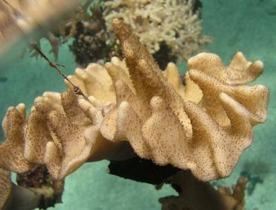
In the investigation of secondary metabolites from marine invertebrates, several terpenoid metabolites have been isolated from cultured octocorals Erythropodium [1], Klyxum simplex [2,3,4], Sinularia flexibilis [5], Sarcophyton trocheliophorum [6], Briareum excavatum [7,8,9,10,11,12,13,14,15] and Briareum sp. [16]. Some of these metabolites have been found to possess several kinds of biological activities, such as cytotoxic [2,4,5,8,16] and anti-inflammatory activities [3,4,11,12,13,14]. The current chemical investigation of cultured octocoral Lobophytum crassum (Figure 1) led to the discovery of three new cembranoids, culobophylins A–C (13), and two known compounds lobophylin B (4), and lobophylin A (5) [17]. The structures of 15 were established by detailed spectroscopic analysis, including extensive examination of 2D NMR (1H–1H COSY, HMQC and HMBC) correlations. The cytotoxicity of metabolites 15 against human promyelocytic leukemia (HL60), human breast carcinoma (MDA-MB-231) and human colon adenocarcinoma (HCT-116 and DLD-1) cell lines was studied, and the ability of 15 to inhibit the expression of the pro-inflammatory iNOS (inducible nitric oxide synthase) and COX-2 (cyclooxygenase-2) proteins in lipopolysaccharide (LPS)-stimulated RAW264.7 macrophage cells was also evaluated.
Figure 1. Soft coral Lobophytum crassum.
Marinedrugs 09 02526 g001

2. Results and Discussion

The EtOAc extract of the freeze-dried specimen was fractionated by silica gel column chromatography and the eluted fractions were further separated utilizing normal phase HPLC to yield metabolites 15 (Chart 1).
Chart 1. Structures of metabolites 15.
Marinedrugs 09 02526 g002
The new metabolite culobophylin A (1) had a molecular formula of C20H30O3, which was determined by HRESIMS and NMR spectroscopic data. The IR spectrum of 1 showed absorption bands at 3458 and 1694 cm−1, suggesting the presence of hydroxy and carbonyl groups. The 13C NMR data of 1 showed the presence of 20 carbons (Table 1): three methyls, six sp3 methylenes, one sp2 methylene, three sp3 methines (including two oxygenated carbons at δ 76.6 and 75.6), two sp2 methines, and one sp3 quaternary carbon. The remaining three signals appearing in the lower field region of the spectrum are due to the quaternary carbons of three olefinic carbons (δ 148.2, 132.9 and 131.8) and one aldehyde carbonyl (δ 194.7). From the 1H NMR (Table 1) spectrum of 1, the presence of one aldehyde proton resonating as a singlet at δH 9.56 was observed. Moreover, the 1H NMR data revealed the presence of two olefinic methylene protons (δ 6.33, J = 1.5 Hz; 6.14, d, J = 1.5 Hz) and two olefinic methine protons (δ 5.18, dd, J = 5.0, 5.0 Hz; 4.84, d, J = 7.5 Hz). Furthermore, two oxygenated methines (δ 4.66, ddd, J = 11.0, 6.0, 5.0 Hz; 3.96, dd, J =9.5, 4.0 Hz) and three methyls (δ 1.61, s; 1.56, s; 1.10, s) were also designated from the 1H NMR signals. The planar structure and all of the 1H and 13C chemical shifts of 1 were elucidated by 2D NMR spectroscopic analysis, in particular 1H–1H COSY and HMBC experiments (Figure 2). From the 1H–1H COSY correlations (Figure 2), it was possible to establish three partial structures of consecutive proton spin systems extending from H2-5 to H-7; H-9 to H-11; H2-13 to H-3. The following key HMBC correlations permitted connection of the carbon skeleton: H2-5 to C-3 and C-4; H2-13 to C-11 and C-12; H2-16 to C-1, C-15 and C-17; H-17 to C-1 and C-15; H3-18 to C-3, C-4 and C-5; H3-19 to C-7, C-8 and C-9; and H3-20 to C-11, C-12 and C-13. Thus, 1 was found to possess three double bonds at C-7/C-8, C-11/C-12 and C-15/C-16 and an aldehyde group at C-15. Furthermore, the HMBC cross-peak from H-14 to C-3 suggested that C-3 and C-14 were linked through an oxygen to form a tetrahydrofuran ring. The relative configuration of 1 elucidated mainly from the NOESY spectrum was compatible with that of 1 offered by using the MM2 force field calculations which suggested the most stable conformations as shown in Figure 3. In the NOESY spectrum, both H3-18 and H-14 showed NOEs with H-1 but not with H-3. Thus, assuming the β-orientation of H-1, H3-18 and H-14 should be positioned on the β face. Moreover, H-3 should be positioned on the α face. Also, the NOE correlations of H3-19 with H2-6 but not with H-7 and H3-20, with H-10a (δ 2.33) but not with H-11, indicated the E configuration of the double bonds between C-7/C-8 and C-11/C-12. Furthermore, the relative stereochemistry of 1 was mostly confirmed to be the same as that of 4 by comparison of the proton chemical shifts and coupling constants [17]. On the basis of the above findings and other detailed NOE correlations, the structure of 1 was established unambiguously.
Table 1. 1H and 13C NMR data for 13.
Position123
δH (J in Hz) aδc (mult.) bδH (J in Hz) aδc (mult.) bδH (J in Hz) aδc (mult.) b
13.12 dt (10.0, 8.5) c 41.1 (CH) d2.53 m46.1 (CH)2.75 dt (6.0, 5.5) 49.8 (CH)
22.12 m; 2.04 m27.3 (CH2)1.70 m24.7 (CH2)2.08 m31.3 (CH2)
33.96 dd (9.5, 4.0)76.6 (CH) 3.91 dd (9.5, 4.0) 77.3 (CH) 4.13 dd (7.5, 7.5) 82.5 (CH)
4 74.2 (C) 74.2 (C) 74.4 (C)
52.00 m; 1.54 m 38.7 (CH2) 1.94 m; 1.52 m 38.6 (CH2) 2.18 dd (13.5, 3.5); 1.55 m 46.2 (CH2)
62.22 m; 2.06 m 21.4 (CH2) 2.17 m; 2.04 m 21.4 (CH2) 3.46 ddd (8.5, 2.5, 2.0) 54.5 (CH)
75.18 dd (5.0, 5.0) 126.4 (CH) 5.15 dd (5.5, 5.5) 126.2 (CH) 3.10 d (2.0) 61.6 (CH)
8 132.9 (C) 133.1 (C) 146.5 (C)
92.14 m; 1.98 m 38.1 (CH2) 2.15 m; 2.02 m 38.1 (CH2) 2.14 m; 1.82 m 27.9 (CH2)
102.33 m; 2.04 m 24.4 (CH2) 2.35 m; 2.04 m 24.4 (CH2) 2.27 m 28.6 (CH2)
114.84 d (7.5) 127.2 (CH) 4.92 d (8.5) 127.5 (CH) 5.16 dd (7.5, 7.5) 125.0 (CH)
12 131.8 (C) 131.5 (C) 132.5 (C)
131.71 m; 1.51 m 40.1 (CH2) 2.28 m; 2.08 m 39.9 (CH2) 1.92 d (6.5) 39.4 (CH2)
144.66 ddd (11.0, 6.0, 5.0) 75.6 (CH) 4.42 ddd (11.0, 5.5, 5.5) 76.8 (CH) 4.05 dd (7.0, 7.0) 79.0 (CH)
15 148.2 (C) 54.2 (C) 144.4 (C)
166.33 d (1.5); 6.14 d (1.5) 134.9 (CH2) 2.51 d (4.5); 2.43 d (5.0) 50.8 (CH2) 4.83 s; 4.73 s 112.3 (CH2)
179.56 s 194.7 (CH) 1.37 s 22.1 (CH3) 1.77 s 22.1 (CH3)
181.10 s 23.1 (CH3) 1.07 s 22.9 (CH3) 1.15 s 21.9 (CH3)
191.56 s 16.4 (CH3) 1.58 s 16.4 (CH3) 5.28 s; 5.12 s 115.0 (CH2)
201.61 s 15.3 (CH3) 1.67 s 15.3 (CH3) 1.62 s 17.1 (CH3)
a 500 MHz in CDCl3; b 125 MHz in CDCl3; c J values (Hz) are given in parentheses; d Numbers of attached protons were deduced by DEPT experiments.
Figure 2. Selected 1H−1H COSY (▬) and HMBC (→) correlations of 13.
Marinedrugs 09 02526 g003
Figure 3. Computer-generated model of 1 using MM2 force field calculations and key NOE correlations.
Marinedrugs 09 02526 g004
Culobophylin B (2) was isolated as a colorless oil with the molecular formula C20H32O3, which possesses five degrees of unsaturation, as indicated by HRESIMS (m/z 343.2251, [M + Na]+) and NMR spectroscopic data (Table 1). In addition, 1H and 13C NMR spectroscopic data (Table 1) of 2 showed the structural unit of a 3,14-oxa-bridged tetrahydrofuran. By comparison of the NMR data of 2 with that of 4, it was found that the 1H and 13C NMR data of 2 were very similar to those of 4 [17]. However, the 1H and 13C NMR spectroscopic data revealed that the signals corresponding to one 1,1-disubstituted carbon–carbon double bond in 4 were not present and were replaced by one 1,1-disubstituted epoxide in 2H 2.51, 1H, d, J = 4.5 Hz and δH 2.43, 1H, d, J = 5.0 Hz; δC 54.2, C and δC 50.8 CH2) (Table 1). 1H–1H COSY and HMBC (Figure 2) further revealed that 2 possesses one 1,1-disubstituted epoxide at C-15. On the basis of the above observations, and with the assistance of additional 2D NMR (1H–1H COSY and HMBC) correlations, it was possible to establish the planar structure of 2, as illustrated in Figure 2. The relative stereochemistries of all stereocenters except C-15 of 2 were confirmed to be the same as those of 1 and 4 by comparison of the proton shifts, coupling constants, and NOE correlations of 2 with those of 1 and 4.
Culobophylin C (3) was obtained as a colorless oil and showed a [M + Na]+ ion peak in the HRESIMS spectrum corresponding to the molecular formula C20H30O3, the same as that of 2. IR absorptions were observed at 3425 cm−1, suggesting the presence of a hydroxy group in 3. The 13C NMR spectrum of 3 showed twenty signals accounting for three methyls, five sp3 methylenes, two sp2 methylenes, five sp3 methines , one sp2 methine and four quaternary carbons (including one oxygenated carbon at δ 74.4 and three olefinic carbons with resonances at δ 146.5, 144.4 and 132.5). The 1H NMR data revealed the presence of four olefinic methylene protons (δ 5.28, 5.12, 4.83 and 4.73, each a singlet). Two proton signals at δ 3.46 ddd (1H, 8.5, 2.5, 2.0) and 3.10 (1H, d, J = 2.0 Hz) correlated with two carbon signals at δ 54.5 and 61.6 and in the HMQC spectrum of 3 were attributed to the proton of one 1,2-disubstituted epoxide. The planar structure and all of the 1H and 13C chemical shifts of 3 were elucidated by 2D NMR spectroscopic analysis, in particular 1H–1H COSY and HMBC experiments (Figure 2). Thus, 3 was found to possess three double bonds at C-8/C-19, C-11/C-12 and C-15/C-16, one hydroxy group at C-4, one 1,2-disubstituted epoxide at C-6/C-7, and an oxa-bridged ether linkage at C-3/C-14. The relative configurations of the five chiral centers at C-3, C-4, C-6, C-7 and C-14 in 3 were elucidated by detailed NOE analysis, as shown in Figure 4. In these experiments, it was found that H3-18 showed NOE interactions with H-14 and H-7. Thus, assuming the β-orientation of H3-18, H-7 and H-14 should be positioned on the β face. The NOE correlation observed between H-14 and H-1 also reflected the β-orientation of H-1. Furthermore, the NOESY spectrum showed NOE interaction of H3-20 with H-10, but not with H-11, revealing the E geometry of the C-11/C-12 double bond. On the basis of these results and other detailed NOE correlations, the structure of 3 was established unambiguously.
Figure 4. Computer-generated model of 3 using MM2 force field calculations and key NOE correlations.
Marinedrugs 09 02526 g005
The cytotoxicities of compounds 15 against HL60, MDA-MB-231, DLD-1 and HCT-116 cancer cells are shown in Table 2. The results show that compound 1, the most potent of compounds 15, exhibited cytotoxicity against the HL60, MDA-MB-231, DLD-1 and HCT-116 cancer cell lines with IC50s of 3.0, 16.8, 4.6 and 16.3 μg/mL, respectively. Furthermore, compound 2 exhibited moderate to weak cytotoxic activity against HL60, DLD-1 and HCT-116 cancer cell lines (the IC50 values were 6.8, 16.2 and 16.7 μg/mL for HL60, DLD-1 and HCT-116, respectively). The other tested compounds were not cytotoxic (IC50 > 20 μg/mL) toward the above four cancer cell lines. The in vitro anti-inflammatory effects of 15 were also tested. Furthermore, the anti-inflammatory activity of 15 against the accumulation of pro-inflammatory iNOS and COX-2 proteins in RAW264.7 macrophage cells stimulated with LPS was evaluated using immunoblot analysis. At a concentration of 10 µM, compounds 15 did not inhibit COX-2 and iNOS proteins expression relative to the control cells stimulated with LPS only (Figure 5).
Table 2. Cytotoxicity (IC50 μg/mL) of compounds 15.
CompoundCell LinesHCT-116
HL60MDA-MB-231DLD-1
1316.84.616.3
26.8a16.216.7
3aaaa
4aaaa
5aaaa
Doxorubicin C0.056.35.70.5
a IC50 > 20 μg/mL.
Figure 5. Effect of compounds 15 at 10 μM on the expression of iNOS and COX-2 proteins of RAW264.7 macrophage cells examined by immunoblot analysis. (A) Immunoblots of iNOS and β-actin; (B) immunoblots of COX-2 and β-actin. Values represent mean ± SEM (n = 6). The relative intensity of the LPS-only-stimulated group was taken as 100%. * Significantly different from the LPS-only-stimulated group (* P < 0.05). a Stimulated with LPS; b stimulated with LPS in the presence of 15.
Marinedrugs 09 02526 g006

3. Experimental Section

3.1. General Experimental Procedures

Optical rotation values were measured using a Jasco P-1010 digital polarimeter. IR spectra were recorded on a Varian Digilab FTS 1000 Fourier transform infrared spectrophotometer. The NMR spectra were recorded on a Varian Unity INOVA 500 FT-NMR instrument at 500 MHz for 1H NMR and 125 MHz for 13C NMR, respectively, in CDCl3. ESIMS and HRESIMS data were recorded with a Bruker APEX II mass spectrometer. Gravity column chromatography was performed on silica gel (230–400 mesh, Merck). TLC was carried out on precoated Kieselgel 60 F254 (0.2 mm, Merck) and spots were visualized by spraying with 10% H2SO4 solution followed by heating. High-performance liquid chromatography was performed using a system comprised of a Hitachi L-7100 pump and a Rheodyne 7725 injection port. A preparative normal phase column (250 × 21.2 mm, 5 μm) was used for HPLC.

3.2. Animal Material

Specimens of the soft coral Lobophytum crassum were collected off the coast of Pingtung, southern Taiwan, and transplanted to a 120-ton cultivating tank equipped with a flow-through sea water system in July 2003. The cultured soft coral was harvested in December 2010. A voucher specimen (specimen no. 2010CSC-1) was deposited in the National Museum of Marine Biology and Aquarium, Taiwan.

3.3. Extraction and Separation

The frozen bodies of soft coral (5.0 kg, fresh wt.) were collected and freeze-dried. The freeze-dried material was minced and extracted exhaustively with EtOAc (5 × 2 L). The organic extract was evaporated to yield a residue (60.5 g), which was fractionated by open column chromatography on silica gel using n-hexane–EtOAc and EtOAc–acetone mixtures of increasing polarity to yield 15 fractions. Fraction 6, eluting with n-hexane-EtOAc (15:1), was further separated by silica gel column chromatography with gradient elution (n-hexane-EtOAc, 15:1 to 10:1) to yield five subfractions (6A–6E). Subfraction 6C was subjected to normal phase HPLC (n-hexane-EtOAc, 15:1) to obtain compound 4 (3.0 mg). Fraction 8, eluting with n-hexane-EtOAc (5:1), was further separated by silica gel column chromatography with gradient elution (n-hexane-EtOAc, 5:1 to 2:1) to give six subfractions (8A–8F). Subfraction 8B was separated by normal phase HPLC using n-hexane-EtOAc (5:1) to afford 5 (2.5 mg). In the same manner, compound 3 (2.0 mg) was obtained from subfraction 8 D normal phase HPLC (n-hexane-EtOAc, 3:1). Fraction 9, eluting with n-hexane-EtOAc (3:1), was further separated by silica gel column chromatography with gradient elution (n-hexane-EtOAc, 3:1 to 1:1) to yield five subfractions (9A–9E). Subfraction 9C was further purified by was subjected to normal phase HPLC (n-hexane-EtOAc, 2:1) to obtain compounds 1(1.2 mg) and 2 (3.0 mg).
Culobophylin A (1): colorless oil; Marinedrugs 09 02526 i001 = −50 (c 0.1, CHCl3); IR (neat) νmax 3458, 2924, 2853, 1694, 1458 and 1377 cm1; 1H and 13C NMR data, see Table 1; ESIMS m/z 341 [100, (M + Na)+]; HRESIMS m/z 341.2091 (calcd. for C20H30O3Na, 341.2093).
Culobophylin B (2): colorless oil; Marinedrugs 09 02526 i001 = −24 (c 0.3, CHCl3); IR (neat) νmax 3499, 2925, 2853, 1457, 1382 and 1264 cm1; 1H and 13C NMR data, see Table 1; ESIMS m/z 343 [100, (M + Na)+]; HRESIMS m/z 343.2251 (calcd. for C20H32O3Na, 341.2249).
Culobophylin C (3): colorless oil; Marinedrugs 09 02526 i001 = −83 (c 0.3, CHCl3); IR (neat) νmax 3425, 2923, 1638, and1459 cm1, 1H and 13C NMR data, see Table 1; ESIMS m/z 341 [100, (M + Na)+]; HRESIMS m/z 341.2095 (calcd. for C20H30O3Na, 341.2093).
Lobophylin B (4): colorless oil; Marinedrugs 09 02526 i001 = −30 (c 0.5, CHCl3); Marinedrugs 09 02526 i001 [lit. = −35 (c 0.3, CHCl3) [17]].
Lobophylin A (5): colorless oil; Marinedrugs 09 02526 i001 = −45 (c 0.3, CHCl3); [lit. Marinedrugs 09 02526 i001 = −39 (c 0.3, CHCl3) [17]].

3.4. Cytotoxicity Testing

Cell lines were purchased from the American Type Culture Collection (ATCC). Cytotoxicity assays of compounds 15 were performed using the MTT [3-(4,5-dimethylthiazol-2-yl)-2,5-diphenyltetrazolium bromide] colorimetric method [18,19].

3.5. In Vitro Anti-Inflammatory Assay

Macrophage (RAW264.7) cell line was purchased from ATCC. In vitro anti-inflammatory activities of compounds 1-5 were measured by examining the inhibition of lipopolysaccharide (LPS) induced upregulation of iNOS (inducible nitric oxide synthetase) and COX-2 (cyclooxygenase-2) proteins in macrophages cells using western blotting analysis [20,21].

3.6. Molecular Mechanics Calculations

Implementation of the MM2 force filed in Chem3D Pro software [22], was used to calculate the molecular models.

4. Conclusions

In previous reports, several 3,14-ether linkage-related cembranoids were identified from the marine soft corals Sinularia gibberosa [23,24], Sarcophyton infundibuliforme [25] and Lobophytum sp. [17]. Among these compounds, only one (3,14-epoxy-1(E),7(E),11(E)-cembratrien-4,15-diol) has been found to possess moderate cytotoxicity toward three cancer cells (A-549, HT-29 and P-388) [24]. In the present study, only compound 1 exhibited significant cytotoxicity against the growth of HL60 and DLD-1 cancer cell lines. According to the structures of 15, it seems that the aldehyde group in compound 1 is critical for the cytotoxic activity of metabolites 15. It is worth noting that metabolite 2 is rarely found in cembranoids possessing an isopropyl moiety with an epoxide group [26].

Acknowledgements

This research was supported by grants from the National Museum of Marine Biology & Aquarium and the National Science Council (NSC 100-2325-B-291-001), Taiwan, awarded to J.-H. Su.
  • Samples Availability: Not available.

Supplementary Files

  • Supplementary File 1::

    PDF-Document (PDF, 1263 KB)

  • References

    1. Taglialatela-Scafati, O.; Deo-Jangra, U.; Campbell, M.; Roberge, M.; Andersen, R.J. Diterpenoids from cultured Erythropodium caribaeorum. Org. Lett. 2002, 4, 4085–4088. [Google Scholar]
    2. Chen, B.-W.; Wu, Y.-C.; Chiang, M.Y.; Su, J.-H.; Wang, W.-H.; Fang, T.-Y.; Sheu, J.-H. Eunicellin-based diterpenoids from the cultured soft coral Klyxum simplex. Tetrahedron 2009, 65, 7016–7022. [Google Scholar]
    3. Chen, B.-W.; Chao, C.-H.; Su, J.-H.; Wen, Z.-H.; Sung, P.-J.; Sheu, J.-H. Anti-inflammatory eunicellin-based diterpenoids from the cultured soft coral Klyxum simplex. Org. Biomol. Chem. 2010, 8, 2363–2366. [Google Scholar]
    4. Chen, B.-W.; Chao, C.-H.; Su, J.-H.; Tsai, C.-W.; Wang, W.-H.; Wen, Z.-H.; Hsieh, C.-H.; Sung, P.-J.; Wu, Y.-C.; Sheu, J.-H. Klysimplexins I–T, eunicellin-based diterpenoids from the cultured soft coral Klyxum simplex. Org. Biomol. Chem. 2011, 9, 834–844. [Google Scholar]
    5. Su, J.-H.; Lin, Y.-F.; Lu, Y.; Huang, C.-Y.; Wang, W.-H.; Fang, T.-Y.; Sheu, J.-H. Oxygenated cembranoids from the cultured and wild-type soft corals Sinularia flexibilis. Chem. Pharm. Bull. 2009, 57, 1189–1192. [Google Scholar]
    6. Su, J.-H.; Lu, Y.; Lin, W.-Y.; Wang, W.-H.; Sung, P.-J.; Sheu, J.-H. A cembranoid, trocheliophorol, from the cultured soft coral Sarcophyton trocheliophorum. Chem. Lett. 2010, 39, 172–173. [Google Scholar]
    7. Sung, P.-J.; Lin, M.-R.; Su, Y.-D.; Chiang, M.Y.; Hu, W.-P.; Su, J.-H.; Cheng, M.-C.; Hwang, T.-L.; Sheu, J.-H. New briaranes from the octocoral Briareum excavatum (Briareidae) and Junceella fragilis (Ellisellidae). Tetrahedron 2008, 64, 2596–2604. [Google Scholar]
    8. Hwang, T.-L.; Lin, M.-R.; Tsai, W.-T.; Yeh, H.-C.; Hu, W.-P.; Sheu, J.-H.; Sung, P.-J. New polyoxygenated briaranes from octocorals Briareum excavatum and Ellisella robusta. Bull. Chem. Soc. Jpn. 2008, 81, 1638–1646. [Google Scholar]
    9. Sung, P.-J.; Lin, M.-R.; Hwang, T.-L.; Fan, T.-Y.; Su, W.-C.; Ho, C.-C.; Fang, L.-S.; Wang, W.-H. Briaexcavatins M–P, four new briarane-related diterpenoids from cultured octocoral Briareum excavatum (Briareidae). Chem. Pharm. Bull. 2008, 56, 930–935. [Google Scholar]
    10. Sung, P.-J.; Lin, M.-R.; Chiang, M.Y. The structure and absolute stereochemistry of briaexcavatin U, a new chlorinated briarane from a cultured octocoral Briareum excavatum. Chem. Lett. 2009, 38, 154–155. [Google Scholar]
    11. Sung, P.-J.; Lin, M.-R.; Chiang, M.Y.; Hwang, T.-L. Briaexcavatins V–Z, discovery of new briaranes from a cultured octocoral Briareum excavatum. Bull. Chem. Soc. Jpn. 2009, 82, 987–996. [Google Scholar]
    12. Sung, P.-J.; Chen, B.-Y.; Lin, M.-R.; Hwang, T.-L.; Wang, W.-H.; Sheu, J.-H.; Wu, Y.-C. Excavatoids E and F, discovery of two new briaranes from the cultured octocoral Briareum excavatum. Mar. Drugs 2009, 7, 472–482. [Google Scholar]
    13. Su, J.-H.; Chen, B.-Y.; Hwang, T.-L.; Chen, Y.-H.; Huang, I.-C.; Lin, M.-R.; Chen, J.-J.; Fang, L.-S.; Wang, W.-H.; Li, J.-J.; Sheu, J.-H.; Sung, P.-J. Excavatoids L–N, new 12-hydroxy- briaranes from the cultured octocoral Briareum excavatum (Briareidae). Chem. Pharm. Bull. 2010, 58, 662–665. [Google Scholar]
    14. Sung, P.-J.; Chen, B.-Y.; Chiang, M.Y.; Hou, C.-H.; Su, Y.-D.; Hwang, T.-L.; Chen, Y.-H.; Chen, J.-J. Excavatoids G–K, new 8,17-epoxybriarane from the cultured octocoral Briareum excavatum (Briareidae). Bull. Chem. Soc. Jpn. 2010, 83, 539–545. [Google Scholar]
    15. Sung, P.-J.; Li, G.-Y.; Su, Y.-D.; Lin, M.-R.; Chang, Y.-C.; Kung, T.-H.; Lin, C.-S.; Chen, Y.-H.; Su, J.-H.; Lu, M.-C.; Kuo, J.; Weng, C.-F.; Hwang, T.-L. Excavatoids O and P, new 12-hydroxdybriaranes from the octocoral Briareum excavatum. Mar. Drugs 2010, 8, 2639–2646. [Google Scholar]
    16. Sung, P.-J.; Lin, M.-R.; Chiang, M.Y.; Syu, S.-M.; Fang, L.-S.; Wang, W.-H.; Sheu, J.-H. Briarenolide D, a new hydroperoxybriarane diterpenoid from a cultured octocoral Briareum sp. Chem. Lett. 2010, 39, 1030–1032. [Google Scholar]
    17. Hegazy, M.E.F.; Su, J.-H.; Sung, P.-J.; Sheu, J.-H. Cembranoids with 3,14-ether linkage and a secocembrane with bistetrahydrofuran from the Dongsha Atoll soft coral Lobophytum sp. Mar. Drugs 2011, 9, 1243–1253. [Google Scholar]
    18. Alley, M.C.; Scudiero, D.A.; Monks, A.; Hursey, M.L.; Czerwinski, M.J.; Fine, D.L.; Abbott, B.J.; Mayo, J.G.; Shoemaker, R.H.; Boyd, M.R. Feasibility of drug screening with panels of human tumor cell lines using a microculture tetrazolium assay. Cancer Res. 1988, 48, 589–601. [Google Scholar]
    19. Scudiero, D.A.; Shoemaker, R.H.; Paull, K.D.; Monks, A.; Tierney, S.; Nofziger, T.H.; Currens, M.J.; Seniff, D.; Boyd, M.R. Evaluation of a soluble tetrazolium/formazan assay for cell growth and drug sensitivity in culture using human and other tumor cell lines. Cancer Res. 1988, 48, 4827–4833. [Google Scholar]
    20. Jean, Y.-H.; Chen, W.-F.; Sung, C.-S.; Duh, C.-Y.; Huang, S.-Y.; Lin, C.-S.; Tai, M.-H.; Tzeng, S.-F.; Wen, Z.-H. Capnellene, a natural marine compound derived from soft coral, attenuates chronic constriction injury-induced neuropathic pain in rats. Br. J. Pharmacol. 2009, 158, 713–725. [Google Scholar]
    21. Jean, Y.-H.; Chen, W.-F.; Duh, C.-Y.; Huang, S.-Y.; Hsu, C.-H.; Lin, C.-S.; Sung, C.-S.; Chen, I.-M.; Wen, Z.-H. Inducible nitric oxide synthase and cyclooxygenase-2 participate in anti-inflammatory and analgesic effects of the natural marine compound lemnalol from Formosan soft coral Lemnalia cervicorni. Eur. J. Pharmacol. 2008, 578, 323–331. [Google Scholar]
    22. Chem3D Ultra, version 9.0.1. CambridgeSoft Corporation: Cambridge, MA, USA, 2005.
    23. Ahmed, A.F.; Wen, Z.-H.; Su, J.-H.; Hsieh, Y.-T.; Wu, Y.-C.; Hu, W.-P.; Sheu, J.-H. Oxygenated cembranoids from a Formosan soft coral Sinularia gibberosa. J. Nat. Prod. 2008, 71, 179–185. [Google Scholar]
    24. Duh, C.-Y.; Hou, R.-S. Cytotoxic cembranoids from the soft corals Sinularia gibberosa and Sarcophyton trocheliophorum. J. Nat. Prod. 1996, 59, 595–598. [Google Scholar]
    25. Wang, C.-Y.; Chen, A.-N.; Shao, C.-L.; Li, L.; Xu, Y.; Qian, P.-Y. Chemical constituents of soft coral Sarcophyton infundibuliforme from the south China sea. Biochem. Syst. Ecol. 2011, 39, 853–856. [Google Scholar]
    26. Wei, X.; Rodríguez, A.D.; Baran, P.; Raptis, R.G.; Sánchez, J.A.; Ortega-Barria, E.; González, J. Antiplasmodial cembradiene diterpenoids from a Southwestern Caribbean gorgonian octocoral of the genus Eunicea. Tetrahedron 2004, 60, 11813–11819. [Google Scholar]

    Share and Cite

    MDPI and ACS Style

    Lee, N.-L.; Su, J.-H. Tetrahydrofuran Cembranoids from the Cultured Soft Coral Lobophytum crassum. Mar. Drugs 2011, 9, 2526-2536. https://doi.org/10.3390/md9122526

    AMA Style

    Lee N-L, Su J-H. Tetrahydrofuran Cembranoids from the Cultured Soft Coral Lobophytum crassum. Marine Drugs. 2011; 9(12):2526-2536. https://doi.org/10.3390/md9122526

    Chicago/Turabian Style

    Lee, Nai-Lun, and Jui-Hsin Su. 2011. "Tetrahydrofuran Cembranoids from the Cultured Soft Coral Lobophytum crassum" Marine Drugs 9, no. 12: 2526-2536. https://doi.org/10.3390/md9122526

    APA Style

    Lee, N. -L., & Su, J. -H. (2011). Tetrahydrofuran Cembranoids from the Cultured Soft Coral Lobophytum crassum. Marine Drugs, 9(12), 2526-2536. https://doi.org/10.3390/md9122526

    Article Metrics

    Back to TopTop