Article

Rough Titanium Oxide Coating Prepared by Micro-Arc Oxidation Causes Down-Regulation of hTERT Expression, Molecular Presentation, and Cytokine Secretion in Tumor Jurkat T Cells

1
Department of Experimental Physics, National Research Tomsk Polytechnic University, Tomsk 634050, Russia
2
Laboratory of Immunology and Cell Biotechnology, Immanuel Kant Baltic Federal University, Kaliningrad 236029, Russia
3
Department of Pathophysiology, Siberian State Medical University, Tomsk 634050, Russia
4
Institute of Strength Physics and Materials Science of SB RAS, Tomsk 634055, Russia
*
Authors to whom correspondence should be addressed.
Materials 2018, 11(3), 360; https://doi.org/10.3390/ma11030360
Submission received: 16 January 2018 / Revised: 15 February 2018 / Accepted: 24 February 2018 / Published: 28 February 2018
(This article belongs to the Special Issue Metals Functionalization via Plasma Electrolytic Oxidation)

Abstract

:
The response of the human Jurkat T cell leukemia-derived cell line (Jurkat T cells) after 24 h of in vitro exposure to a titanium substrate (12 × 12 × 1 mm3) with a bilateral rough (Ra = 2.2–3.7 μm) titanium oxide coating (rTOC) applied using the micro-arc method in a 20% orthophosphoric acid solution was studied. A 1.5-fold down-regulation of hTERT mRNA expression and decreases in CD3, CD4, CD8, and CD95 presentation and IL-4 and TNFα secretion were observed. Jurkat T cell inactivation was not correlated with the generation of intracellular reactive oxygen species (ROS) and was not mediated by TiO2 nanoparticles with a diameter of 14 ± 8 nm at doses of 1 mg/L or 10 mg/L. The inhibitory effect of the rTOC (Ra = 2.2–3.7 μm) on the survival of Jurkat T cells (Spearman’s coefficient rs = −0.95; n = 9; p < 0.0001) was demonstrated by an increase in the necrotic cell count among the cell population. In turn, an elevation of the Ra index of the rTOC was accompanied by a linear increase (r = 0.6; p < 0.000001, n = 60) in the magnitude of the negative electrostatic potential of the titanium oxide surface. Thus, the roughness of the rTOC induces an electrostatic potential and decreases the viability of the immortalized Jurkat T cells through mechanisms unrelated to ROS generation. This may be useful for replacement surgery applications of rough TiO2 implants in cancer patients.

Graphical Abstract

1. Introduction

The highly proliferative Jurkat line of human leukemic T lymphoblast-like cells (Jurkat T cells) is widely used in toxicological research for the in vitro modeling of normal blood Т lymphocyte reactions [1] and of the cells that are involved in acute T lymphoblastic leukemia and lymphoma [2]. Because of largely similar immunophenotype and cytokine profile with human blood CD45CD3+ primary cells, Jurkat T cells are actively used to study immune and cytotoxic reactions to anticancer drugs [3] toxicants [4], and biomaterials [5], as well as ions [6] and titanium dioxide nanoparticles [7]. The last two substrates are released during the biodegradation of endoprostheses and during triggered immunopathological reactions.
Titanium is a modern biomaterial with bio-inert properties that is broadly used in various areas of biomedicine [8], primarily due to the ability of titanium dioxide to form surface metal-ceramic films [9]. Recent publications provide evidence of the physicochemical properties of Ti surfaces transcending the bio-inert concept [10]. Titanium dioxide is very insoluble, and biologically stable. It cannot pass undamaged skin, and even when inhaled or ingested, TiO2 is not thought to have serious effects on humans. Moreover, zinc doped-titanium oxide nanoparticles are thought to be promising candidates for therapeutic angiogenic applications in cases of cardiovascular pathology [11]. However, there are also reports indicating that TiO2 particles may be considered a biohazard [7].
For instance, there is a yet unsolved problem (up to partial or complete destruction) related to wear particles produced by implants and prostheses. The latter manifests itself in the accumulation of wear debris (ions, nanoparticles) in tissues and their massive escape from tissues into blood [12,13], with continual release of particles of titanium alloys into the tissues [14]. The generated metal particle (wear debris) sizes range between 40 and 50 nm [12] and are distributed throughout the vascular system [15].
For all this, blood concentrations of metal wear debris may reach the toxic value of 6.5 mg/L to induce systemic wear-related complications [16]. The particles remain in the soft tissues; local concentrations of titanium in patients may be 304 mg/kg of dry tissue [17]. Therefore, the experimental research of the oxide metal nanoparticles toxicity in vitro is very significant for spine and dental implant surgery, orthopedics, and traumatology.
Furthermore, Curtis and Varde [18] hypothesized that the topography of natural and artificial matrices plays a crucial role in determining cell behavior. Indeed, various types of cells, including Jurkat T cells [19], react directly to surface micro- [20] and nanotopography [21].
Indeed, a series of works focused on determination of the relation between either smooth or grooved titanium and its alloys surface roughness and adhesion [22] or proliferation [23] of adherent cells, mainly osteoblasts. A close relation between the surface roughness amplitude and cell adhesion was not found. As a result, a new parameter, called the “adhesion power”, was developed [24]. However, the underlying processes and potential mechanisms regulating tumor cell reactions on rough metal surface of implants and endoprosthesis remain insufficiently investigated.
This study aimed to examine the morphofunctional reaction of Jurkat T cells to short-term in vitro exposure to a rough titanium oxide coating and their possible wear debris, such as TiO2 nanoparticles.

2. Materials and Methods

2.1. Substrate Preparation and Coating Deposition and Characterization

Commercially pure titanium (99.58 Ti, 0.12 O, 0.18 Fe, 0.07 C, 0.04 N, 0.01 H wt. %) plates (12 × 12 × 1 mm3) were used as substrates for the deposition of titanium oxide coatings. The samples were cleaned ultrasonically with an Elmasonic S10 (Elma Schmidbauer GmbH, Sigen, Germany) for 10 min in distilled water immediately before deposition. The bilateral coating was prepared according to the anodal regime as described previously [25] using a Micro-Arc 3.0 apparatus (Institute of Strength Physics and Materials Science of SB RAS, Tomsk, Russia). The setup consists of a pulse power source, a computer to control the deposition process, a galvanic bath with water cooling, and the electrodes. An aqueous solution of 20% phosphoric acid was employed to obtain the coating. The coating was formed in pulse mode with the following parameters: pulse time, 100 µs; pulse frequency, 100 Hz; deposition time, 20 min; voltage, 250 V. The specimens were dried using dry heat with a Binder FD53 (Binder GmbH, Tuttlingen, Germany) at 453 K for 1 h.
The surface roughness of the substrate was assessed with a Talysurf 5-120 profilometer (Taylor Hobson Ltd., Leicester, UK). Ten randomly selected traces were recorded for each specimen. The average roughness (Ra), peak-to-valley roughness (Rz) and maximum roughness (Rmax) were estimated. A strong linear correlation (r = 0.95; significance 99%) was identified between Ra, Rz and Rmax. Therefore, only Ra was used for further roughness characterization. Samples with an Ra of 1.5–4 μm were used. To obtain samples with an Ra > 2 μm, the substrate surface was pretreated by Al2O3 sandblasting (particle diameter 250–380 μm, HITK, Dresden, Germany) and chemical etching. The chemical etching in acid etch on the basis of solutions of 30% hydrochloric and 60% sulfuric acids heated to a boiling temperature allows for the titanium surface to be clearer of the abrasive powder and generates multilevel surfaces.
The coating thicknesses of five witness samples were measured before and after the coating deposition (GOST 9.302-88 ESZKS) using a Russian-produced MK-25 micrometer (Micron manufactory, Moscow, Russia). The average thickness was 9 ± 2 μm.
The morphology and elemental composition of the coating surface were studied using a scanning electron microscope (SEM; Phillips SEM 515, Philips, Amsterdam, The Netherlands), equipped with an energy-dispersive X-ray spectroscope (EDAX ECON IV, Phillips, Amsterdam, The Netherlands).
According to the SEM, the topography of the TiO2 coating consists of a micropore and microrough structure (Figure 1a). The size of the coating pores was measured using the standard intercept method. The general porosity was calculated as the ratio between the total length of intervals between the pores and the total length of the intercepts [25]. The surface porosity reached 20%, with an average pore diameter of 2.1 ± 0.4 μm, which corresponded with our previous results [26].
Investigation of the morphology of the titanium substrate that was subjected to sandblasting with corundum particles and chemical etching showed that the surface had a strongly pronounced relief (Figure 1b); therefore, a roughness of Ra > 2 μm was reached. When TiO2 coating was applied to the relief of the titanium surface, groups of porous (2–5 μm in diameter) surfaces, with average Ras of 2.20 ± 0.12 μm, 2.28 ± 0.11 μm, and 3.70 ± 0.14 μm, were achieved.
The key elements of the oxide coating included oxygen (56 at %) and titanium (44 at %), according to EDX analysis (Figure 1c). X-ray diffraction analysis was performed with a DRON-7 diffractometer (Burevestnik, Nizhny Novgorod, Russia) in the angular range 2θ = 10–90°. The obtained diffractograms were interpreted using the International Center for Diffraction Data (ICDD) database. The phase composition of the micro-arc oxide (MAO) coating included titanium oxides (TiO and TiO2), predominantly TiO2 in the form of anatase.
An Olympus GX-71 inverted reflected light microscope (Olympus Corporation, Tokyo, Japan) equipped with an Olympus DP 70 digital camera was used to obtain dark field images of the coating relief and to locate cells.
The lifting electrode method (the Eguchi method) [27] was used to measure the surface EP on a macroscale. The measurements were conducted under ambient conditions. The homemade device has been previously described in detail [28]. This device measures the electric field potential of weakly charged bodies. The longitudinal resolution of the device was 5 mm, and the measured potentials ranged from tens of millivolts to hundreds of volts. The measuring electrode that was installed on the surface of the coating was used to measure the charge. The potential induced at the measuring electrode ( V i n ) is related to the surface potential ( V L ) by the following expression:
V L = C i n + C l C l V i n
where Cin is the input capacitance of the measuring instrument and Cl is the measuring capacitance.

2.2. Titanium Dioxide Nanoparticles

Titanium dioxide nanopowder was produced using the electric conductor explosion method at the Institute of High Current Electronics of the Siberian Branch of the Russian Academy of Sciences (Tomsk, Russia). Transmission electron microscopy (TEM) using a JEM1400 instrument (JEOL, Akishima, Japan) showed a monocentric distribution of cube-shaped particles with an average diameter of 14 nm and a standard deviation of 8 nm. The detailed features of the nanoparticles used have been described [26].
Nanoparticle suspensions were prepared immediately prior to cell loading via ultrasonic treatment (Elmasonic S10, Sigen, Germany) of a nanoparticle suspension in an isotonic sodium chloride solution for 5 min. Ten maximum allowable concentrations (MACs, 1 mg/L) or 100 MACs (10 mg/L) of nanoparticles served as the final concentrations in the cell suspension and were mixed carefully with a pipette. As a comparison test, MACs (3 mg/L) of Fe3O4 nanoparticles with an average diameter of 40 ± 23 nm were produced using the electric conductor explosion method at the Institute of High Current Electronics of the Siberian Branch of the Russian Academy of Sciences (Tomsk, Russia). The physical-chemical features of Fe3O4 nanoparticles are described in Khlusov et al. [29].

2.3. Cell Culture

The immortalized Jurkat 5332 cell line of human leukemic T lymphoblast-like cells that were received from the cell bank of the Institute of Cytology (Institute of Cytology, Russian Academy of Sciences, Saint Petersburg, Russia) was used at a density of 1 × 10⁶ living mononuclear cells per 1 mL of nutrient medium to assess the in vitro molecular characteristics of the TiO2 coating and the biocompatibility of the TiO2 nanoparticles. Calculations of cell concentration and viability prior to culturing were conducted with a Countess TM Automated Cell Counter (Invitrogen, Carlsbad, CA, USA) using 0.4% trypan blue solution (Invitrogen). The cultures consisted of 98% living cells.
The Jurkat T cells were resuspended in complete culture medium consisting of 90% RPMI-1640 (Sigma-Aldrich, St. Louis, MO, USA), 10% inactivated (for 30 min at 56 °С) fetal bovine serum (Sigma), and 0.3 mg/mL l-glutamine (Sigma). The wells of a 24-well test plate (Orange Scientific, Braine-l’Alleud, Belgium; well area 1.86 cm2) were filled with one TiO2-coated substrate or with a TiO2 nanoparticle suspension. The cell culture was then incubated for 24 h in a humidified atmosphere of 95% air and 5% CO2 at 37 °C. A cell suspension without three-dimensional or nanoscale samples was used as a control.
After incubation, the cell suspension was centrifuged at 500 g for 15 min. The obtained cell pellet was used to measure hTERT gene expression, apoptosis, necrosis, reactive oxygen species, and membrane antigen presentation. The cell culture supernatants were employed to measure cytokine concentrations. In vitro manipulation was conducted with the approval of Local Ethics Committee of Innovation Park of Immanuel Kant Baltic Federal University (Permission No. 4 from 23 October 2013).

2.4. hTERT Expression in Jurkat T Cells

The expression of the hTERT (human telomerase reverse transcriptase) gene in Jurkat T cells was analyzed via SYBR Green-based quantitative polymerase chain reaction (PCR), as previously described [30]. mRNA was extracted from Jurkat T cells using the RNeasy Mini Kit (Qiagen, Germantown Rd, MD, USA) according to the manufacturer’s instructions. Total RNA (100 ng) was subsequently reverse-transcribed into cDNA using the QuantiTect Reverse Transcription Kit (Qiagen). The thermal cycling conditions were as follows: denaturation at 94 °C for 6 s, alignment at 60 °C for 6 s, and elongation at 72 °C for 6 s (45 cycles). The hTERT primers are shown in Table 1. A second pair of 18S rRNA primers was used as an internal control. All of the reactions were performed in duplicate. Reaction mixtures without RNA were employed as negative controls in each run. The fold change in the expression of genes of interest was calculated using the ddCt method. The results are expressed in arbitrary units (the ratio between the relative amount of hTERT cDNA and the relative amount of 18S rRNA cDNA). 18S rRNA cDNA may have utility as a control for experiments using these cells, as the gene did not exhibit significant changes in expression in Malathi Banda’s study “Evaluation and validation of housekeeping genes” [31].

2.5. Reactive Oxygen Species and Measurement of Cell Death

The intracellular concentration of reactive oxygen species (ROS) and the proportion of dead cells were measured with a MACS Quant FL7 flow cytometer (Miltenyi Biotec, Bergisch Gladbach, Germany) ROS production in live cells was detected using H2DCFDA (Sigma Aldrich, St. Louis, MO, USA) (Figure 2). Levels of intracellular ROS were quantified by subtracting the background ratio of MFI (mean fluorescence intensity) to the number of cells in this gate of the non-fluorescent sample (negative control) from the same ratio of fluorescent samples in flow cytometry analysis with MACS Quant FL7 (Miltenyi Biotec): X = MFIs/ns–MFInc/nnc, (X—level of intracellular ROS, MFIs—mean fluorescence intensity of the fluorescent sample, ns—number of cells in the fluorescent sample, MFInc—mean fluorescence intensity of the negative control, and nnc—number of cells in the negative control).
The ratio of living and dead (apoptotic and necrotic) cells and the overall number of cells that were used in the assay were measured via flow cytofluorometry using propidium iodide (Sigma Aldrich, USA) and Annexin V-FITC (Abcam, Cambridge, UK) with a MACS Quant FL7 (Miltenyi Biotec) (Figure 3).

2.6. Cytokine Profile in the Cell Culture

To measure the spontaneous secretion of interleukins (IL-1b, IL-2, IL-4, IL-6, IL-8, and IL-10) and the tumor necrosis factor alpha (TNFα) in the supernatants, a solid-phase sandwich immunoassay (SIA) was performed. The SIA procedure was conducted according to the instructions of the manufacturer of the cytokine assay system (Vektor Best, Novosibirsk, Russia) using an automated EPLISA processing system (Dynex Technologies, Chantilly, VA, USA). The concentration of each cytokine was expressed in pg/mL.

2.7. Cellular Immunophenotype Detection

The cellular antigen profile was analyzed using a method based on the interaction between specific monoclonal antibodies (mAbs) and clustering determinants on the cell surface, according to the manufacturer’s instructions.
After culturing, the cells were washed with phosphate-buffered saline (рН 7.2), and a single-cell suspension was mixed with a single standard mAb. Propidium iodide (Sigma Aldrich) and Annexin V-FITC (Abcam) were used to exclude dead cells, with identification using CD3-VioBlue (e-Bioscience, San Diego, CA, USA). Live Jurkat T cell subpopulations were identified using allophycocyanin (APC)-labeled anti-CD16, CD25, CD95, CD8 and anti-CD45RO (BD Pharmingen); PE-labeled anti-CD56, CD71, CD4 and anti-CD45RA (Figure 3) (Table 2).
After 30 min of incubation at 4 °C with the labeled mAb, the cells were washed in PBS, centrifuged (300× g for 5 min), resuspended in PBS (200 µL) and assayed using a MACS Quant FL7 (Miltenyi Biotec). The cytometric results were examined using KALUZA Analysis Software (Beckman Coulter, Brea, CA, USA).

2.8. Statistical Analysis

The results were analyzed using STATISTICA software for Windows 6.0. The following distribution parameters were calculated: the median (Me), the 25% quartile (Q1) and the 75% quartile (Q3). The Mann-Whitney U-test (PU) was performed, and the differences were considered significant at p < 0.05. The relationship between the studied parameters was established via regression and correlation (Spearman) analyses. The coefficients (r) were kept at a significance level greater than 95%.

3. Results

TiO2-coated titanium samples did not cause an increase in apoptosis or necrosis in the selected cell system in vitro. The intracellular level of ROS, which is known to be related to the processes of cellular activation and death, remained at 0.087 (0.078–0.094) arbitrary units. This did not differ statistically from that in the control culture without TiO2-coated titanium samples (0.099 (0.088–0.112) arbitrary units). Nevertheless, a negative correlation was established between the TiO2 surface topography and the viable Jurkat T cell count after 24 h of culture (rs = −0.95; n = 9; p < 0.0001), which was predominantly demonstrated by an increase in necrosis. A regression analysis showed a linear reduction in the proportion of viable T cells in the culture, whereas the roughness index, Ra, which is associated with the sample topography increased (Figure 4). When exposed to a TiO2 surface with an Ra of 2.28–3.7 μm, the median Jurkat T cell survival rate decreased significantly (PU < 0.05) from 79% to 68% viable cells (compared with control cell growth on plastics, which yielded 82.9% viable cells).
According to the obtained data (Table 3), short-term exposure of Jurkat T cells to the titanium samples with rough TiO2 coatings resulted in a 1.5-fold down-regulation of hTERT expression. In addition, the correlation analysis showed a close relationship (r = −0.9; n = 7; p < 0.006) between the reduced hTERT expression in Jurkat Т cells and the increase in the average roughness index, Ra, which is associated with the complexity of the TiO2 coating topography.
The human T cell lymphoblast-like cells were characterized by a wide range of cell surface markers (Table 4). In the control cell culture, 95% of the cells were CD45RA+ naïve (not activated by the antigen) immortalized T cells. The majority of the cells (27–98%) in the 24-h culture displayed the CD3+CD4+CD71+CD45RA+ immunophenotype. Markers of cell differentiation, maturation, and death (CD8, CD16, CD56, CD25, and CD95) were observed in 0–2.5% of tumor immune-competent cells (Table 4).
Investigation of the Jurkat T cell cytokine profile revealed the secretion of IL-2, IL-4, IL-8, IL-10, and TNFα, but not IL-1b or IL-6 (Table 5). In this study, we did not use mitogens, cytokines, or chemical activators, many of which usually lead to 100–1000-fold enhancement of cytokine production [32], promote in vitro cell survival, and prevent the toxic influence of irritants.
Notably, the detection of IL-2 in the supernatants (Table 5) was not accompanied by the expression of its receptor (the CD25 antigen) (Table 4). It appears that the IL-2-dependent pathway (the pathway involving the cytokine and the corresponding receptor) does not serve as the key mechanism underlying the autocrine activation of T cell proliferation and the in vitro survival of the tumor clone. However, IL-2-independent activation occurred and was conditioned by cell surface expression of the CD71 proliferation molecule (the transferrin receptor) and the CD95 apoptotic cue (Table 4). As a result, after 24 h of culture, the total karyocyte count increased from 1 × 10⁶/mL to 1.12 (1.1–1.4) × 106/mL, with 82.9% of the T cells surviving. According to Table 4, a subset of the nonadherent Jurkat T cells died through apoptosis (median of 5.2%) and necrosis (median of 8.2%).
The addition of TiO2-coated titanium samples to the cell culture changed the morphofunctional state of the human leukemic T lymphoblast-like cells (Table 4 and Table 5). Exposure of the cells to the artificial surface did not increase the expression of the low- molecular-weight isoform of CD45 (CD45RO), which is specific to stimulated T cells. Nevertheless, flow cytofluorometry showed statistically significant decreases (Table 4) in the proportion of nonadherent cells expressing the following T cell differentiation and maturation antigens: CD3, by 0.5%; CD8, by 1.85%; and, CD4, by 2.1%, when compared with the control.
The short-term contact of Jurkat T cells with the TiO2 coating was not accompanied by changes in the spontaneous secretion of IL-2, the primary lymphokine produced by the Jurkat cell line. However, the secretion of IL-4, which is also capable of facilitating T cell growth and the generation of cytotoxic Т-lymphocytes [33], was completely abolished (Table 5).
The reduction in Jurkat T cell survival was not related to the examined TNF superfamily members (CD95 (Fas/APO-1) or TNFα). TNFα secretion and CD95 expression decreased to 90% (PU < 0.01) and 56% (PU < 0.002), respectively, of the levels in control cell cultures (Table 4 and Table 5).
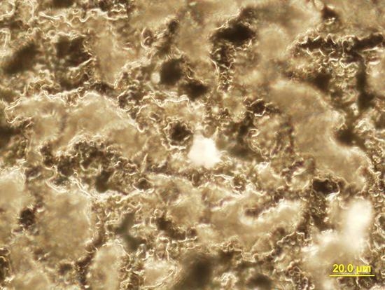
The singular molecular genetic hypoergy of the Jurkat T cells induced by the TiO2 coating can be accounted for by both direct and indirect effects of the artificial material via the products of its destruction and biodegradation (including nanoparticles). Moreover, a subset of the Jurkat T cells adhered to the TiO2 surface (Figure 5).
Changes in surface roughness had a pronounced effect on the morphofunctional parameters of the leukemic T lymphoblast-like cells (Table 6).
The results obtained from the Jurkat T cell cultures (Table 7) showed that titanium dioxide, unlike a Fe3O4 nanosuspension, as well the TiO2 surface itself (see above) at a dosage range of 10–100 MACs had no significant effect on the apoptotic death of leukemic T lymphoblast-like cells. The well-known nano irritant Fe3O4 depressed apoptotic cell outcome. The intracellular concentrations of ROS did not change in cells exposed to either type of nanoparticles (Table 7).
Conversely, the Jurkat T cells reacted to the nanoscale stimuli by increasing the TNFα concentration in the extracellular medium (up to 22%, regardless of the nanosuspension dose) and, to a lesser extent, the IL-2 concentration (up to 18% at a nanosuspension dose of 100 MACs). It is worth noting that the inverse cytokine effect of the titanium dioxide nanosuspension compared with the influence of the bulk samples (Table 5 and Table 8). In contrast, the suppressive effect of the TiO2 nanoparticles on the secretion of IL-4 (11–18% as compared with the control, Table 8) corresponded to that of the TiO2 coating (Table 5). Surprisingly, the TiO2 nanoparticles at a dose of 10 MACs stimulated higher cytokine levels than the corresponding dose of Fe3O4 nanoparticles (Table 8).
The obtained results do not allow an unambiguous connection to be established between the effects of the TiO2 surface on Jurkat T cells and the nanoscale products of their biodestruction for the following reasons: (1) TiO2 nanoparticles influenced the cellular secretion of cytokines ambiguously; (2) tumor Jurkat T cells may have low sensitivity to TiO2 or Fe3O4 nanoparticles at the doses tested despite the marked toxicity of these particles to normal bone marrow cells [34]; (3) high nanoparticle concentrations (1–10 mg/L) can be achieved only through biomechanical impact on the ceramic/metallic TiO2 coating, and this is impossible in short-term cell culture; and, (4) TiO2 nanoparticles did not trigger intracellular oxidation processes (Table 7).
Measurement of the electrostatic properties of the TiO2 coating on the titanium substrate showed a negative surface charge and an average magnitude of potential of V = −147 ± 35 mV (61 runs). The magnitude of the negative surface potential increased with the roughness of the samples (r = 0.6; p < 0.000001, n = 60).

4. Discussion

After 24 h of culture on a plastic surface (2D culture), the Jurkat T cells that were used in these experiments expressed a catalytic telomerase subunit with reverse transcriptase function (Table 3). These cells also displayed the predominant CD3+CD4+CD71+CD45RA+ immunophenotype (Table 4), which, to a great extent, functionally corresponds to naïve/resting T helper/inductor cells [35,36] with expression of the transferrin receptor (CD71), a T-lymphocyte mitogen [37]. Only 0.55% of the T cells in the 24-h culture displayed the CD45RO isoform of the transmembrane antigen that is expressed in vitro by activated T lymphocytes and/or memory T cells [38,39].
The Jurkat T cells secreted a wide range of immunomodulatory cytokines and chemokines (IL-2, IL-4, IL-8, IL-10, and TNFα) into the extracellular medium, but did not secrete IL-1b or IL-6 (Table 3). The cells displayed pro-inflammatory (IL-2, IL-8, and TNFα) and anti-inflammatory (IL-4 and IL-10) activities [39] IL-2, IL-4 [32], and IL-8 [40] can promote the proliferation and survival of tumor cells by means of autocrine/paracrine signaling pathways. In addition, while IL-1 is considered an autocrine co-stimulator of the growth of T cells [32] and leukemic lymphoblasts [41], including the Jurkat line [42], it is also an endogenous inhibitor of CD95(Fas)-mediated apoptosis [43]. IL-6 also has an anti-apoptotic effect on T cells [36]. The Fas (CD95+) transmembrane receptor was observed on only 1% of the Jurkat T cell population (Table 4); therefore, an absence of IL-1b and IL-6 secretion in the Jurkat cell culture (Table 5) could facilitate the cell death processes reported in Table 4. Moreover, IL-10 (Table 5) is capable of inducing anergy in T cells [44], suppressing their proliferation and activation and inhibiting IL-2 expression, as described in [39]. In the absence of co-stimulatory autocrine factors (such as IL-1 and IL-6), the observed imbalance in IL-2 secretion (Table 5) and the absence of its receptor (CD25 antigen, Table 4) can explain the slow in vitro growth of the Jurkat T cells. To a certain extent, these data corroborate the hypothesis regarding the necessity of several ‘survival signals’ for suppressing the genetic cell death program [45].
For a long time, titanium was considered a bio-inert material [9,10] because of the formation of a thin titanium dioxide film on its surface through self-passivation. Micro-arc oxidation of titanium substrates made it possible to increase the average thickness of the TiO2 coating to up to 9 μm, implying an increase in corrosion resistance and in the dielectric properties of the sample surface. On average, the metal-ceramic TiO2 film did not trigger apoptosis or necrosis of the Jurkat T cells (Table 4). Moreover, exposure of the cell cultures to a potential stimulus was accompanied by the suppression of cell death signaling pathways that were potentiated by the TNFα superfamily receptor CD95 (Fas/APO-1) (Table 4 and Table 5). However, in cells exposed to samples of the TiO2 coating with an Ra greater than 2.2 μm, the morphofunctional inactivation of the Jurkat T cells was accompanied by a progressive decrease in the viability of the cells in the culture (Figure 4) in the context of complete suppression of IL-4-dependent mechanisms of T cell survival (Table 5). The underlying molecular genetic mechanism could involve the observed 1.5-fold down-regulation of hTERT expression following exposure (Table 3), which correlated with increased TiO2 surface roughness. Telomerase forms a ribonucleic acid complex that is crucial for supporting cell proliferation; it becomes increasingly active in most tumor cells, including Jurkat T cells [46,47]. Expression of the telomerase gene (hTERT) contributes to the viability of Jurkat T cells [46] and results in elevated production of cytokines [48]. The data obtained in the present study suggest a certain toxic effect of the TiO2 coating that is mediated by inhibition of the genetic (preserving the length of telomeric DNA sequences) and secretory (IL-4) mechanisms supporting the proliferation of tumor Jurkat T cells.
Taken together, the observed effects of the TiO2 nanosuspensions (Table 7 and Table 8) do not allow for it to be concluded that nanoparticles are a proven mediator of the negative cellular and molecular effects of the TiO2 coating on cell viability. Some researchers [7] argue that titanium oxide nanoparticles, unlike the ions and oxides of other metals, do not display cytotoxicity against Jurkat T cells [1,6]. Other reports claim that TiO2 nanoparticles are capable of causing in vitro damage to and apoptosis of lymphocytes [49] and leukemic cells, not through their endocytosis but through direct contact with the cytoplasmic cell membrane [50]. Nanoparticles can be adsorbed on the surface of Jurkat cells [51].
In the present study, the interaction of the Jurkat T cells with the topography of TiO2 particles, but not with the products of their destruction, appears to cause the observed decrease in cell viability and functional activity. A close connection between the Ra and the genetic and molecular features of Jurkat T cells was established (Figure 4 and Table 6). The correlations of Ra with the CD antigen profile and the proportions of apoptotic and necrotic cells (Table 6) suggest that the cell surface is a target of the impact of the physical properties of the TiO2 surface. However, the mechanisms underlying the down-regulation of hTERT expression associated with TiO2 surface roughness have not yet been established.
The process of necrosis begins with the loss of membrane integrity. Membrane channels are sensitive to the topography of biomaterials [52]. For instance, the entry of extracellular calcium and the release of calcium from intracellular stores are triggers of T cell activation [53] and/or programmed cell death [54]. However, non-activated Jurkat T cells are capable of adhering to the TiO2 surface, primarily at zero-charged segments, through the electrostatic interaction of negatively charged glycoproteins on their surface membranes (zeta potential) [5]. The micro-arc TiO2 coating has a negative charge and a surface electrostatic potential that increases linearly with the surface roughness. A subset of T lymphoblast-like cells adheres to the TiO2 surface (Figure 4). These cells are exposed to the surface charge carriers. The electric field of the surface appears to be a physical factor that is capable of modulating the activity of intracellular signaling pathways and molecules. The lifted electrode of the electric potential measuring device registers the electric field at a distance of 500 μm (approximately 50 cell diameters) from the measured surface [28]. This suggests that, over a period of 24 h, the electrostatic potential of the TiO2 sample coating covering 77% of the well surface in the culture plate impacts the majority of the Jurkat T cell population. A favorable impact of negatively charged calcium phosphate coatings (with a comparable surface roughness) on the differentiation and maturation of normal human stromal cells has been previously described [55]. A change in the value and sign of the transmembrane potential that is related to the zeta potential would significantly affect cell fate [56].
The obtained results suggest that the roughness of the TiO2 dielectric surface induces an electrostatic potential that is capable of altering the molecular genetic features and viability of immortalized human leukemic T lymphoblast-like cells through mechanisms unrelated to ROS generation. The functional hypoergy of immune-competent cells can explain the high survival rate of titanium implants in the human body and can impact the choice of materials for endoprosthetics and osteosynthesis in patients suffering from hematopoietic and lymphoid malignancies.

5. Conclusions

When compared with other surface modification techniques, MAO is one of the most applicable methods to deposit a bioceramic layer on Ti and its alloys and to improve the biological properties of titanium [57]. Titanium substrates with TiO2 coatings are widely used in orthopedics, traumatology, dental implantology, and oral surgery. This study established down-regulating expression of membrane antigens that are indicative of the activation, differentiation, and maturation of tumor Jurkat T cells (CD3, CD4, CD8, and CD95) and reducing the secretion of IL-4 and TNFα. The inactivation of Jurkat T cells caused by short-term exposure to the rough TiO2 coating is not related to the generation of intracellular ROS and is not mediated by TiO2 nanoparticles released from the biodegradation of titanium implants and endoprosthetics. Nevertheless, the inhibitory effect of a TiO2 Ra of 2.2–3.7 μm on the viability of Jurkat T cells primarily occurred through a progressive increase in the proportion of necrotic cells in the cell population. In turn, increased Ra was accompanied by an increase in the magnitude of the negative electrostatic potential of the TiO2 surface.
Thus, rough titanium oxide coating (rTOC) inhibits Jurkat T cell survival in 24-h culture. rTOC causes in vitro morphofunctional inactivation of Jurkat T cells. The impact of rTOC is not related to intracellular ROS generation. Jurkat T cells have low sensitivity to TiO2 nanoparticles. Negative surface charge induced by rTOC might mediate its cellular effects.
We suggest that the roughness of the dielectric TiO2 coating induces a negative charge that is likely capable of structural (down-regulation of antigen expression) and functional (down-regulation of hTERT expression and cytokine secretion) regulation of tumor immune cells by means of biological mechanisms and metabolic pathways unrelated to ROS generation. The in vitro inactivation of Jurkat T cells caused by a rough TiO2 coating suggests that it is important to study the effects of such coatings on other tumor cell lines and to identify intracellular signaling pathways that are susceptible to physicochemical factors. Hereafter, this may be useful for replacement surgery applications of rough TiO2 implants in cancer patients.

Acknowledgments

This investigation was supported by the Russian Science Foundation (grant number 16-15-10031). The authors thank Elena V. Legostaeva from the Institute of Strength Physics and Materials Science of SB RAS (Tomsk, Russia) for assistance in the design of the titanium samples and assistance with titanium oxide deposition. Material testing was conducted as a part of the fundamental research program of the Russian Academy of Science for 2015–2016, No. 23.2.5.

Author Contributions

Igor Khlusov, Larisa Litvinova, Valeria Shupletsova, Vladimir Leitsin, Marina Khlusova, Vladimir Pichugin, and Yurii Sharkeev conceived and designed the experiments; Valeria Shupletsova, Olga Khaziakhmatova, Elena Melashchenko, Kristina Yurova, Vladimir Pichugin, and Yurii Sharkeev performed the experiments; Igor Khlusov, Larisa Litvinova, Valeria Shupletsova, Olga Khaziakhmatova, Elena Melashchenko, Kristina Yurova, Vladimir Leitsin, Marina Khlusova, and Vladimir Pichugin analyzed the data; Igor Khlusov, Larisa Litvinova, Vladimir Leitsin, Vladimir Pichugin, and Yurii Sharkeev contributed reagents/materials/analysis tools; Igor Khlusov, Larisa Litvinova, and Yurii Sharkeev wrote the paper.

Conflicts of Interest

The authors declare no conflicts of interest.

References

  1. Au, A.; Ha, J.; Hernandez, M.; Polotsky, A.; Hungerford, D.S.; Frondoza, C.G. Nickel and vanadium metal ions induce apoptosis of T-lymphocyte Jurkat cells. J. Biomed. Mater. Res. A 2006, 79, 512–521. [Google Scholar] [CrossRef] [PubMed]
  2. Chen, X.; Su, J.; Chang, J.; Kanekura, T.; Li, J.; Kuang, Y.H.; Peng, S.; Yang, F.; Lu, H.; Zhang, J.L. Inhibition of CD147 gene expression via RNA interference reduces tumor cell proliferation, activation, adhesion, and migration activity in the human Jurkat T-lymphoma cell line. Cancer Investig. 2008, 26, 689–697. [Google Scholar] [CrossRef] [PubMed]
  3. Liao, H.F.; Chen, Y.J.; Chou, C.H.; Wang, F.W.; Kuo, C.D. Norcantharidin induces cell cycle arrest and inhibits progression of human leukemic Jurkat T cells through mitogen-activated protein kinase-mediated regulation of interleukin-2 production. Toxicol. In Vitro 2011, 25, 206–212. [Google Scholar] [CrossRef] [PubMed]
  4. Luongo, D.; Russo, R.; Balestrieri, A.; Marzocco, S.; Bergamo, P.; Severino, L. In vitro study of AFB1 and AFM1 effects on human lymphoblastoid Jurkat T-cell model. J. Immunotoxicol. 2014, 11, 353–358. [Google Scholar] [CrossRef] [PubMed]
  5. Stevens, M.J.; Donato, L.J.; Lower, S.K.; Sahai, N. Oxide-dependent adhesion of the Jurkat line of T lymphocytes. Langmuir 2009, 25, 6270–6278. [Google Scholar] [CrossRef] [PubMed]
  6. Caicedo, M.; Jacobs, J.J.; Reddy, A.; Hallab, N.J. Analysis of metal ion-induced DNA damage, apoptosis, and necrosis in human (Jurkat) T-cells demonstrates Ni2+ and V3+ are more toxic than other metals: Al3+, Be2+, Co2+, Cr3+, Cu2+, Fe3+, Mo5+, Nb5+, Zr2+. J. Biomed. Mater. Res. A 2008, 86, 905–913. [Google Scholar] [CrossRef] [PubMed]
  7. Tuomela, S.; Autio, R.; Buerki-Thurnherr, T.; Arslan, O.; Kunzmann, A.; Andersson-Willman, B.; Wick, P.; Mathur, S.; Scheynius, A.; Krug, H.F.; et al. Gene expression profiling of immune-competent human cells exposed to engineered zinc oxide or titanium dioxide nanoparticles. PLoS ONE 2013, 8, e68415. [Google Scholar] [CrossRef] [PubMed]
  8. Ratner, B.D.; Hoffman, A.S. Biomaterials Science: An Introduction to Materials in Medicine, 2nd ed.; Elsevier Academic Press: San Diego, CA, USA, 2004. [Google Scholar]
  9. Thull, R. Titan in der Zahnheilkunde-Grundlagen. Z. Mitteilungen 1992, 82, 39–45. [Google Scholar]
  10. Ogawa, T. Ultraviolet photofunctionalization of titanium implants. Int. J. Oral Maxillofac. Implants 2014, 29, e95–e102. [Google Scholar] [CrossRef] [PubMed]
  11. Nethi, S.K.; Neeraja Aparna Anand, P.; Rico-Oller, B.; Rodríguez-Diéguez, A.; Gómez-Ruiz, S.; Patra, C.R. Design, synthesis and characterization of doped-titanium oxide nanomaterials with environmental and angiogenic applications. Sci. Total Environ. 2017, 599–600, 1263–1274. [Google Scholar] [CrossRef] [PubMed]
  12. Lavigne, M.; Belzile, E.L.; Roy, A.; Morin, F.; Amzica, T.; Vendittoli, P.A. Comparison of whole-blood metal ion levels in four types of metal-on-metal large-diameter femoral head total hip arthroplasty: The potential influence of the adapter sleeve. J. Bone Jt. Surg. Am. 2011, 93, 128–136. [Google Scholar] [CrossRef] [PubMed]
  13. Matusiewicz, H. Potential release of in vivo trace metals from metallic medical implants in the human body: From ions to nanoparticles—A systematic analytical review. Acta Biomater. 2014, 10, 2379–2403. [Google Scholar] [CrossRef] [PubMed]
  14. Agins, H.J.; Alcock, N.W.; Bansal, M.; Salvati, E.A.; Wilson, P.D.; Pellicci, P.M.; Bullough, P.G. Metallic wear in failed titanium-alloy total hip replacements. J. Bone Jt. Surg. A 1988, 70, 347–356. [Google Scholar] [CrossRef]
  15. Case, C.P.; Langkamer, V.G.; James, C.; Palmer, M.R.; Kemp, A.J.; Heap, P.F.; Solomon, L. Widespread dissemination of metal debris from implants. J. Bone Jt. Surg. 1994, 76, 701–712. [Google Scholar] [CrossRef]
  16. Bradberry, S.M.; Wilkinson, J.M.; Ferner, R.E. Systemic toxicity related to metal hip prostheses. Clin. Toxicol. 2014, 52, 837–847. [Google Scholar] [CrossRef] [PubMed]
  17. Wang, J.C.; Yu, W.D.; Sandhu, H.S.; Betts, F.; Bhuta, S.; Delamarter, R.B. Metal debris from titanium spinal implants. Spine 1999, 24, 899–903. [Google Scholar] [CrossRef] [PubMed]
  18. Curtis, A.S.G.; Wilkinson, C. Control of cell behavior: Topological factors. J. Natl. Cancer Inst. 1964, 33, 15–26. [Google Scholar] [CrossRef] [PubMed]
  19. Khlusov, I.A.; Litvinova, L.S.; Shupletsova, V.V.; Dunets, N.A.; Khaziakhmatova, O.G.; Yurova, K.A.; Yu, M.; Khlusova, Y.; Sharkeev, P. Morphofunctional changes of Jurkat T lymphoblasts upon short-term contact with a relief calcium phosphate surface. Cell Tissue Biol. 2017, 11, 59–64. [Google Scholar] [CrossRef]
  20. Curtis, A.S.G.; Wilkinson, C. Topographical control of cells. Biomaterials 1997, 18, 1573–1583. [Google Scholar] [CrossRef]
  21. Ravichandran, R.; Liao, S.; Ng, C.C.; Chan, C.K.; Raghunath, M.; Ramakrishna, S. Effects of nanotopography on stem cell phenotypes. World J. Stem Cells 2009, 1, 55–66. [Google Scholar] [CrossRef] [PubMed]
  22. Anselme, K.; Bigerelle, M. Topography effects of pure titanium substrates on human osteoblast long-term adhesion. Acta Biomater. 2005, 1, 211–222. [Google Scholar] [CrossRef] [PubMed]
  23. Ponsonnet, L.; Reybier, K.; Jaffrezic, N.; Comte, F.; Lagneau, C.; Lissac, M.; Martelet, C. Relationship between surface properties (roughness, wettability) of titanium and titanium alloys and cell behavior. Mater. Sci. Eng. C 2003, 23, 551–560. [Google Scholar] [CrossRef]
  24. Anselme, K.; Bigerelle, M.; Noel, B.; Dufresne, E.; Judas, D.; Iost, A.; Hardouin, P. Qualitative and quantitative study of human osteoblast adhesion on materials with various surface roughnesses. J. Biomed. Mater. Res. Part A 2000, 49, 155–166. [Google Scholar] [CrossRef]
  25. Legostaeva, E.V.; Kulyashova, K.S.; Komarova, E.G.; Epple, M.; Sharkeev, Y.P.; Khlusov, I.A. Physical, chemical and biological properties of micro-arc deposited calcium phosphate coatings on titanium and zirconium-niobium alloy. Materialwissenschaft und Werkstofftechnik 2013, 44, 188–197. [Google Scholar] [CrossRef]
  26. Khlusov, I.A.; Sharkeev, Y.P.; Pichugin, V.F.; Legostaeva, E.V.; Litvinova, L.S.; Shupletsova, V.V.; Sokhonevich, N.A.; Khaziakhmatova, O.G.; Khlusova, M.Y.; Gutor, S.S.; et al. Influence of the structure of the titanium oxide coating surface on immunocompetent tumor cells. Russ. Phys. J. 2016, 58, 1527–1533. [Google Scholar] [CrossRef]
  27. Eguchi, M. On the permanent electret. Philos. Mag. 1925, 49, 178–192. [Google Scholar] [CrossRef]
  28. Gostischev, E.A. A Noncontacting Manner to Determine the Potentials of Charged Surface of Objects and the Device for Its Realization: Russian Federation. Patent 2,223,511, 2004. [Google Scholar]
  29. Khlusov, I.A.; Zagrebin, L.V.; Shestov, S.S.; Itin, V.I.; Sedoi, V.S.; Feduschak, T.A.; Terekhova, O.G.; Magaeva, A.A.; Naiden, E.P.; Antipov, S.A.; et al. Colony-forming activity of unipotent hemopoietic precursors under the effect of nanosized ferrites in a constant magnetic field in vitro. Bull. Exp. Biol. Med. 2008, 145, 151–157. [Google Scholar] [CrossRef] [PubMed]
  30. Yakushina, V.D.; Vasil’eva, O.A.; Ryazantseva, N.V.; Novitsky, V.V.; Tashireva, L.A. The effects of galectin-1 on the gene expression of the transcription factors TBX21, GATA-3, FOXP3 and RORC. Mol. Cell. Biochem. 2015, 398, 245–249. [Google Scholar] [CrossRef] [PubMed]
  31. Banda, M.; Bommineni, A.; Thomas, R.A.; Luckinbill, L.S.; Tucker, J.D. Evaluation and validation of housekeeping genes in response to ionizing radiation and chemical exposure for normalizing RNA expression in real-time PCR. Mutat. Res. 2008, 649, 126–134. [Google Scholar] [CrossRef] [PubMed]
  32. Terui, Y.; Ikeda, M.; Tomizuka, H.; Kasahara, T.; Ohtsuki, T.; Uwai, M.; Mori, M.; Itoh, T.; Tanaka, M.; Yamada, M.; et al. Activated endothelial cells induce apoptosis in leukemic cells by endothelial interleukin-8. Blood 1998, 92, 2672–2680. [Google Scholar] [PubMed]
  33. Devita, V.T.; Lawrence, T.S.; Rosenberg, S.A. Cancer: Principles & Practice of Oncology, 8th ed.; Wolters Kluwer/Lippincott Williams & Wilkins: Philadelphia, PA, USA, 2008. [Google Scholar]
  34. Novitsky, V.A.; Khlusov, I.A.; Ryazantseva, N.V.; Chechina, O.E.; Feduschak, T.A.; Ermakov, A.E.; Uimin, M.A. Structural and functional state of the bone marrow during its in vitro interaction with ferromagnetic nanoparticles. Bull. Exp. Biol. Med. 2011, 151, 473–476. [Google Scholar] [CrossRef] [PubMed]
  35. Gallagher, P.F.; Fazekas de St Groth, B.; Miller, J.F. CD4 and CD8 molecules can physically associate with the same T-cell receptor. Proc. Natl. Acad. Sci. USA 1989, 86, 10044–10048. [Google Scholar] [CrossRef] [PubMed]
  36. Law, H.K.W.; Tu, W.; Liu, E.; Lau, Y.L. Insulin-like growth factor I promotes cord blood T cell maturation through monocytes and inhibits their apoptosis in part through interleukin-6. BMC Immunol. 2008, 9, 74. [Google Scholar] [CrossRef] [PubMed] [Green Version]
  37. Bi, B.Y.; Lefebvre, A.M.; Duś, D.; Spik, G.; Mazurier, J. Effect of lactoferrin on proliferation and differentiation of the Jurkat human lymphoblastic T cell line. Arch. Immunol. Ther. Exp. (Warsz.) 1997, 45, 315–320. [Google Scholar] [PubMed]
  38. Akbar, A.N.; Terry, L.; Timms, A.; Beverley, P.C.; Janossy, G. Loss of CD45R and gain of UCHL1 reactivity is a feature of primed T cells. J. Immunol. 1988, 140, 2171–2178. [Google Scholar] [PubMed]
  39. Khalaf, H.; Jass, J.; Olsson, P.E. Differential cytokine regulation by NF-kappa B and AP-1 in Jurkat T-cells. BMC Immunol. 2010, 11, 26. [Google Scholar] [CrossRef] [PubMed]
  40. Waugh, D.J.J.; Wilson, C. The interleukin-8 pathway in cancer. Clin. Cancer Res. 2008, 14, 6735–6741. [Google Scholar] [CrossRef] [PubMed]
  41. Jasmin, C.; Georgoulias, V.; Smadja-Joffe, F.; Boucheix, C.; Le Bousse-Kerdiles, C.; Allouche, M.; Cibert, C.; Azzarone, B. Autocrine growth of leukemic cells. Leuk. Res. 1990, 14, 689–693. [Google Scholar] [CrossRef]
  42. Arana-Argáez, V.E.; Delgado-Rizo, V.; Pizano-Martínez, O.E.; Martínez-Garcia, E.A.; Martín-Márquez, B.T.; Muñoz-Gómez, A.; Petri, M.H.; Armendáriz-Borunda, J.; Espinosa-Ramírez, G.; Zúñiga-Tamayo, D.A.; et al. Inhibitors of MAPK pathway ERK1/2 or p38 prevent the IL-1{beta}-induced up-regulation of SRP72 autoantigen in Jurkat cells. J. Biol. Chem. 2010, 285, 32824–32833. [Google Scholar] [CrossRef] [PubMed]
  43. Tatsuta, T.; Cheng, J.; Mountz, J.D. Intracellular IL-1beta is an inhibitor of Fas-mediated apoptosis. J. Immunol. 1996, 157, 3949–3957. [Google Scholar] [PubMed]
  44. Groux, H.; Bigler, M.; de Vries, J.E.; Roncarolo, M.G. Interleukin-10 induces a long-term antigen-specific anergic state in human CD4+ T cells. J. Exp. Med. 1996, 184, 19–29. [Google Scholar] [CrossRef] [PubMed]
  45. Raff, M.C.; Barres, B.A.; Burne, J.F.; Coles, H.S.; Ishizaki, Y.; Jacobson, M.D. Programmed cell death and the control of cell survival: Lessons from the nervous system. Science 1993, 262, 695–700. [Google Scholar] [CrossRef] [PubMed]
  46. Lin, Z.; Lim, S.; Viani, M.A.; Sapp, M.; Lim, M.S. Down-regulation of telomerase activity in malignant lymphomas by radiation and chemotherapeutic agents. Am. J. Pathol. 2001, 159, 711–719. [Google Scholar] [CrossRef]
  47. Soares, J.; Lowe, M.M.; Jarstfer, M.B. The catalytic subunit of human telomerase is a unique caspase-6 and caspase-7 substrate. Biochemistry 2011, 50, 9046–9055. [Google Scholar] [CrossRef] [PubMed]
  48. Kanzaki, Y.; Onoue, F.; Sakurai, H.; Ide, T. Telomerase upregulates expression levels of interleukin (IL)-1alpha, IL-1beta, IL-6, IL-8, and granulocyte-macrophage colony-stimulating factor in normal human fibroblasts. Biochem. Biophys. Res. Commun. 2003, 305, 150–154. [Google Scholar] [CrossRef]
  49. Ghosh, M.; Chakraborty, A.; Mukherjee, A. Cytotoxic, genotoxic and the hemolytic effect of titanium dioxide (TiO2) nanoparticles on human erythrocyte and lymphocyte cells in vitro. J. Appl. Toxicol. 2013, 33, 1097–1110. [Google Scholar] [CrossRef] [PubMed]
  50. Takaki, K.; Higuchi, Y.; Hashii, M.; Ogino, C.; Shimizu, N. Induction of apoptosis associated with chromosomal DNA fragmentation and caspase-3 activation in leukemia L1210 cells by TiO2 nanoparticles. J. Biosci. Bioeng. 2014, 117, 129–133. [Google Scholar] [CrossRef] [PubMed]
  51. Montet-Abou, K.; Montet, X.; Weissleder, R.; Josephson, L. Transfection agent induced nanoparticle cell loading. Mol. Imaging 2005, 4, 165–171. [Google Scholar] [PubMed]
  52. Tobasnick, G.; Curtis, A.S.G. Chloride channels and the reactions of cells to topography. Eur. Cells Mater. 2001, 2, 49–61. [Google Scholar] [CrossRef]
  53. Dadsetan, S.; Zakharova, L.; Molinski, T.F.; Fomina, A.F. Store-operated Ca2+ influx causes Ca2+ release from the intracellular Ca2+ channels that is required for T cell activation. J. Biol. Chem. 2008, 283, 12512–12519. [Google Scholar] [CrossRef] [PubMed]
  54. Nohara, L.L.; Stanwood, S.R.; Omilusik, K.D.; Jefferies, W.A. Tweeters, woofers and horns: The complex orchestration of calcium currents in T lymphocytes. Front. Immunol. 2015, 6, 234. [Google Scholar] [CrossRef] [PubMed]
  55. Khlusov, I.A.; Khlusova, M.Y.; Pichugin, V.F.; Sharkeev, Y.P.; Legostaeva, E.V. Artificial niches for stromal stem cells as a potential instrument for the design of the surface of biomimetic osteogenic materials. Russ. Phys. J. 2014, 56, 1206–1211. [Google Scholar] [CrossRef]
  56. Sundelacruz, S.; Levin, M.; Kaplan, D.L. Role of membrane potential in the regulation of cell proliferation and differentiation. Stem Cell Rev. 2009, 5, 231–246. [Google Scholar] [CrossRef] [PubMed]
  57. Wang, Y.; Yu, H.; Chen, C.; Zhao, Z. Review of the biocompatibility of micro-arc oxidation coated titanium alloys. Mater. Des. 2015, 85, 640–652. [Google Scholar] [CrossRef]
Figure 1. SEM-images of the titanium oxides (TiO2) coating before sandblasting and subsequent acid etching (a), the Ti surface after acid etching (b), EDX spectrum (c) and X-ray diffraction pattern of the TiO2 coating (d).
Materials 11 00360 g001
Figure 2. Algorithm for determining intracellular levels of ROS after 24 h in culture with the addition of a nanoparticle suspension. (a) Jurkat gating based on a forward scatter (FSC) vs. side scatter (SSC) plot; (b) The analysis was carried out using gating of Jurkat cells. The abscissa axis is the fluorescence intensity of antibodies against Viability Fixable Dye; the ordinate is the number of cells; (c) Evaluation of the production of reactive oxygen species.
Materials 11 00360 g002
Figure 3. Gating strategy for Jurkat T cell CD staining. (a) Jurkat cell gating based on a forward scatter (FSC) vs. side scatter (SSC) plot; (b) The analysis was carried out using gating of Jurkat cells. The abscissa axis is the fluorescence intensity of the antibodies against Annexin; the ordinate is the fluorescence intensity of the antibodies against propidium iodide. Area “III” contains viable cells; (c) The analysis was carried out using gating on the “III” area. Histogram of the distribution of Jurkat cells; the axis of abscissas is the fluorescence intensity of the antibodies against CD3; the ordinate is the number of cells; (d1) The analysis was carried out using gating of the “III” area. Histogram of the distribution of Jurkat cells; the abscissa axis is the fluorescence intensity of the antibodies against CD56; the ordinate axis is the fluorescence intensity of the antibodies against CD16, (d2d8) The analysis was carried out using gating of the "III" area. Histogram of the distribution of Jurkat cells; the abscissa axis is the fluorescence intensity of the antibodies against CD3; the ordinate axis is the fluorescence intensity of the antibodies against CD25, CD95, CD71, CD8, CD4, CD45RA, or CD45RO.
Materials 11 00360 g003
Figure 4. Regression of Jurkat T cell viability after 24 h of culture in the presence of rough TiO2-coated titanium samples.
Materials 11 00360 g004
Figure 5. Location of Jurkat T cells on the rough surface of the TiO2 coating. Reflecting optical microscopy, dark field. Magnification, 1000×.
Materials 11 00360 g005
Table 1. Sequences and probes used for polymerase chain reaction (PCR) to measure hTERT expression in Jurkat T cells.
GenePrimers and ProbesAmplicon Mass
hTERTForward: 5′-ACCGTCTGCGTGAGGAGATC-3′
Reverse: 5′-CCGGTAGAAAAAGAGCCTGTTC-3′
131 base pairs
18S rRNAForward: 5′-CTGAGAAACGGCTACCACATC-3′
Reverse: 5′-GCCTCGAAAGAGTCCTGTATTG-3′
153 base pairs
Table 2. Monoclonal antibody panel used for Jurkat T cell analysis.
Studied SubpopulationFluorochromes and Labeled Monoclonal Antibodies
PEFITCVioBluePEAPC
Jurkat T cellsPropidium iodideAnnexin VCD3CD56CD16
CD4CD8
CD71CD95
CD45RACD45RO
-CD25
Table 3. hTERT expression levels in Jurkat T cells after 24 h of culture in the presence of a model matrix consisting of a TiO2 coating on a titanium substrate, Me (Q1–Q3).
Group Studied (n = 7)hTERT, a.u.
Control cell culture9.81 (8.90–11.18)
Cell culture in contact with the TiO2-coated titanium samples 6.00 (4.48–6.95) *
Note: Here and below, n = the number of observations (samples) in each group; * = statistical significance (p < 0.05) in comparison with the control according to the Mann-Whitney U-test; a.u. = arbitrary units.
Table 4. Membrane molecular markers and death indices of Jurkat T cells after 24 h of culture in the presence of the TiO2 coating Me (Q1–Q3).
Dead or Necrotic Cells, %Cells Expressing Specific Surface Markers, %
ApoptosisNecrosisCD3CD4CD8CD71CD45ROCD45RACD16CD56CD25CD95
Cells cultured on plastic surface (control), n = 15
5.2
(5.2–8.9)
8.2
(8.1–14.2)
98
(97.9–98.85)
27.5
(26.54–28.43)
2.16
(1.9–3.0)
94.0
(93.5–95.0)
0.55
(0.49–0.60)
94.9
(94.0–95.22)
2.46
(2.34–2.83)
2.5
(2.3–2.97)
0
(0–0.16)
1.0
(0.9–1.2)
Cells in contact with the TiO2-coated titanium samples, n = 15
5.9
(5.6–6.9)
15.3
(8.2–19.3)
97.5
(97–98) *
<0.03
25.4
(24.76–26.70) *
<0.002
0.31
(0.2–0.43) *
<0.000003
94.1
(93.49–95.0)
0.50
(0.41–0.50)
94.0
(93.15–94.3)
2.33
(2.17–2.34)
2.36
(2.22–2.80)
0
(0–0.12)
0.56
(0.3–0.6) *
<0.002
* = statistical significance (p < 0.05) in comparison with the control according to the Mann-Whitney U-test.
Table 5. Cytokine concentrations (pg/mL) in the supernatants of Jurkat T cells after 24 h of culture in the presence of a TiO2 coating, Me (Q1–Q3).
Cytokine Concentration
IL-1bIL-2IL-4IL-6IL-8IL-10TNFα
Control cell culture on plastic surface, n = 12
0
(0–0.23)
5.67
(5.46–5.98)
1.10
(0.74–1.24)
011.12
(9.18–12.59)
6.10
(5.40–6.96)
14.95
(13.97–15.51)
Cell culture in contact with the TiO2-coated titanium samples, n = 12
0
(0–0.10)
6.10
(3.30–8.10)
0 (0–0.79) *
<0.007
012.78
(11.71–13.22)
7.80
(4.87–9.86)
13.48
(8.0–14.15) *
<0.01
* = statistical significance (p < 0.05) in comparison with the control according to the Mann-Whitney U-test.
Table 6. Spearman’s correlation coefficient between the Ra of the TiO2 coating and the morphofunctional parameters of the Jurkat T cell cultures.
Index% Viable Cells% Apoptotic Cells% Necrotic CellsCD8CD56IL-2IL-4IL-10TNFa
Ra−0.95
n = 9
0.0001
−0.69
n = 9
0.042
0.95
n = 9
0.0001
0.71
n = 12
0.004
−0.95
n = 12
0.0001
−0.95
n = 12
0.000003
−0.69
n = 12
0.013
−0.92
n = 12
0.00003
0.68
n = 12
0.015
Table 7. Proportions of apoptotic Jurkat T cells and intracellular levels of ROS after 24 h of culture with nanoparticle suspensions, Me (Q1–Q3).
No.GroupResults
Number of Apoptotic Cells, %ROS, a.u.
1Cells cultured on plastic surface without nanoparticle suspension, n = 65.87
(5.32–6.99)
0.130
(0.123–0.150)
Concentration of TiO2 nanoparticle samples, n = 3
2100 MACs4.82
(4.24–8.50)
0.131
(0.129–0.194)
310 MACs4.76
(3.65–6.39)
0.139
(0.130–0.162)
Concentration of Fe3O4 nanoparticle samples, n = 3
410 MACs4.39
(3.05–5.10)
P1 < 0.02
0.123
(0.103–0.150)
Note: MACs = maximum allowable concentrations; a.u. = arbitrary units; P1 significant difference compared with the corresponding group according to the Mann-Whitney U-test.
Table 8. Cytokine levels (pg/mL) in the culture medium after 24 h of culture of Jurkat T cells with nanoparticle suspensions, Me (Q1–Q3).
NoGroupCytokine Concentration
TNFαIL-2IL-4
1Cells cultured on plastic surface without nanoparticle samples, n = 627.41
(26.62–28.17)
4.02
(3.97–4.04)
4.14
(4.06–4.24)
Concentration of TiO2 nanoparticle samples, n = 3
2100 MACs33.52
(32.77–35.0)
P1 < 0.05
4.73
(4.61–5.0)
P1 < 0.05
3.67
(3.60–3.67)
P1 < 0.045
310 MACs33.51
(33.17–35.49)
P1, P4 < 0.05
3.61
(3.56–4.55)
3.40
(3.27–3.60)
P1, P4 < 0.045
Concentration of Fe3O4 nanoparticle samples, n = 3
410 MACs26.62
(24.72–27.34)
4.23
(4.0–4.29)
4.39
(4.10–4.52)
Note: P1, P4 = statistically significant differences compared with the corresponding group according to the Mann-Whitney U-test.