
Greater Osseointegration Potential with Nanostructured Surfaces on TiZr: Accelerated vs. Real-Time Ageing

 ,
,  and
and
Abstract
:1. Introduction
2. Materials and Methods
- Group 1: Roxolid, sandblasted, large-grit, acid-etched (Rxd SLA—Control 1)—hydrophobic, no nanostructures → dry/micro
- Group 2: Roxolid sandblasted, large-grit, acid-etched, nano structures, accelerated aged (Rxd SLA nano AA—Test 1)—hydrophobic, with nanostructures → dry/nano/AA
- Group 3: Roxolid SLActive non-aged (Rxd SLActive ‘fresh’—Control 2)—hydrophilic, without nanostructures → wet/micro
- Group 4: Roxolid SLActive, nano structures, accelerated aged (Rxd SLActive nano AA—Test 3)—hydrophilic, with nanostructures → wet/nano/AA
- Group 5: Roxolid SLActive real-time aged (Rxd SLActive RTA—Control 2)—hydrophilic, with nanostructures → wet/nano/RTA
2.1. Pre-Surgical and Surgical Phase
2.2. Post-Surgical Phase
2.3. Biomechanical Pull-Out Measurements
2.4. Statistical Analysis
2.5. Surface Textural, Chemical, and Area Evaluation
Static contact angle measurements
Surface roughness measurements
X-ray photoelectron spectroscopy (XPS)
Scanning electron microscopy (SEM)
Surface area analysis
3. Results
3.1. Surface Properties Characterization
3.1.1. Surface Morphology, Roughness, and Chemistry
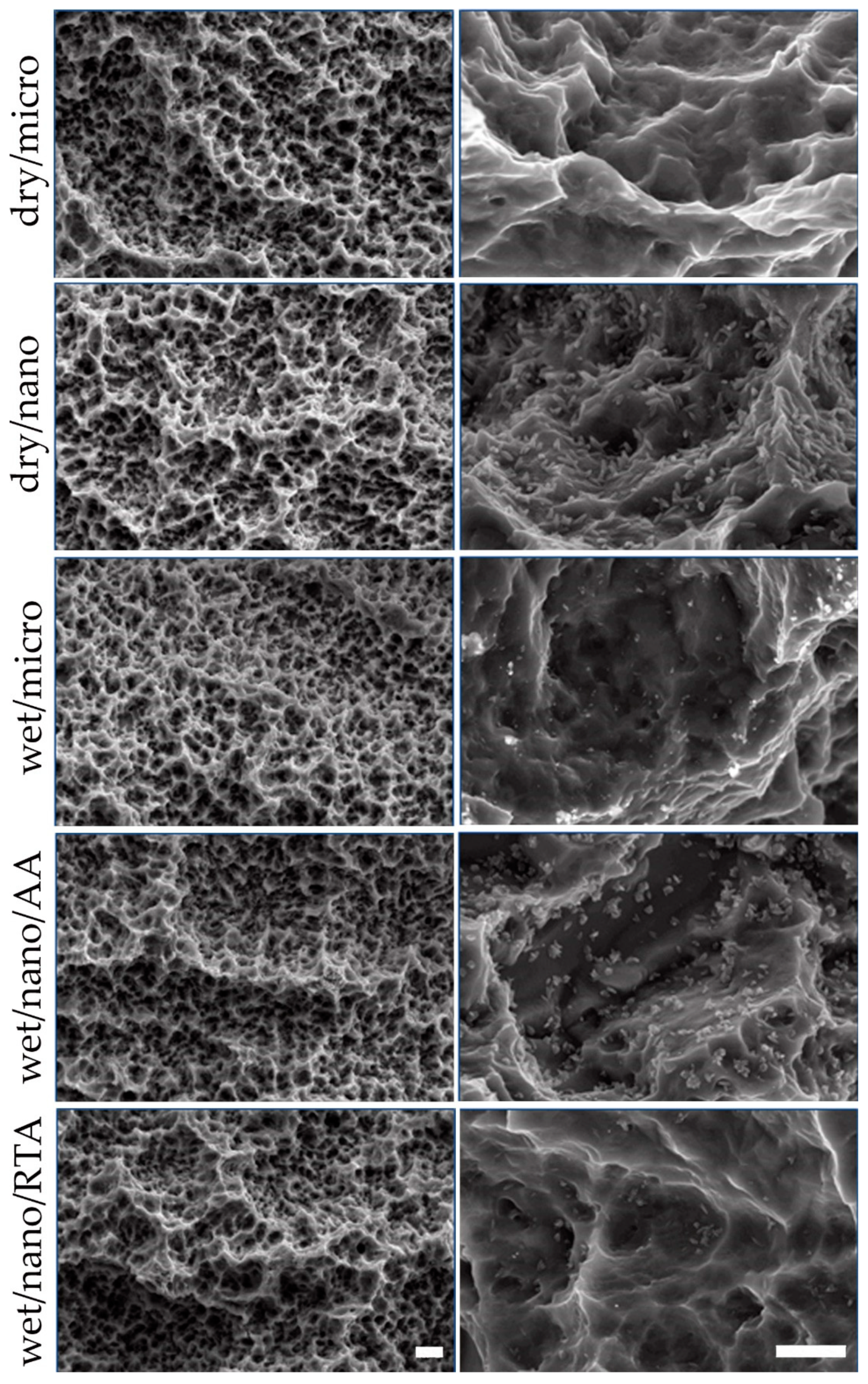
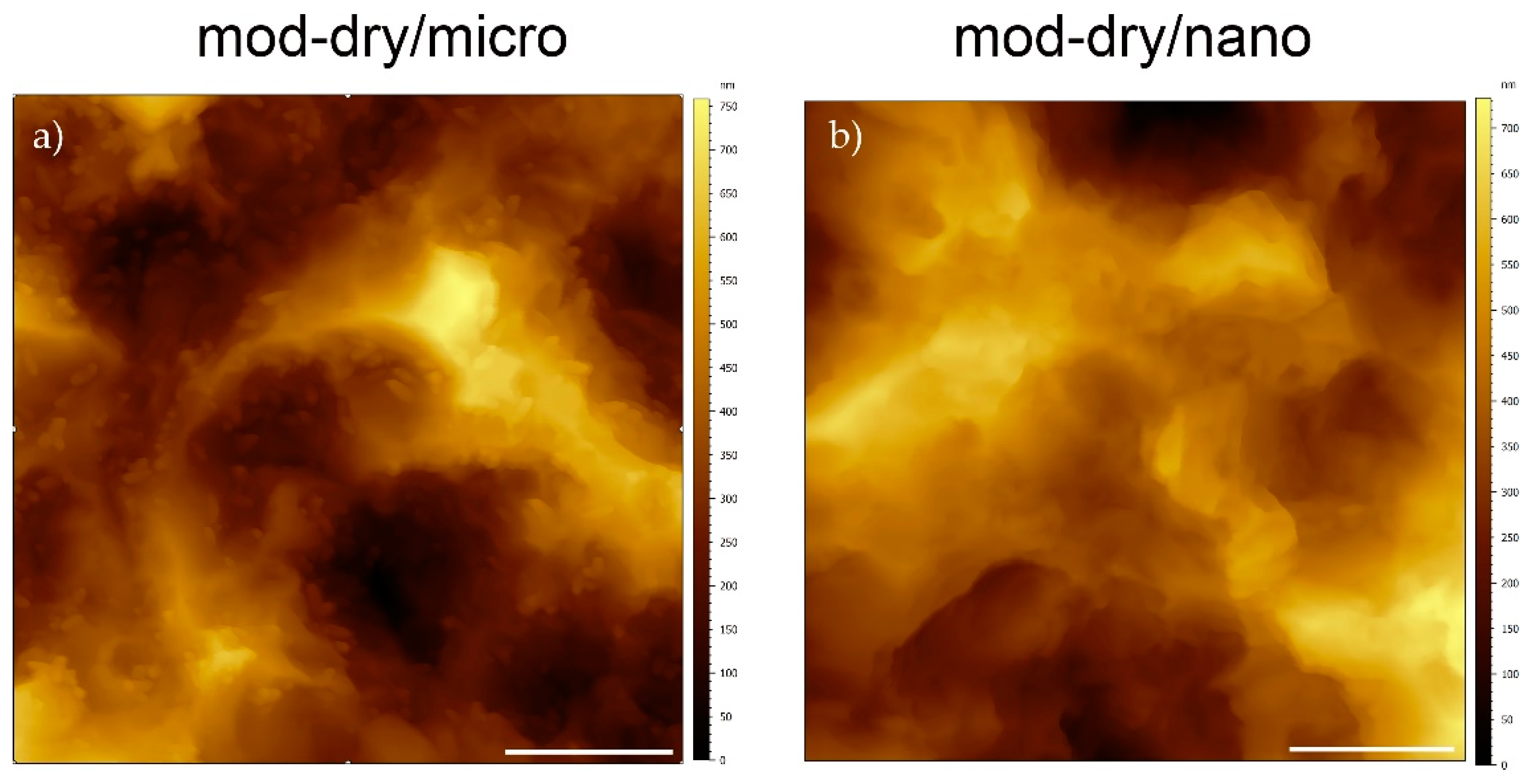
Surface morphology:
Surface roughness
Surface chemistry
Wettability
3.1.2. Surface Area Evaluation
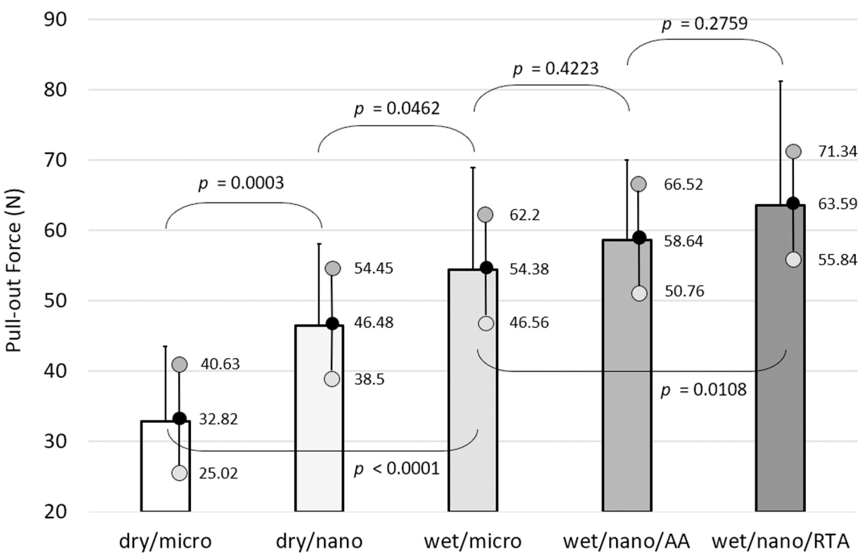
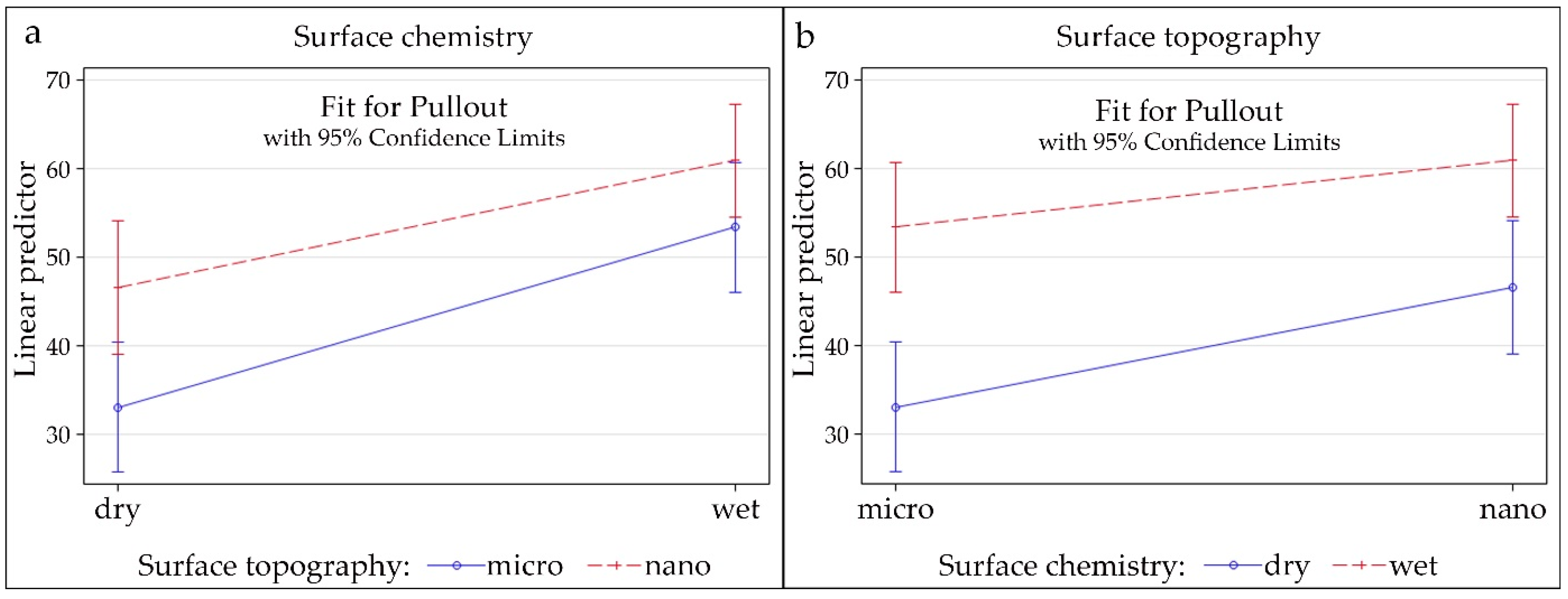
3.2. Biomechanical Pull-Out Measurements
4. Discussion
Supplementary Materials
Author Contributions
Funding
Institutional Review Board Statement
Informed Consent Statement
Data Availability Statement
Acknowledgments
Conflicts of Interest
References
- Fischer, K.; Stenberg, T. Prospective 10-year cohort study based on a randomized controlled trial (rct) on implant-supported full-arch maxillary prostheses. Part 1: Sandblasted and acid-etched implants and mucosal tissue. Clin. Implant. Dent. Relat. Res. 2012, 14, 808–815. [Google Scholar] [CrossRef] [PubMed]
- Roccuzzo, M.; Bonino, L.; Dalmasso, P.; Aglietta, M. Long-term results of a three arms prospective cohort study on implants in periodontally compromised patients: 10-year data around sandblasted and acid-etched (sla) surface. Clin. Oral. Implant. Res. 2014, 25, 1105–1112. [Google Scholar] [CrossRef]
- Van Velzen, F.J.; Ofec, R.; Schulten, E.A.; Ten Bruggenkate, C.M. 10-year survival rate and the incidence of peri-implant disease of 374 titanium dental implants with a sla surface: A prospective cohort study in 177 fully and partially edentulous patients. Clin. Oral. Implant. Res. 2015, 26, 1121–1128. [Google Scholar] [CrossRef]
- Mamalis, A.A.; Markopoulou, C.; Vrotsos, I.; Koutsilirieris, M. Chemical modification of an implant surface increases osteogenesis and simultaneously reduces osteoclastogenesis: An in vitro study. Clin. Oral. Implant. Res. 2011, 22, 619–626. [Google Scholar] [CrossRef]
- Wall, I.; Donos, N.; Carlqvist, K.; Jones, F.; Brett, P. Modified titanium surfaces promote accelerated osteogenic differentiation of mesenchymal stromal cells in vitro. Bone 2009, 45, 17–26. [Google Scholar] [CrossRef]
- Hong, J.; Kurt, S.; Thor, A. A hydrophilic dental implant surface exhibits thrombogenic properties in vitro. Clin. Implant. Dent. Relat. Res. 2013, 15, 105–112. [Google Scholar] [CrossRef] [PubMed]
- Alfarsi, M.A.; Hamlet, S.M.; Ivanovski, S. Titanium surface hydrophilicity enhances platelet activation. Dent. Mater. J. 2014, 33, 749–756. [Google Scholar] [CrossRef] [Green Version]
- Hamlet, S.; Alfarsi, M.; George, R.; Ivanovski, S. The effect of hydrophilic titanium surface modification on macrophage inflammatory cytokine gene expression. Clin. Oral. Implant. Res. 2012, 23, 584–590. [Google Scholar] [CrossRef]
- Donos, N.; Hamlet, S.; Lang, N.P.; Salvi, G.E.; Huynh-Ba, G.; Bosshardt, D.D.; Ivanovski, S. Gene expression profile of osseointegration of a hydrophilic compared with a hydrophobic microrough implant surface. Clin. Oral. Implant. Res. 2011, 22, 365–372. [Google Scholar] [CrossRef] [PubMed]
- Qu, Z.; Rausch-Fan, X.; Wieland, M.; Matejka, M.; Schedle, A. The initial attachment and subsequent behavior regulation of osteoblasts by dental implant surface modification. J. Biomed. Mater. Res. Part A 2007, 82, 658–668. [Google Scholar] [CrossRef] [PubMed]
- Shanbhag, S.; Shanbhag, V.; Stavropoulos, A. Genomic analyses of early peri-implant bone healing in humans: A systematic review. Int. J. Implant. Dent. 2015, 1, 5. [Google Scholar] [CrossRef] [PubMed] [Green Version]
- Bornstein, M.M.; Valderrama, P.; Jones, A.A.; Wilson, T.G.; Seibl, R.; Cochran, D.L. Bone apposition around two different sandblasted and acid-etched titanium implant surfaces: A histomorphometric study in canine mandibles. Clin. Oral. Implant. Res. 2008, 19, 233–241. [Google Scholar] [CrossRef] [PubMed]
- Buser, D.; Broggini, N.; Wieland, M.; Schenk, R.K.; Denzer, A.J.; Cochran, D.L.; Hoffmann, B.; Lussi, A.; Steinemann, S.G. Enhanced bone apposition to a chemically modified sla titanium surface. J. Dent. Res. 2004, 83, 529–533. [Google Scholar] [CrossRef]
- Schwarz, F.; Herten, M.; Sager, M.; Wieland, M.; Dard, M.; Becker, J. Bone regeneration in dehiscence-type defects at chemically modified (slactive) and conventional sla titanium implants: A pilot study in dogs. J. Clin. Periodontol. 2007, 34, 78–86. [Google Scholar] [CrossRef] [PubMed]
- Schwarz, F.; Sager, M.; Ferrari, D.; Herten, M.; Wieland, M.; Becker, J. Bone regeneration in dehiscence-type defects at non-submerged and submerged chemically modified (slactive) and conventional sla titanium implants: An immunohistochemical study in dogs. J. Clin. Periodontol. 2008, 35, 64–75. [Google Scholar] [CrossRef]
- El Chaar, E.; Zhang, L.; Zhou, Y.; Sandgren, R.; Fricain, J.C.; Dard, M.; Pippenger, B.; Catros, S. Osseointegration of superhydrophilic implants placed in defect grafted bones. Int. J. Oral. Maxillofac. Implant. 2019, 34, 443–450. [Google Scholar] [CrossRef] [PubMed]
- Lai, H.C.; Zhuang, L.F.; Zhang, Z.Y.; Wieland, M.; Liu, X. Bone apposition around two different sandblasted, large-grit and acid-etched implant surfaces at sites with coronal circumferential defects: An experimental study in dogs. Clin. Oral. Implant. Res. 2009, 20, 247–253. [Google Scholar] [CrossRef]
- Lang, N.P.; Salvi, G.E.; Huynh-Ba, G.; Ivanovski, S.; Donos, N.; Bosshardt, D.D. Early osseointegration to hydrophilic and hydrophobic implant surfaces in humans. Clin. Oral. Implant. Res. 2011, 22, 349–356. [Google Scholar] [CrossRef] [PubMed]
- Rossi, F.; Lang, N.P.; Ricci, E.; Ferraioli, L.; Baldi, N.; Botticelli, D. Long-term follow-up of single crowns supported by short, moderately rough implants-a prospective 10-year cohort study. Clin. Oral. Implant. Res. 2018, 29, 1212–1219. [Google Scholar] [CrossRef]
- Rossi, F.; Lang, N.P.; Ricci, E.; Ferraioli, L.; Marchetti, C.; Botticelli, D. Early loading of 6-mm-short implants with a moderately rough surface supporting single crowns—A prospective 5-year cohort study. Clin. Oral. Implant. Res. 2015, 26, 471–477. [Google Scholar] [CrossRef]
- Rossi, F.; Ricci, E.; Marchetti, C.; Lang, N.P.; Botticelli, D. Early loading of single crowns supported by 6-mm-long implants with a moderately rough surface: A prospective 2-year follow-up cohort study. Clin. Oral. Implant. Res. 2010, 21, 937–943. [Google Scholar] [CrossRef]
- Morton, D.; Bornstein, M.M.; Wittneben, J.G.; Martin, W.C.; Ruskin, J.D.; Hart, C.N.; Buser, D. Early loading after 21 days of healing of nonsubmerged titanium implants with a chemically modified sandblasted and acid-etched surface: Two-year results of a prospective two-center study. Clin. Oral. Implant. Res. 2010, 12, 9–17. [Google Scholar] [CrossRef] [PubMed]
- Donos, N.; Horvath, A.; Calciolari, E.; Mardas, N. Immediate provisionalization of bone level implants with a hydrophilic surface. A five-year follow-up of a randomized controlled clinical trial. Clin. Oral. Implant. Res. 2019, 30, 139–149. [Google Scholar] [CrossRef] [PubMed]
- Slotte, C.; Gronningsaeter, A.; Halmoy, A.M.; Ohrnell, L.O.; Stroh, G.; Isaksson, S.; Johansson, L.A.; Mordenfeld, A.; Eklund, J.; Embring, J. Four-millimeter implants supporting fixed partial dental prostheses in the severely resorbed posterior mandible: Two-year results. Clin. Oral. Implant. Res. 2012, 14 (Suppl. 1), e46–e58. [Google Scholar] [CrossRef] [PubMed]
- Nicolau, P.; Korostoff, J.; Ganeles, J.; Jackowski, J.; Krafft, T.; Neves, M.; Divi, J.; Rasse, M.; Guerra, F.; Fischer, K. Immediate and early loading of chemically modified implants in posterior jaws: 3-year results from a prospective randomized multicenter study. Clin. Oral. Implant. Res. 2013, 15, 600–612. [Google Scholar] [CrossRef] [PubMed]
- Nedir, R.; Nurdin, N.; Khoury, P.; Perneger, T.; Hage, M.E.; Bernard, J.P.; Bischof, M. Osteotome sinus floor elevation with and without grafting material in the severely atrophic maxilla. A 1-year prospective randomized controlled study. Clin. Oral. Implant. Res. 2013, 24, 1257–1264. [Google Scholar] [CrossRef]
- Nack, C.; Raguse, J.D.; Stricker, A.; Nelson, K.; Nahles, S. Rehabilitation of irradiated patients with chemically modified and conventional sla implants: Five-year follow-up. J. Oral. Rehabil. 2015, 42, 57–64. [Google Scholar] [CrossRef]
- Sener-Yamaner, I.D.; Yamaner, G.; Sertgoz, A.; Canakci, C.F.; Ozcan, M. Marginal bone loss around early-loaded sla and slactive implants: Radiological follow-up evaluation up to 6.5 years. Implant. Dent. 2017, 26, 592–599. [Google Scholar] [CrossRef] [PubMed] [Green Version]
- Filippi, A.; Higginbottom, F.L.; Lambrecht, T.; Levin, B.P.; Meier, J.L.; Rosen, P.S.; Wallkamm, B.; Will, C.; Roccuzzo, M. A prospective noninterventional study to document implant success and survival of the straumann bone level slactive dental implant in daily dental practice. Quintessence Int. 2013, 44, 499–512. [Google Scholar] [PubMed]
- Luongo, G.; Oteri, G. A noninterventional study documenting use and success of implants with a new chemically modified titanium surface in daily dental practice. J. Oral. Implantol. 2010, 36, 305–314. [Google Scholar] [CrossRef]
- Wallkamm, B.; Ciocco, M.; Ettlin, D.; Syfrig, B.; Abbott, W.; Listrom, R.; Levin, B.P.; Rosen, P.S. Three-year outcomes of straumann bone level slactive dental implants in daily dental practice: A prospective non-interventional study. Quintessence Int. 2015, 46, 591–602. [Google Scholar] [PubMed]
- Wennerberg, A.; Svanborg, L.M.; Berner, S.; Andersson, M. Spontaneously formed nanostructures on titanium surfaces. Clin. Oral. Implant. Res. 2013, 24, 203–209. [Google Scholar] [CrossRef] [PubMed]
- Park, J.W.; Han, S.H.; Hanawa, T. Effects of surface nanotopography and calcium chemistry of titanium bone implants on early blood platelet and macrophage cell function. BioMed Res. Int. 2018, 2018, 1362958. [Google Scholar] [CrossRef]
- Schwartz-Filho, H.O.; Morandini, A.C.; Ramos-Junior, E.S.; Jimbo, R.; Santos, C.F.; Marcantonio, E., Jr.; Wennerberg, A.; Marcantonio, R.A. Titanium surfaces with nanotopography modulate cytokine production in cultured human gingival fibroblasts. J. Biomed. Mater. Res. Part A 2012, 100, 2629–2636. [Google Scholar] [CrossRef] [PubMed]
- Gittens, R.A.; Olivares-Navarrete, R.; Cheng, A.; Anderson, D.M.; McLachlan, T.; Stephan, I.; Geis-Gerstorfer, J.; Sandhage, K.H.; Fedorov, A.G.; Rupp, F.; et al. The roles of titanium surface micro/nanotopography and wettability on the differential response of human osteoblast lineage cells. Acta Biomater. 2013, 9, 6268–6277. [Google Scholar] [CrossRef] [PubMed] [Green Version]
- Mendonca, G.; Mendonca, D.B.; Simoes, L.G.; Araujo, A.L.; Leite, E.R.; Duarte, W.R.; Aragao, F.J.; Cooper, L.F. The effects of implant surface nanoscale features on osteoblast-specific gene expression. Biomaterials 2009, 30, 4053–4062. [Google Scholar] [CrossRef] [PubMed]
- Lavenus, S.; Trichet, V.; Le Chevalier, S.; Hoornaert, A.; Louarn, G.; Layrolle, P. Cell differentiation and osseointegration influenced by nanoscale anodized titanium surfaces. Nanomedicine 2012, 7, 967–980. [Google Scholar] [CrossRef]
- Maximiano, W.M.A.; Marino Mazucato, V.; de Oliveira, P.T.; Jamur, M.C.; Oliver, C. Nanotextured titanium surfaces stimulate spreading, migration, and growth of rat mast cells. J. Biomed. Mater. Res. Part A 2017, 105, 2150–2161. [Google Scholar] [CrossRef]
- Berglundh, T.; Stavropoulos, A.; Berglundh, T.; Stavropoulos, A.; on behalf of Working Group 1 of the VIII European Workshop on Periodontology. Preclinical in vivo research in implant dentistry. Consensus of the eighth european workshop on periodontology. J. Clin. Periodontol. 2012, 39 (Suppl. 12), 1–5. [Google Scholar] [CrossRef] [PubMed]
- Pippenger, B.E.; Rottmar, M.; Kopf, B.S.; Stubinger, S.; Dalla Torre, F.H.; Berner, S.; Maniura-Weber, K. Surface modification of ultrafine-grained titanium: Influence on mechanical properties, cytocompatibility, and osseointegration potential. Clin. Oral. Implant. Res. 2019, 30, 99–110. [Google Scholar] [CrossRef] [PubMed]
- Ronold, H.J.; Ellingsen, J.E. The use of a coin shaped implant for direct in situ measurement of attachment strength for osseointegrating biomaterial surfaces. Biomaterials 2002, 23, 2201–2209. [Google Scholar] [CrossRef]
- Wennerberg, A.; Jimbo, R.; Stubinger, S.; Obrecht, M.; Dard, M.; Berner, S. Nanostructures and hydrophilicity influence osseointegration: A biomechanical study in the rabbit tibia. Clin. Oral. Implant. Res. 2014, 25, 1041–1050. [Google Scholar] [CrossRef] [PubMed]
- Liddell, R.S.; Liu, Z.M.; Mendes, V.C.; Davies, J.E. Relative contributions of implant hydrophilicity and nanotopography to implant anchorage in bone at early time points. Clin. Oral. Implant. Res. 2020, 31, 49–63. [Google Scholar] [CrossRef]
- Nishimura, T.; Ogino, Y.; Ayukawa, Y.; Koyano, K. Influence of the wettability of different titanium surface topographies on initial cellular behavior. Dent. Mater. J. 2018, 37, 650–658. [Google Scholar] [CrossRef] [Green Version]
- Lotz, E.M.; Olivares-Navarrete, R.; Berner, S.; Boyan, B.D.; Schwartz, Z. Osteogenic response of human mscs and osteoblasts to hydrophilic and hydrophobic nanostructured titanium implant surfaces. J. Biomed. Mater. Res. Part A 2016, 104, 3137–3148. [Google Scholar] [CrossRef]


| Sample | Sa (µm) | Sa (SD) | St (µm) | St (SD) | Ssk | Ssk (SD) |
|---|---|---|---|---|---|---|
| dry/micro | 1.35 | 0.04 | 8.89 | 0.32 | 0.37 | 0.02 |
| dry/nano | 1.32 | 0.06 | 8.64 | 0.44 | 0.38 | 0.03 |
| wet/micro | 1.33 | 0.06 | 8.95 | 0.38 | 0.40 | 0.03 |
| wet/nano/AA | 1.31 | 0.07 | 8.93 | 0.29 | 0.38 | 0.03 |
| wet/nano/RTA | 1.35 | 0.04 | 8.90 | 0.21 | 0.40 | 0.03 |
| Sample | O (At. %) | Ti (At. %) | C (At. %) | Zr (At. %) |
|---|---|---|---|---|
| SLA | 38.1 ± 3.03 | 12.9 ± 0.97 | 46.6 ± 4.24 | 2.4 ± 0.26 |
| SLAnano AA | 51.0 ± 1.93 | 16.9 ± 1.61 | 27.7 ± 3.22 | 4.4 ± 0.33 |
| SLActive Fresh | 58.2 ± 0.89 | 21.9 ± 0.54 | 16.3 ± 2.08 | 3.6 ± 0.71 |
| SLActive Nano AA | 58.3 ± 0.19 | 20.5 ± 0.52 | 16.6 ± 0.37 | 4.6 ± 0.36 |
| SLActive Nano RTA | 56.8 ± 2.79 | 20.5 ± 1.20 | 18.3 ± 4.19 | 4.4 ± 0.20 |
Publisher’s Note: MDPI stays neutral with regard to jurisdictional claims in published maps and institutional affiliations. |
© 2021 by the authors. Licensee MDPI, Basel, Switzerland. This article is an open access article distributed under the terms and conditions of the Creative Commons Attribution (CC BY) license (https://creativecommons.org/licenses/by/4.0/).
Share and Cite
Stavropoulos, A.; Sandgren, R.; Bellon, B.; Sculean, A.; Pippenger, B.E. Greater Osseointegration Potential with Nanostructured Surfaces on TiZr: Accelerated vs. Real-Time Ageing. Materials 2021, 14, 1678. https://doi.org/10.3390/ma14071678
Stavropoulos A, Sandgren R, Bellon B, Sculean A, Pippenger BE. Greater Osseointegration Potential with Nanostructured Surfaces on TiZr: Accelerated vs. Real-Time Ageing. Materials. 2021; 14(7):1678. https://doi.org/10.3390/ma14071678
Chicago/Turabian StyleStavropoulos, Andreas, Rebecca Sandgren, Benjamin Bellon, Anton Sculean, and Benjamin E. Pippenger. 2021. "Greater Osseointegration Potential with Nanostructured Surfaces on TiZr: Accelerated vs. Real-Time Ageing" Materials 14, no. 7: 1678. https://doi.org/10.3390/ma14071678
APA StyleStavropoulos, A., Sandgren, R., Bellon, B., Sculean, A., & Pippenger, B. E. (2021). Greater Osseointegration Potential with Nanostructured Surfaces on TiZr: Accelerated vs. Real-Time Ageing. Materials, 14(7), 1678. https://doi.org/10.3390/ma14071678