Article

Pd-Ceria/CNMs Composites as Catalysts for CO and CH4 Oxidation

1
Boreskov Institute of Catalysis, Pr. Lavrentieva 5, 630090 Novosibirsk, Russia
2
Nikolaev Institute of Inorganic Chemistry, Pr. Lavrentieva 3, 630090 Novosibirsk, Russia
*
Author to whom correspondence should be addressed.
Materials 2023, 16(12), 4257; https://doi.org/10.3390/ma16124257
Submission received: 15 May 2023 / Revised: 1 June 2023 / Accepted: 2 June 2023 / Published: 8 June 2023

Abstract

:
The application of composite materials as catalysts for the oxidation of CO and other toxic compounds is a promising approach for air purification. In this work, the composites comprising palladium and ceria components supported on multiwall carbon nanotubes, carbon nanofibers and Sibunit were studied in the reactions of CO and CH4 oxidation. The instrumental methods showed that the defective sites of carbon nanomaterials (CNMs) successfully stabilize the deposited components in a highly-dispersed state: PdO and CeO2 nanoparticles, subnanosized PdOx and PdxCe1−xO2−δ clusters with an amorphous structure, as well as single Pd and Ce atoms, are formed. It was shown that the reactant activation process occurs on palladium species with the participation of oxygen from the ceria lattice. The presence of interblock contacts between PdO and CeO2 nanoparticles has an important effect on oxygen transfer, which consequently affects the catalytic activity. The morphological features of the CNMs, as well as the defect structure, have a strong influence on the particle size and mutual stabilization of the deposited PdO and CeO2 components. The optimal combination of highly dispersed PdOx and PdxCe1−xO2−δ species, as well as PdO nanoparticles in the CNTs-based catalyst, makes it highly effective in both studied oxidation reactions.

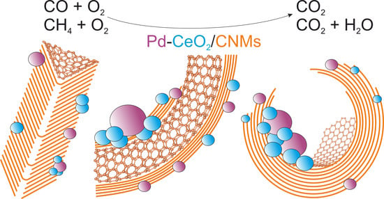
Graphical Abstract

1. Introduction

Metal–oxide catalysts are widely used in a variety of heterogeneous catalytic reactions required to purify the atmosphere from harmful pollutants, such as CO and hydrocarbons [1]. This is of importance both to neutralize the hazardous components of gasoline and natural gas vehicle exhausts and to clean indoor air [2,3,4].
Cerium oxide (ceria) serves as an indispensable support for platinum group metals (PGM) in CO and hydrocarbon oxidation catalysts due to its high oxygen capacity and oxygen mobility, and its ability to accumulate and release oxygen under redox conditions [5,6]. The strong interaction between PGM and CeO2 provides the ability to stabilize deposited PGMs in a highly dispersed state. Thus, due to the synergism between the components, PGM and ceria, the high activity of Pd/CeO2 and Pt/CeO2 catalysts in CO oxidation at low temperatures can be achieved [7,8]. Active CO oxidation catalysts comprise metal ionic forms embedded in the fluorite structure of the CeO2 support, as well as subnanosized clusters/species attached to its surface [8,9,10]. Among PGM/CeO2 catalysts, palladium-based catalysts show the highest activity in the methane oxidation reaction [11,12]. To provide the activity of these catalysts in methane oxidation, it is important to maintain the active palladium species over a wide temperature range. The deactivation of Pd/CeO2 catalysts occurring at elevated temperatures is associated with a decrease in dispersion due to the sintering of active particles [11,13,14]. To improve the stability of methane oxidation catalysts, the authors of [15] managed to achieve the stability of the active component through the fabrication of core-shell structures, fixing PdO inside the CeO2 shell. The authors of [16] used the one-step dry ball-milling method, which also allowed them to synthesize core-shell structures, in which the shell contains Pd embedded in an amorphous ceria layer while the core consists of CeO2. In this catalyst, the active components are PdOx species embedded into the CeO2 surface layer, the strong interaction between which ensures an easy redox exchange between Pd and CeO2. The comprehensive review [12] discusses that by forming a certain combination of Pd in different oxidation states (Pd0, Pd2+, Pd4+, sub-stoichiometric PdOx, etc.), higher catalyst activity can be achieved compared to PdO-based systems. The possibility to stabilize certain forms of palladium on the support surface and their stability under the conditions of oxidative reactions are determined by the metal–support interaction, which means that the structure and, in particular, the defective structure of the support play a key role.
The fabrication of a defective highly dispersed CeO2-based support is the key to creating highly active oxidation catalysts. The pure Pd/CeO2 system does not possess sufficient thermal stability, and for use at elevated temperatures, CeO2 is doped with transition metal ions [17]. Moreover, the fixation of components on the surface of a thermostable support, such as Al2O3, is widely used, allowing for the stabilization of the dispersed deposited components [18,19].
Carbon materials are widely used in catalysis both independently as catalysts and as supports of heterogeneous catalysts [20,21]. The rather high surface area of carbon materials and their tunable porosity allow for the efficient dispersion of metals and oxides on their surface, thus forming supported metal–carbon catalysts [22], as well as more complex multi-component composite catalysts [23,24,25].
In our previous work [23], we tested carbon nanotubes as a support to stabilize Pd or Pt and CeO2 in the dispersed state. We supposed that a carbon support could increase catalysts’ stability in a humid atmosphere, which is important for actual catalyst applications [26]. Of particular interest is the use of various nanostructured graphene-based carbon materials, namely different carbon nanofibers that differ in the packaging of the graphene layers (herring-bone or deck of cards). Of special note are carbon nanotubes, in the structure of which the graphene layers are arranged almost parallel to the growth axis of tube. Various nanostructured carbon materials can differ significantly in their structural characteristics and physicochemical surface properties. Consequently, the nature and structure of the carbon support can have a significant influence on the catalytic properties of the samples. Defects in carbon materials, including point, and extended defects with the presence of various functional groups serve as the centers of stabilization of the highly dispersed supported components up to the single-atom state [27]. Moreover, the dispersion and the nature of the supported components on the carbon surface are determined by the morphology, the structure of the carbon material, the porous structure and the hydrophobic/hydrophilic properties of the surface. The hydrophobic/hydrophilic properties of carbon materials affects the deposition of the active component, but the surface chemistry of carbon materials can easily be modified, for example, by oxidation, to increase their hydrophilicity and favor ionic exchange [20].
For application in catalytic reactions, it is preferable to use carbon materials with a high surface area and a pronounced mesoporous structure. The absence of microporosity in nanostructured carbon materials eliminates diffusion and intraparticle mass transfer in the reaction medium [20].
The main catalytic characteristics of composite catalysts are primarily determined by the chemical state of the active Pd/CeO2 components. However, the role of the support in the dispersion and local distribution of the active components, as well as the interaction between the active phase and the support, are also important [28]. The aim of this work is to study the structural, physical and chemical properties of the composite catalysts Pd-CeO2/CNMs. We used highly ordered carbon nanofibers and nanotubes as well as disordered carbon material of the Sibunit family. The latter material is of great interest for use in heterogeneous catalytic reactions due to its high resistance to attrition and mechanical crushing, as well as its chemical inertness [29]. All CNMs are characterized by a developed mesoporosity in the absence of micropores. For comparison, the conventional Pd-CeO2/Al2O3 was also synthesized and studied. The catalysts were tested in CO and CH4 oxidation reactions. These industrially important reactions are also widely used for testing the active surface centers. The catalysts were characterized by a complex of physicochemical methods, including XRD, TEM and XPS.

2. Materials and Methods

2.1. Synthesis

Carbon nanotubes (CNTs) and nanofibers (CNFs) were synthesized by the decomposition of 100% ethylene [30]. The catalysts of 62%Fe-8%Ni-30%Al2O3 and 65%Ni-25%Cu-10%Al2O3 composition were used as the growth catalysts for CNTs and CNFs, respectively. The synthesized CNTs and CNFs were washed from the growth catalyst by boiling in 2 M HCl for 6 h. The samples were then washed of chlorine ions by repeated boiling in distilled water until the negative reaction when AgNO3 was added to the washed water. The washed samples were dried in an Ar flow at 170 °C for 2 h, and then stored in sealed containers. Sibunit mesoporous material was chosen as a commercial carbon support. A broad class of commercial carbon materials of the Sibunit family includes carbon black and pyrolytic carbon composites, which are produced by the decomposition of natural gas on carbon black grains with subsequent activation, leading to the partial or complete removal of carbon black particles and the formation of a porous structure [31]. A carbon support of the Sibunit family (batch no. 4) was provided by the developer’s company—Center of New Chemical Technologies of the Boreskov Institute of Catalysis (Omsk affiliated department), Russia.
Pd-CeO2/CNMs catalysts, as well as CeO2/CNTs comparison support, were obtained using a previously developed method of co-impregnation of CNMs with active component precursors from acetone solutions [25]. Pd(NO3)2·2H2O palladium nitrate (synthesized following the procedure described by Khranenko et al. [32]) and ammonium cerium(IV) nitrate (NH4)2[Ce(NO3)6] (CAN, Rare metals plant, Novosibirsk, Russia) were used as precursors. The weight of the precursors was calculated to achieve the desired composition in the resulting samples, 6 wt.% Pd and 20 wt.% CeO2. The reagents were dissolved in acetone and added to the CNMs. The mixture was treated with ultrasound for 10 min and kept in a closed container for 12 h. The acetone was evaporated for 12 h in air and then for 24 h in a desiccator at a pressure of p~0.1 atm. The synthesized samples were heated in He to 200 °C at a rate of 10 °C/min, then to 350 °C at a rate of 1 °C/min and kept for 30 min at 350 °C, followed by cooling to room temperature. Further thermal activation involved heating the samples in 1% O2 to 350 °C in the reactor and keeping at this temperature for 2 h.
The Pd-CeO2/Al2O3 sample was prepared by the incipient wetness impregnation method. For 0.50 g Al2O3, 0.40 g CAN and 0.100 g Pd(NO3)2·2H2O were taken. The cerium and palladium precursors were dissolved in 3 mL of acetone. The impregnation was performed in several iterations with an intermediate rapid drying of the support in a flow of warm air. The impregnated sample was calcined similarly as described above. Further thermal activation involved heating the Pd-CeO2/Al2O3 sample in 1% O2 to 450 °C in the reactor and keeping at 450 °C for 2 h.
The catalysts synthesized were designated as the Pd-CeO2/support, where “support” denotes the carbon or oxide support: CNTs, CNFs, Sibunit or Al2O3. The text also uses the designation Pd-CeO2/CNMs to summarize all the catalysts deposited on various carbon nanomaterials. The reference support, which does not contain palladium, was designated as CeO2/CNTs.

2.2. Samples Characterization

2.2.1. X-ray Diffraction Analysis (XRD)

X-ray diffraction patterns were obtained on a STOE STADI MP diffractometer (STOE, Darmstadt, Germany) using MoKα1 radiation (λ = 0.70926 Å). To generate the primary beam, a curved Ge(111) monochromator was used. The analysis was carried out in the transmission mode. A DECTRIS MYTHEN multichannel detector was used to record the signal. The scanning was performed in the 2Θ range of 5–40° with a step of 0.0015° and an accumulation time of 10 s.
The phase analysis was performed using the ICDD PDF-2 (2009) powder database. Full-profile modeling by the Rietveld method to refine the cerium oxide lattice parameter as well as to estimate the size of the coherent scattering regions (CSR) and the lattice microstrain parameter was performed in the Topas v4.2 program (Bruker, Germany). An external standard, LaB6, analysed under similar conditions, was used to account for the instrumental broadening.

2.2.2. X-ray Photoelectron Spectroscopy (XPS)

XPS spectra were obtained on an ES300 (KRATOS Analytical, Manchester, U.K.) using MgKα X-rays (hν = 1253.6 eV). The calibration of the spectrometer energy scale was performed using the spectra of internal levels of gold (Au4f7/2) and copper (Cu2p3/2), with binding energies (Еb) of their peaks taken equal to 84.0 eV and 932.7 eV, respectively. The quantitative surface composition was estimated from the spectral area of the internal levels, taking into account the atomic sensitivity factors (ASF) for the corresponding elements [33]. The Pd3d and Ce3d spectra were decomposed into individual components using a combination of Gauss and Laurentz functions. The scattered electrons background was substracted by the Shirley method. The Ce3d spectra were decomposed into individual components related to the Ce3+ states (v0, v′ and u0, u′) and to the characteristics of the Ce4+ ions (v, v″, v‴ and u, u″, u‴) [34]. The percentage of Ce3+ ions was determined as the ratio of the area of the v0, v′ and u0, u′ components to the total Ce3d spectrum area.

2.2.3. Transmission Electron Microscopy (TEM)

The samples were studied by transmission electron microscopy (TEM) using a double aberration-corrected Themis Z electron microscope (Thermo Fisher Scientific, Waltham, MA, USA) operated at an accelerating voltage of 200 kV. Dark-field images were obtained in the scanning (STEM) mode using a HAADF (High-Angle Annular Dark-Field) detector. The instrument is equipped with a Super-X (Thermo Fisher Scientific) EDX detector to perform local elemental analysis of the samples. The crystal lattices on the obtained (S)TEM images were analyzed using the Fourier method. Copper grids (d = 3 mm) covered with a holey carbon film were used as a substrate for the samples. Alcohol suspensions of the samples were dispersed by ultrasound at a frequency of 35 kHz and deposited on the substrates.

2.3. Catalytic Properties

2.3.1. Catalytic Activity Measurements

The catalytic properties of the samples in the CO and CH4 oxidation reactions were tested using a flow reactor in the temperature-programmed reaction mode (TPR-CO+O2 and TPR-CH4+O2). A RGA 200 (SRS) quadrupole mass spectrometer was used to analyze the gas mixture. The weight and grain size of the Pd-CeO2/CNMs catalysts were 0.1 g and 0.25–0.5 mm, respectively. For the Pd-CeO2/Al2O3 catalyst, the weight and grain size were 0.1 g and 0.14–0.25 mm, respectively.
The initial reaction mixture contained 0.2 vol% CO, 1.0 vol% O2, 0.5 vol% Ne and He as a balance gas for the CO oxidation reaction. The flow rate was 1000 cm3/min, which corresponds to a GHSV of 600 L × g−1 × h−1. For the CH4 oxidation reaction, the initial reaction mixture contained 0.1 vol% CH4, 1.0 vol% O2, 0.5 vol% Ne and He as a balance gas. The flow rate was 100 cm3/min, which corresponds to a GHSV of 60 L × g−1 × h−1. The reaction mixture was admitted to the reactor at −40 °C or 50 °C for CO and CH4 oxidation reactions, respectively. After the steady-state concentrations of the reagents were established, the catalyst was heated at a heating rate of 10 °C/min to 350 °C. The experiments included three consecutive heating cycles with intermediate cooling in the reaction mixture.

2.3.2. Temperature Programmed Reduction

The TPR-CO curves were obtained using the reaction mixture containing 1.0 vol.% CO, 0.5 vol.% Ne and He. The gas mixture was introduced at a flow rate of 100 cm3/min to the catalyst sample (0.1 g), pre-cooled in the reactor to −40 °C. As the steady-state concentrations of CO and CO2 were established, the sample was heated to 450 °C at a 10 °C/min heating rate. The concentrations of CO, CO2, O2, H2 and H2O were monitored during the reaction by a mass-spectrometer at a frequency of 0.27 Hz. Before each TPR-CO experiment, the catalysts were pre-treated with a 1%O2/He gas mixture at 350 °C for 2 h, with subsequent cooling in this mixture and purging in helium afterwards.

3. Results and Discussion

3.1. Textural Properties and XRD Data

The textural characteristics of the used supports are provided in Table 1.
Figure 1 shows the X-ray diffraction patterns of the catalysts and of the corresponding supports. The main phases detected in the Pd-CeO2/CNMs catalysts are cerium oxide CeO2 with a fluorite-type structure (ICDD PDF-2 #00-034-0394), and a carbon material phase whose diffraction pattern does not vary much for different types of supports. In addition, a highly dispersed palladium oxide PdO phase (ICDD PDF-2 #00-41-1107) is detected on all XRD patterns.
To calculate and refine the structural and microstructural parameters of the detected phases, a full-profile modeling of the experimental diffraction data was carried out. The diffraction pattern of the carbon support or aluminum oxide was described by a set of individual peaks. The positions, widths and ratios of the intensities between these peaks were refined relative to the diffraction pattern of the pure support. The obtained parameters of individual diffraction maxima were then fixed and used to describe the diffraction pattern of the catalysts. The scattering from cerium oxide and palladium oxide was modeled using data on the crystal structure of these phases. In the case of the Pd-CeO2/Al2O3 sample, the precise calculation of the CSR size for the PdO phase was rather complicated due to the strong overlap of its lines with the lines of the Al2O3 support. The refined parameters of the cerium oxide and palladium oxide phases are provided in Table 2.
It was found that CeO2 and PdO particles in the composite catalysts are characterized by a small CSR size: 2–7 nm for cerium oxide, and 3–12 nm for palladium oxide. In addition, sufficiently large values of the microstrain parameter (e0) were obtained for these phases. This parameter describes the standard deviation of the interplanar distances from the average value. A high value of the microstrain parameter (above 0.1) is characteristic of defective structures. It can be noted that the highest CSR values for CeO2 were obtained for the catalyst deposited on Al2O3, and the highest CSR value for the PdO phase was obtained for the catalyst deposited on CNTs. An estimation of the phase content from the X-ray diffraction patterns for carbon-based catalysts showed that about 80% of the introduced palladium is in the form of PdO.

3.2. TEM Data

According to TEM data, CNTs have a diameter of 8–20 nm and the tube walls are composed of 6–20 graphene layers (Figure 2a). The CNFs with diameters ranging from 10 to 100 nm have a herring-bone structure (Figure 2b). The Sibunit support comprises the hollow carbon fragments of a spherical or near-spherical shape, ranging in size from 50 to 200 nm (Figure 2c). Aluminum oxide is represented by agglomerates of needle-shaped crystals with sizes d = 4–8 nm and l = 15–50 nm, and the size of agglomerates is 50–200 nm.
TEM images definitely show the presence of both single deposited nanoparticles and the agglomerates of nanoparticles of different sizes on the surface of all carbon and Al2O3 supports. Nevertheless, the morphology and nature of the support affect the agglomeration and distribution features of the deposited components.
For example, when palladium and cerium oxide are deposited on CNTs, both individual nanoparticles of PdO and CeO2 and agglomerates of deposited particles up to 100 nm in size are formed. The extended sections of the tubes contain a small number of supported particles. High-resolution TEM images show that the stabilization of single nanoparticles and agglomerates having a size of about 10–30 nm occurs predominantly near the surface defects of the tubes, such as breaks in the surface graphene layers, as well as in regions with various bends of the tubes (Figure 3a). Earlier, in [23] we observed that nanoparticles can also be found in the inner cavity of the tubes, although the number of such particles visible on TEM images is usually small. Figure 3a–c show HTREM and HAADF-STEM images of the same sample region and the corresponding EDX mapping patterns. It can be seen that the PdO nanoparticles in the agglomerates are about 8–10 nm in size and are somewhat larger than the contacting CeO2 nanoparticles. However, the single PdO nanoparticles located on the surface of CNTs and not contacting with CeO2 have a smaller size of about 2 nm (Figure 3). The size of CeO2 particles also varies depending on their location, and is about 2 nm for single crystallites contacting with CNTs only, and reaches 3–7 nm for crystallites comprising PdO-CeO2 agglomerates. The HAADF-STEM study also revealed the presence of single atoms on the surface of CNTs (Figure 3d). The non-uniformity of the support contrast does not allow us to reliably attribute the bright points observed on the images to Pd or Ce atoms, but the difference in brightness of the two closest dots in the same region of the CNTs suggests that both types of single atoms are present on the surface. Previously, in a study of catalysts with a single active component (Pd/CNTs or CeO2/CNTs), we showed that both Pd and Ce can be stabilized as single atoms on the surface of CNTs [26].
Carbon fibers as a support also provide the effective dispersion of the deposited nanoparticles over the surface. However, the density of nanoparticles on the surface of different fibers varies. This is apparently due to the wide variation in the diameter of the fibers and the heterogeneity of their surface defects. Consequently, the agglomerates of nanoparticles are formed around some fibers, tightly contacting each other and adhering to the fiber surface (Figure 4a), while on the surfaces of other tubes, single nanoparticles are formed (Figure 4d). The inner cavity of the carbon fibers is small, and the bridges of the graphene layers crossing this cavity are visible. Thus, the inner surface of the fibers is inaccessible to the deposited components, so the deposited components in this specimen are located only on the outer fiber surface. The size of the PdO particles observed in the TEM images is 5–8 nm. The size of the CeO2 nanoparticles is 1–5 nm, and larger particles are observed in dense agglomerates. Interestingly, no isolated PdO nanoparticles could be detected in this sample by TEM. As can be seen from Figure 4b,c, the PdO nanoparticles are surrounded and in tight contact with the CeO2 nanocrystallites. Figure 4d–f show a sample region containing a carbon nanofiber with a diameter of 10 nm, on the surface of which multiple CeO2 nanoparticles with the size of 1–2 nm are located. The HAADF-STEM image (Figure 4f) also shows the presence of disordered subnanosized clusters and single atoms on the surface. EDX data show the presence of both Pd and Ce signals in this region, with an atomic ratio of Ce:Pd ≈ 3:2. Since palladium can be incorporated into the fluorite structure of CeO2 [35,36], we assume the formation of highly dispersed and highly defective PdxCe1−xO2−δ species in the form of nanoparticles (1–2 nm) with a fluorite structure and amorphous subnanosized clusters in such areas of the sample.
In the Sibunit-based catalyst, the morphology of the support has a significant influence on the distribution of the agglomerates of PdO-CeO2 nanoparticles. The HAADF-STEM images and the corresponding EDX mapping patterns (Figure 5a–d) show that the agglomerates of deposited particles are located preferentially in the inner cavities of the Sibunit “globules”. Since the “globules” have open sections, the components in the inner cavity remain accessible to the reaction medium, but the activity of the catalyst can be limited by diffusion. The agglomerates in the inner cavities of the Sibunit contain PdO nanoparticles with a size of approximately 5–7 nm and CeO2 nanoparticles with a size of about 2–4 nm (Figure 5c–e). The PdO nanoparticles are in contact with the carbon support and CeO2 nanoparticles, but almost do not contact each other.
The surface of the Sibunit is represented by curved graphene sheets, which can come to the surface at different angles, especially in the areas where the wall thickness changes, and at the open edges of the “globules” (Figure 5e). Thus, the surface structure of the Sibunit combines areas similar to the surface of CNTs (extended graphene sheets) and the surface of CNFs (inclined graphene sheets). The latter, due to their defectiveness, are more preferable for fixing single nanoparticles, which determines some heterogeneity in the distribution of the deposited components over the outer surface of the support in the Pd-CeO2/Sibunit catalyst. Similar to the Pd-CeO2/CNFs catalyst, the Sibunit surface contains both single deposited atoms and PdxCe1−xO2−δ clusters or nanoparticles with an amorphous or fluorite structure (Figure 5f).
Therefore, according to TEM data, the following forms of the deposited components in Pd-CeO2/CNMs composite catalysts can be distinguished: (1) the deposited components anchored on the carbon surface in the form of single nanoparticles (PdO or CeO2) not interacting with each other; (2) agglomerates of PdO and CeO2 nanoparticles of different sizes, in which interblock boundaries between PdO and CeO2 crystallites are formed; (3) subnanosized PdOx or PdxCe1−xO2−δ clusters, characterized by an amorphous structure due to their small size, and fixed on the carbon support surface; and (4) Pd and Ce single atoms stabilized on the carbon surface, or embedded in the structure of oxide phases (Pd can be incorporated into the fluorite structure of CeO2).
The defectiveness of the carbon surface determines the number of deposited species that can be stabilized in atomically dispersed forms. In the case of the less defective CNTs, the number of single Pd and Ce atoms is visually low, while the defective surface of carbon nanofibers and the Sibunit contains multiple single atoms and subnanosized clusters with a disordered structure. It should also be noted that, for nanostructured carbon materials, the inner and outer surfaces on which the supported component can be fixed are often distinguished [37,38]. If the deposited component is located on the inner surface of the carbon material, which occurs in the case of the Sibunit, and is also possible for CNTs, the access of the reagents to it may be restricted, which can affect the catalytic activity.
The TEM data for the Pd-CeO2/Al2O3 reference catalyst are shown in Figure A1. This catalyst is characterized by a non-uniform distribution of the deposited components over the Al2O3 support. A part of the deposited component forms agglomerates up to 100 nm in size (Figure A1a,b), weakly interacting with the Al2O3 support, while another part is fixed on Al2O3 in the form of small agglomerates and single nanoparticles (Figure A1d,e). CeO2 nanoparticles in the agglomerates have a size of 3–10 nm and are located both on the Al2O3 support and on the surface of larger PdO nanoparticles. The size of PdO particles located on the Al2O3 support in the absence of contact with CeO2 is about 2 nm. Larger, 10–20 nm, single PdO nanoparticles are present in some areas of the sample. Large PdO particles are characterized by the presence of contacts with CeO2 crystallites (Figure A1a–c).
Thus, in Pd-CeO2/CNMs and Pd-CeO2/Al2O3 composite catalysts, the stabilization of various forms of the deposited component is determined by the structure and defectiveness of the support surface. The nanoparticles and clusters of the active components are anchored as separate species on the local defects of the support surface, which provides their spatial distribution without agglomeration. The formation of single atoms also increases the dispersion of the deposited components. This is more characteristic of CNFs and Sibunit supports due to the stronger interaction of Pd and Ce species with the carbon surface. In addition, the structure and morphology of the carbon materials influence the agglomeration of PdO and CeO2 nanoparticles. The nanoparticles comprising the agglomerates are larger than the single ones stabilized on the support.

3.3. XPS Data

Table 3 summarizes the surface composition data obtained by XPS for Pd-CeO2/CNMs and Pd-CeO2/Al2O3 catalysts. Analysis of the data shows that the concentrations of Pd and Ce in the samples deposited on different carbon nanomaterials do not differ significantly. In the case of CNFs and the Sibunit, the amount of palladium and cerium on the surface is somewhat higher. According to XRD and TEM data, the crystallite size of PdO and CeO2 is smaller when deposited on the CNFs and Sibunit supports, which leads to a higher intensity of palladium and cerium lines in the XPS spectra. For the Pd-CeO2/Al2O3 catalyst, the amount of palladium and cerium observed in the XPS spectra is about two times lower than in the case of CNMs-based samples. This fact is obviously related to the stabilization of larger PdO and CeO2 particles on the aluminum oxide surface compared to CNMs.
Figure 6 presents the Pd3d spectra for Pd-CeO2/CNTs and Pd-CeO2/Al2O3 samples. The Pd3d spectra for Pd-CeO2/CNFs and Pd-CeO2/Sibunit catalysts are presented in Figure A2. The Pd3d spectra for all studied catalysts can be described by a single spin-orbit Pd3d5/2-Pd3d3/2 doublet with an Eb(Pd3d5/2) value of about 337.1–337.4 eV. A small doublet with Eb(Pd3d5/2)~340 eV can be attributed to the PdO satellite structure [39,40]. The value Eb(Pd3d5/2) = 337.1–337.4 eV is typical of the palladium in a 2+ oxidation state. For palladium oxide PdO, the binding energy is usually observed in the region of 336.6–336.9 eV [39,40,41]. For Pd2+ ionic species stabilized in the CeO2 matrix, the Eb value is about 338.0 eV [36,42,43]. According to structural investigation and TPR-CO data, which will be discussed below, highly dispersed forms of Pd2+ and PdO particles of various sizes are observed in the studied catalysts. Apparently, the presence of several palladium states with close binding energy values does not allow us to reliably distinguish them in the Pd3d spectra. However, based on the position of the maximum of the Pd3d5/2 peak, we can conclude that among the studied samples, the fraction of palladium oxide PdO is maximal on the surface of the Pd-CeO2/Al2O3 catalyst and minimal in the case of the Pd-CeO2/CNFs sample.
Analysis of Ce3d spectra shows that the fraction of Ce3+ in the samples based on carbon nanomaterials is close to 20%. For the Pd-CeO2/Al2O3 sample, the Ce3+ fraction is slightly lower—15%. The Ce3+ content above 15% indicates the formation of defective CeO2 particles, with an increased concentration of oxygen vacancies [23,26]. This is consistent with the XRD data indicating the formation of small CeO2 nanocrystallites with a high value of the microstrain parameter. The lattice microdistortions induce the formation of Ce3+ ions and oxygen vacancies.

3.4. TPR-CO+O2, TPR-CH4+O2: Catalytic Data

The influence of the support nature was studied for Pd-CeO2/CNMs and Pd-CeO2/ Al2O3 catalysts in the CO and CH4 oxidation reactions. Figure 7 shows the temperature dependencies of CO and CH4 conversion. Table 4 summarizes catalytic data on the behavior of the studied catalysts in CO and CH4 oxidation reactions, indicating reference temperatures at which conversions of 10% (T10), 50% (T50) and 90% (T90) are achieved.
The catalytic study showed that the CNMs-based catalysts are active in the CO oxidation reaction at temperatures below 0 °С and are characterized by a close low-temperature activity. The Pd-CeO2/Al2O3 catalyst manifests activity only at temperatures above 25 °C. In the mid-temperature region, the T50 values are close to 78 °C for all CNMs-based samples. At temperatures above 80 °C, the CO conversion dependencies are different. Pd-CeO2/CNTs and Pd-CeO2/CNFs catalysts are characterized by lower temperatures of achieving a high CO conversion, and T90 for these catalysts is 115 and 121 °С, which is about 20 °С lower than for the Pd-CeO2/Sibunit catalyst (138 °С). The CO conversion curve for the Pd-CeO2/Al2O3 catalyst is shifted by 70 °С to higher temperatures, relative to the curves for CNMs-based catalysts.
In the case of the CH4 oxidation reaction, the lowest temperature to achieve 10% conversion of CH4 (T10) is observed for the Pd-CeO2/CNTs (235 °C) and Pd-CeO2/Sibunit (235 °C) catalysts. The methane conversion curves for the Pd-CeO2/CNFs and Pd-CeO2/Al2O3 catalysts are shifted toward higher temperatures.
To investigate the activity of the catalyst oxygen with respect to CO, and to identify the forms of oxidized Pd on the catalyst surface, the temperature-programmed reaction with CO in the absence of oxygen in the gas phase (TPR-CO method) was applied.

3.5. TPR-CO Data

Figure 8 shows the temperature dependencies of CO2 evolution during the interaction of the catalysts with CO. The evolution of CO2 for the CeO2/CNTs reference support starts around 100 °C, and no peaks are observed in the range of 100–300 °C. In agreement with our previous results [44], CO2 evolution occurs at the temperature range of 100–250 °C due to the small activation energy of the interaction of CO and oxygen on the surface of CeO2, while the evolution of CO2 at temperatures above 300 °C is due to the interaction of CO with the bulk oxygen of CeO2 [45,46]. For Pd-containing catalysts, several peaks of CO2 evolution are observed in the temperature range up to 300 °C, which allows us to refer them to different forms of oxidized palladium. According to [9,47,48], the peaks of CO2 evolution in the region of −40–70 °C (peak 1), 70–200 °C (peak 2) and 250–300 °C (peak 3) refer to active PdOx surface clusters, PdO nanoparticles and CeO2 nanoparticles modified by Pd2+ ions, respectively.
According to Figure 8 (see inset), the release of CO2 for Pd-CeO2/CNFs and Pd-CeO2/CNTs catalysts starts already at −40 °C (peak 1), while for Pd-CeO2/Al2O3 and Pd-CeO2/Sibunit catalysts, CO2 is registered at −20 and 0 °C. This indicates that the Pd-CeO2/CNTs and Pd-CeO2/CNFs catalysts contain more loosely bound reactive oxygen in the PdOx surface clusters. The second peak for these catalysts, which is responsible for the interaction of CO with PdO, is observed at a close temperature of about 150 °C, while for the Pd-CeO2/Sibunit catalyst, the Tmax of the PdO reduction peak is lower and is about 140 °C. This implies that the Pd-CeO2/Sibunit catalyst contains PdO particles with more reactive oxygen, which may indicate a higher dispersity or defectiveness of PdO crystallites in this sample. The Tmax of the third peak of CO2 evolution is almost the same for all CNMs-based catalysts. For the Pd-CeO2/Al2O3 catalyst, a shift of all peaks to the high-temperature region is observed. Moreover, in the temperature range of 100–270 °C, two poorly resolved peaks responsible for the interaction of CO with PdO are observed. The separation of these two peaks is probably related to the presence of two types of PdO nanoparticles, which differ not only in size, but also in the presence or absence of contacts with CeO2 nanoparticles, according to TEM data.
Table 5 presents quantitative data on the evolved CO2 for the second peak, calculated as the area under the corresponding peak. For peak 1, the amount of CO2 released in all the catalysts is close and equals 5–10 μmol/g, and the CO2/Pd ratio is in the range of 0.009–0.017, which corresponds to ~1–2% of all the Pd in the catalyst. For peak 2, corresponding to the interaction of CO with PdO, the amounts of evolved CO2 differ significantly. The CO2-2/Pd ratio exceeds 1 for the Pd-CeO2/CNTs catalyst and the total amount of evolved CO2 for this catalyst is the highest. According to XRD and TEM data, the Pd-CeO2/CNTs catalyst comprises larger and well-crystallized PdO and CeO2 nanoparticles that are efficiently mixed in PdO-CeO2 agglomerates. In this case, during the TPR-CO reaction, the interblock boundaries between PdO and CeO2 crystallites provide additional oxygen transfer and involvement of oxygen from CeO2 nanoparticles in the PdO reduction process, leading to an increased amount of CO reacting with the PdO as a second peak. The variation of the maximum temperature of the second peak for different catalysts is most likely related not only to the size of the PdO particles, but also to the modification of the CeO2 fluorite lattice by Pd2+ ions, which facilitate the mobility of oxygen in the fluorite lattice [9].
Thus, the results of the catalytic testing of the studied Pd-CeO2/CNMs composites showed that all the samples are characterized by similar catalytic activity in the oxidation of CO. For all carbon-based catalysts, the ignition of the CO oxidation reaction occurs at a low temperature near 0 °C, indicating the high activity of these catalysts. This activity is directly related to the stabilization of highly dispersed PdOx clusters and PdxCe1−xO2−δ particles on the surface of carbon materials’ defects, which can react with CO at around-zero temperatures (Figure 9). The observation of low-temperature TPR-CO peaks at subzero temperatures confirms this assumption.
The notable differences in catalytic activity are observed in CH4 oxidation, in which the methane conversion curves have a different temperature shift. CNTs and Sibunit-based catalysts start operating at lower temperatures than the CNFs-based one. The presence of dispersed PdO nanoparticles determines the activity of catalysts in methane oxidation (Figure 9). The possibility of the stabilization of deposited active components in the form of the agglomerates of PdO and CeO2 nanoparticles having the contacting boundaries allows for the efficient oxygen transfer from CeO2 to PdO. The optimal combination of highly dispersed PdxCe1−xO2−δ species and PdO nanoparticles in the CNTs-based catalyst makes it highly effective in both studied oxidation reactions.
It should be noted that when the carbon-based samples interact with oxygen, the oxidation of the carbon support with the evolution of CO2 at temperatures above 300 °C is possible (Figure A3a). It is important to note that CO is not formed until 350 °C. During the reaction of CH4 oxidation, only CO2 and H2O are observed as reaction products, while CO formation does not occur (Figure A3b). At the same time, the evolution of water is observed at a temperature of 280 °C, which is 100 °C higher than the start temperature of CH4 oxidation (180 °C). We associate this with the decomposition of the hydroxide that can form during the interaction of PdO with CH4. Isothermal tests for 4 h at 350 °С show the gradual deactivation of the catalyst (Figure A3c) so that the CH4 conversion decreases from 77 to 30%. Therefore, further studies are needed to improve the stability of Pd-CeO2/CNMs catalysts.

4. Conclusions

Highly dispersed composite catalysts containing palladium and CeO2 supported on nanostructured carbon materials were synthesized and studied in CO and CH4 oxidation reactions. The carbon materials of different structures (CNTs, CNFs, Sibunit) were found to be effective in stabilizing the active forms of the deposited Pd-CeO2 composite components. Cerium is stabilized in the form of small CeO2 nanoparticles ranging in size from 1 to 5 nm, while palladium is in the Pd2+ state, forming subnanosized PdOx clusters and PdO nanoparticles with a size of 2–12 nm. The defective structure of the carbon support surface is responsible for anchoring the highly dispersed nanoparticles and clusters.
Carbon-based catalysts are characterized by close activity in the oxidation of CO, starting to work at around-zero temperatures. The activity in methane oxidation differs, and is determined both by the dispersion of PdO nanoparticles and by the presence of PdO-CeO2 interblock contacts, providing oxygen transfer. The data obtained indicate the good accessibility of active palladium species supported on carbons having a different morphology.
The use of aluminum oxide as a reference support leads to the formation of the same forms of the active component, but their distribution over the support surface differs considerably. Dispersed PdO nanoparticles are stabilized on the Al2O3 surface as separated entities without contact with CeO2 particles. This significantly affects the oxygen transfer from the ceria lattice to the reagents adsorbed on palladium species, so the activity of the Pd-CeO2/Al2O3 catalyst in CO and CH4 oxidation is lower than that of all Pd-CeO2/CNMs systems.
Thus, carbon materials with different structures could be efficiently used as supports in composite catalysts. By using carbon materials with varying structures, defectiveness and morphologies, it is possible to anchor the deposited active components in the desired structural and electronic state.

Author Contributions

Conceptualization, A.B. and O.S.; validation, A.B., A.S. and O.P.; investigation, O.S., L.K., E.S., A.Z., I.G., T.K. and A.S.; writing—original draft preparation, O.S., T.K. and A.Z.; writing—review and editing, A.B., L.K., E.S., O.P. and S.K.; visualization, O.S., L.K., A.S., I.G. and E.S.; supervision, S.K.; project administration, A.B.; funding acquisition, A.B. All authors have read and agreed to the published version of the manuscript.

Funding

This research was funded by the Russian Science Foundation, grant number 21-13-00094.

Institutional Review Board Statement

Not applicable.

Informed Consent Statement

Not applicable.

Data Availability Statement

The data presented in this study are available on request from the corresponding author.

Acknowledgments

The TEM, XRD and XPS studies were carried out using facilities of the shared research center “National center of investigation of catalysts” at the Boreskov Institute of Catalysis.

Conflicts of Interest

The authors declare no conflict of interest. The funders had no role in the design of the study; in the collection, analyses or interpretation of data; in the writing of the manuscript; or in the decision to publish the results.

Appendix A

Figure A1. TEM data for Pd-CeO2/Al2O3: (a,b) HAADF-STEM image and corresponding EDX-mapping pattern; (c) HRTEM image with FTT of the selected region shown in the inset; the interplanar distances corresponding to PdO and CeO2 are indicated; (d,e) HAADF-STEM image and corresponding EDX-mapping pattern. The square mark in the figure (a) indicates the region of the sample shown in the HRTEM image (c). The EDX elemental maps use the following colors: blue for Al, red for Pd and green for Ce. The maps are presented in background-corrected intensities.
Materials 16 04257 g0a1
Figure A2. The Pd3d spectra of (a) Pd-CeO2/CNFs and (b) Pd-CeO2/Sibunit samples.
Materials 16 04257 g0a2
Figure A3. Catalytic data for Pd-CeO2/CNTs catalyst: (a) Temperature dependence of O2 consumption and CO, CO2 and H2O evolution during temperature-programmed oxidation (TPO) in 1%O2/He; (b) Temperature dependences of CH4, O2 conversion and CO, H2O and CO2 evolution during CH4 oxidation; (c) Time dependence of CH4 conversion at 350 °C.
Materials 16 04257 g0a3

References

  1. Yao, Y.-F.Y. The Oxidation of CO and Hydrocarbons over Noble Metal Catalysts. J. Catal. 1984, 87, 152–162. [Google Scholar] [CrossRef]
  2. Farrauto, R.J.; Deeba, M.; Alerasool, S. Gasoline Automobile Catalysis and Its Historical Journey to Cleaner Air. Nat. Catal. 2019, 2, 603–613. [Google Scholar] [CrossRef]
  3. Al Soubaihi, R.; Saoud, K.; Dutta, J. Critical Review of Low-Temperature CO Oxidation and Hysteresis Phenomenon on Heterogeneous Catalysts. Catalysts 2018, 8, 660. [Google Scholar] [CrossRef] [Green Version]
  4. Kim, H.J.; Shin, D.; Jeong, H.; Jang, M.G.; Lee, H.; Han, J.W. Design of an Ultrastable and Highly Active Ceria Catalyst for CO Oxidation by Rare-Earth- and Transition-Metal Co-Doping. ACS Catal. 2020, 10, 14877–14886. [Google Scholar] [CrossRef]
  5. Masui, T.; Ozaki, T.; Machida, K.; Adachi, G. Preparation of Ceria–Zirconia Sub-Catalysts for Automotive Exhaust Cleaning. J. Alloys Compd. 2000, 303–304, 49–55. [Google Scholar] [CrossRef]
  6. He, H.; Dai, H.; Au, C. Defective Structure, Oxygen Mobility, Oxygen Storage Capacity, and Redox Properties of RE-Based (RE = Ce, Pr) Solid Solutions. Catal. Today 2004, 90, 245–254. [Google Scholar] [CrossRef]
  7. Luo, M.-F.; Hou, Z.-Y.; Yuan, X.-X.; Zheng, X.-M. Characterization Study of CeO2 Supported Pd Catalyst for Low-Temperature Carbon Monoxide Oxidation. Catal. Lett. 1998, 50, 205–209. [Google Scholar] [CrossRef]
  8. Boronin, A.I.; Slavinskaya, E.M.; Figueroba, A.; Stadnichenko, A.I.; Kardash, T.Y.; Stonkus, O.A.; Fedorova, E.A.; Muravev, V.V.; Svetlichnyi, V.A.; Bruix, A.; et al. CO Oxidation Activity of Pt/CeO2 Catalysts below 0 °C: Platinum Loading Effects. Appl. Catal. B Environ. 2021, 286, 119931. [Google Scholar] [CrossRef]
  9. Stonkus, O.A.; Kardash, T.Y.; Slavinskaya, E.M.; Zaikovskii, V.I.; Boronin, A.I. Thermally Induced Structural Evolution of Palladium-Ceria Catalysts. Implication for CO Oxidation. ChemCatChem 2019, 11, 3505–3521. [Google Scholar] [CrossRef]
  10. Priolkar, K.R.; Bera, P.; Sarode, P.R.; Hegde, M.S.; Emura, S.; Kumashiro, R.; Lalla, N.P. Formation of Ce1−xPdxO2−δ Solid Solution in Combustion-Synthesized Pd/CeO2 Catalyst: XRD, XPS, and EXAFS Investigation. Chem. Mater. 2002, 14, 2120–2128. [Google Scholar] [CrossRef]
  11. Farrauto, R.J.; Hobson, M.C.; Kennelly, T.; Waterman, E.M. Catalytic Chemistry of Supported Palladium for Combustion of Methane. Appl. Catal. A Gen. 1992, 81, 227–237. [Google Scholar] [CrossRef]
  12. Colussi, S.; Fornasiero, P.; Trovarelli, A. Structure-Activity Relationship in Pd/CeO2 Methane Oxidation Catalysts. Chin. J. Catal. 2020, 41, 938–950. [Google Scholar] [CrossRef]
  13. Ciuparu, D.; Lyubovsky, M.R.; Altman, E.; Pfefferle, L.D.; Datye, A. Catalytic Combustion of Methane over Palladium-Based Catalysts. Catal. Rev. 2002, 44, 593–649. [Google Scholar] [CrossRef]
  14. Farrauto, R.J.; Lampert, J.K.; Hobson, M.C.; Waterman, E.M. Thermal Decomposition and Reformation of PdO Catalysts; Support Effects. Appl. Catal. B Environ. 1995, 6, 263–270. [Google Scholar] [CrossRef]
  15. Cargnello, M.; Delgado Jaén, J.J.; Hernández Garrido, J.C.; Bakhmutsky, K.; Montini, T.; Calvino Gámez, J.J.; Gorte, R.J.; Fornasiero, P. Exceptional Activity for Methane Combustion over Modular Pd@CeO2 Subunits on Functionalized Al2O3. Science 2012, 337, 713–717. [Google Scholar] [CrossRef]
  16. Danielis, M.; Colussi, S.; de Leitenburg, C.; Soler, L.; Llorca, J.; Trovarelli, A. Outstanding Methane Oxidation Performance of Palladium-Embedded Ceria Catalysts Prepared by a One-Step Dry Ball-Milling Method. Angew. Chem. Int. Ed. 2018, 57, 10212–10216. [Google Scholar] [CrossRef]
  17. Vlaic, G.; Di Monte, R.; Fornasiero, P.; Fonda, E.; Kašpar, J.; Graziani, M. Redox Property–Local Structure Relationships in the Rh-Loaded CeO2–ZrO2 Mixed Oxides. J. Catal. 1999, 182, 378–389. [Google Scholar] [CrossRef]
  18. Heck, R.M.; Farrauto, R.J.; Gulati, S.T.; Ronald, M.; Heck, R.J.; Farrauto, S.T.G. Catalytic Air Pollution Control; John Wiley & Sons, Inc.: Hoboken, NJ, USA, 2009; Volume 5, ISBN 9780470275030. [Google Scholar]
  19. Heck, R.M.; Farrauto, R.J. Automobile Exhaust Catalysts. Appl. Catal. A Gen. 2001, 221, 443–457. [Google Scholar] [CrossRef]
  20. Serp, P.; Machado, B. Carbon (Nano)Materials for Catalysis. In Nanostructured Carbon Materials for Catalysis; The Royal Society of Chemistry: London, UK, 2015; pp. 1–45. [Google Scholar]
  21. Melchionna, M.; Marchesan, S.; Prato, M.; Fornasiero, P. Carbon Nanotubes and Catalysis: The Many Facets of a Successful Marriage. Catal. Sci. Technol. 2015, 5, 3859–3875. [Google Scholar] [CrossRef] [Green Version]
  22. Mironenko, R.M.; Belskaya, O.B.; Likholobov, V.A. Synthesis of Pd/C Catalysts: Approaches to Regulating the Structure of Active Sites toward Achieving High Selectivity in Hydrogenation of Organic Compounds. Russ. J. Gen. Chem. 2020, 90, 532–549. [Google Scholar] [CrossRef]
  23. Kibis, L.; Zadesenets, A.; Garkul, I.; Korobova, A.; Kardash, T.; Slavinskaya, E.; Stonkus, O.; Korenev, S.; Podyacheva, O.; Boronin, A. Pd-Ce-Ox/MWCNTs and Pt-Ce-Ox/MWCNTs Composite Materials: Morphology, Microstructure, and Catalytic Properties. Materials 2022, 15, 7485. [Google Scholar] [CrossRef] [PubMed]
  24. Kibis, L.S.; Korobova, A.N.; Zadesenets, A.V.; Romanenko, A.V.; Kardash, T.Y.; Stonkus, O.A.; Korenev, S.V.; Podyacheva, O.Y.; Slavinskaya, E.M.; Boronin, A.I. Catalysts for Low-Temperature CO Oxidation Based on Platinum, CeO2, and Carbon Nanotubes. Dokl. Phys. Chem. 2022, 505, 115–121. [Google Scholar] [CrossRef]
  25. Kibis, L.S.; Korobova, A.N.; Fedorova, E.A.; Kardash, T.Y.; Zadesenets, A.V.; Korenev, S.V.; Stonkus, O.A.; Slavinskaya, E.M.; Podyacheva, O.Y.; Boronin, A.I. Application of N-Doped Carbon Nanotubes for the Preparation of Highly Dispersed PdO–CeO2 Composite Catalysts. J. Struct. Chem. 2022, 63, 407–422. [Google Scholar] [CrossRef]
  26. Kibis, L.S.; Zadesenets, A.V.; Garkul, I.A.; Korobova, A.N.; Kardash, T.Y.; Fedorova, E.A.; Slavinskaya, E.M.; Stonkus, O.A.; Korenev, S.V.; Podyacheva, O.Y.; et al. Pd-Ce-Ox Species on MWCNTs Surface: Probing the Structure-Activity Correlation in Low-Temperature CO Oxidation. Appl. Surf. Sci. 2023, 611, 155750. [Google Scholar] [CrossRef]
  27. Gawande, M.B.; Fornasiero, P.; Zbořil, R. Carbon-Based Single-Atom Catalysts for Advanced Applications. ACS Catal. 2020, 10, 2231–2259. [Google Scholar] [CrossRef]
  28. Machado, B.F.; Oubenali, M.; Rosa Axet, M.; Trang NGuyen, T.; Tunckol, M.; Girleanu, M.; Ersen, O.; Gerber, I.C.; Serp, P. Understanding the Surface Chemistry of Carbon Nanotubes: Toward a Rational Design of Ru Nanocatalysts. J. Catal. 2014, 309, 185–198. [Google Scholar] [CrossRef] [Green Version]
  29. Likholobov, V.A.; Fenelonov, V.B.; Okkel, L.G.; Goncharova, O.V.; Avdeeva, L.B.; Zaikovskii, V.I.; Kuvshinov, G.G.; Semikolenov, V.A.; Duplyakin, V.K.; Baklanova, O.N.; et al. New Carbon-Carbonaceous Composites for Catalysis and Adsorption. React. Kinet. Catal. Lett. 1995, 54, 381–411. [Google Scholar] [CrossRef]
  30. Podyacheva, O.Y.; Simonov, P.A.; Yashnik, S.A.; Kvon, R.I.; Stonkus, O.A.; Sobolev, V.I.; Khairulin, S.R.; Bukhtiyarov, V.I. Concurrent Anchoring of Au NPs and Amine Groups under the Decomposition of Supported L-Arginine-Au(III) Complexes:A Simple Way to Active Au/C Catalyst for Pure Hydrogen Production. ChemistrySelect 2022, 7, e202201110. [Google Scholar] [CrossRef]
  31. Surovikin, V.F.; Plaxin, G.V.; Semikolenov, V.A.; Likholobov, V.A.; Tiunova, I.J. Porous Carbonaceous Material. U.S. Patent 4,978,649, 18 December 1990. [Google Scholar]
  32. Khranenko, S.P.; Baidina, I.A.; Gromilov, S.A. Crystal Structure Refinement for Trans-[Pd(NO3)2(H2O)2]. J. Struct. Chem. 2007, 48, 1152–1155. [Google Scholar] [CrossRef]
  33. Moulder, J.; Stickle, W.; Sobol, W.; Bomben, K.D. Handbook of X-ray Photoelectron Spectroscopy; Perkin-Elmer Corp.: Eden Prairie, MN, USA, 1992. [Google Scholar]
  34. Romeo, M.; Bak, K.; El Fallah, J.; Le Normand, F.; Hilaire, L. XPS Study of the Reduction of Cerium Dioxide. Surf. Interface Anal. 1993, 20, 508–512. [Google Scholar] [CrossRef]
  35. Hiley, C.I.; Fisher, J.M.; Thompsett, D.; Kashtiban, R.J.; Sloan, J.; Walton, R.I. Incorporation of Square-Planar Pd2+ in Fluorite CeO2: Hydrothermal Preparation, Local Structure, Redox Properties and Stability. J. Mater. Chem. A 2015, 3, 13072–13079. [Google Scholar] [CrossRef] [Green Version]
  36. Gulyaev, R.V.; Kardash, T.Y.; Malykhin, S.E.; Stonkus, O.A.; Ivanova, A.S.; Boronin, A.I. The Local Structure of PdxCe1−xO2−x−δ Solid Solutions. Phys. Chem. Chem. Phys. 2014, 16, 13523–13539. [Google Scholar] [CrossRef] [PubMed]
  37. Chizari, K.; Janowska, I.; Houllé, M.; Florea, I.; Ersen, O.; Romero, T.; Bernhardt, P.; Ledoux, M.J.; Pham-Huu, C. Tuning of Nitrogen-Doped Carbon Nanotubes as Catalyst Support for Liquid-Phase Reaction. Appl. Catal. A Gen. 2010, 380, 72–80. [Google Scholar] [CrossRef]
  38. Ersen, O.; Werckmann, J.; Houllé, M.; Ledoux, M.-J.; Pham-Huu, C. 3D Electron Microscopy Study of Metal Particles inside Multiwalled Carbon Nanotubes. Nano Lett. 2007, 7, 1898–1907. [Google Scholar] [CrossRef] [PubMed]
  39. Pillo, T.; Zimmermann, R.; Steiner, P.; Hüfner, S. The Electronic Structure of PdO Found by Photoemission (UPS and XPS) and Inverse Photoemission (BIS). J. Phys. Condens. Matter 1997, 9, 3987–3999. [Google Scholar] [CrossRef]
  40. Kibis, L.S.; Titkov, A.I.; Stadnichenko, A.I.; Koscheev, S.V.; Boronin, A.I. X-ray Photoelectron Spectroscopy Study of Pd Oxidation by RF Discharge in Oxygen. Appl. Surf. Sci. 2009, 255, 9248–9254. [Google Scholar] [CrossRef]
  41. Brun, M.; Berthet, A.; Bertolini, J.C. XPS, AES and Auger Parameter of Pd and PdO. J. Electron Spectrosc. Relat. Phenom. 1999, 104, 55–60. [Google Scholar] [CrossRef]
  42. Xia, Y.; Ye, J.; Cheng, D.G.; Chen, F.; Zhan, X. Identification of a Flattened Pd-Ce Oxide Cluster as a Highly Efficient Catalyst for Low-Temperature CO Oxidation. Catal. Sci. Technol. 2018, 8, 5137–5147. [Google Scholar] [CrossRef]
  43. Muravev, V.; Simons, J.F.M.; Parastaev, A.; Verheijen, M.A.; Struijs, J.J.C.; Kosinov, N.; Hensen, E.J.M. Operando Spectroscopy Unveils the Catalytic Role of Different Palladium Oxidation States in CO Oxidation on Pd/CeO2 Catalysts. Angew. Chem. Int. Ed. 2022, 61, e202200434. [Google Scholar] [CrossRef]
  44. Lashina, E.A.; Slavinskaya, E.M.; Boronin, A.I. Low-Temperature Activity of Pd/CeO2 Catalysts: Mechanism of CO Interaction and Mathematical Modelling of TPR-CO Kinetic Data. Chem. Eng. Sci. 2021, 244, 116812. [Google Scholar] [CrossRef]
  45. Giordano, F.; Trovarelli, A.; De Leitenburg, C.; Giona, M. A Model for the Temperature-Programmed Reduction of Low and High Surface Area Ceria. J. Catal. 2000, 193, 273–282. [Google Scholar] [CrossRef]
  46. Slavinskaya, E.M.; Kardash, T.Y.; Stonkus, O.A.; Gulyaev, R.V.; Lapin, I.N.; Svetlichnyi, V.A.; Boronin, A.I. Metal–Support Interaction in Pd/CeO2 Model Catalysts for CO Oxidation: From Pulsed Laser-Ablated Nanoparticles to Highly Active State of the Catalyst. Catal. Sci. Technol. 2016, 6, 6650–6666. [Google Scholar] [CrossRef]
  47. Slavinskaya, E.M.; Gulyaev, R.V.; Zadesenets, A.V.; Stonkus, O.A.; Zaikovskii, V.I.; Shubin, Y.V.; Korenev, S.V.; Boronin, A.I. Low-Temperature CO Oxidation by Pd/CeO2 Catalysts Synthesized Using the Coprecipitation Method. Appl. Catal. B Environ. 2015, 166–167, 91–103. [Google Scholar] [CrossRef]
  48. Slavinskaya, E.M.; Zadesenets, A.V.; Stonkus, O.A.; Stadnichenko, A.I.; Shchukarev, A.V.; Shubin, Y.V.; Korenev, S.V.; Boronin, A.I. Thermal Activation of Pd/CeO2-SnO2 Catalysts for Low-Temperature CO Oxidation. Appl. Catal. B Environ. 2020, 277, 119275. [Google Scholar] [CrossRef]
Figure 1. X-ray diffraction patterns of Pd-CeO2/CNTs (a), Pd-CeO2/CNFs (b), Pd-CeO2/Sibunit (c) and Pd-CeO2/Al2O3 (d) shown as black curves. X-ray diffraction patterns of the corresponding supports are shown in blue; the scattering contribution from the PdO phase is shown in red. The *-sign indicates the reflexes of the CeO2 phase.
Materials 16 04257 g001
Figure 2. TEM images of (a) Pd-CeO2/CNTs, (b) Pd-CeO2/CNFs, (c) Pd-CeO2/Sibunit and (d) Pd-CeO2/Al2O3. The images are presented at the same magnification.
Materials 16 04257 g002
Figure 3. TEM data for Pd-CeO2/CNTs catalyst: (a) HRTEM image; (b,c) HAADF-STEM image of the region marked in figure (a), and a corresponding EDX-mapping pattern showing the distribution of carbon (blue), palladium (red) and cerium (green) in the selected region; (d) HAADF-STEM image showing an agglomerate of PdO and CeO2 nanoparticles on the CNTs’ surface. The insets in figure d show the images of selected areas with brightness/contrast correction for better visualization of single Pd and Ce atoms present on the surface of CNTs.
Materials 16 04257 g003
Figure 4. TEM data for Pd-CeO2/CNFs catalyst: (a) TEM image; (b) HRTEM image of the region marked in figure (a); (c) HRTEM image and an EDX-mapping pattern (in inset) of the same region; (df) TEM image, corresponding EDX-mapping pattern and a HAADF-STEM image of the marked region. The following colors are used for the EDX elemental maps: blue for carbon, red for palladium and green for cerium. The maps are presented in background-corrected intensities. The yellow circles indicate single Pd and Ce atoms on the CNFs surface.
Materials 16 04257 g004
Figure 5. TEM data for Pd-CeO2/Sibunit: (ad) HAADF-STEM images and corresponding EDX-mapping patterns; (e) I HRTEM image; (f) HAADF-STEM image, the yellow circles indicate single Pd and Ce atoms on Sibunit surface. The EDX elemental maps use the following colors: blue for carbon, red for palladium and green for cerium. The maps are presented in background-corrected intensities.
Materials 16 04257 g005
Figure 6. The Pd3d spectra of (a) Pd-CeO2/CNTs and (b) Pd-CeO2/Al2O3 samples.
Materials 16 04257 g006
Figure 7. Temperature dependencies of (a) CO and (b) CH4 conversion for Pd-CeO2/CNTs, Pd-CeO2/CNFs, Pd-CeO2/Sibunit and Pd-CeO2/Al2O3 catalysts.
Materials 16 04257 g007
Figure 8. CO2 evolution during TPR-CO for Pd-CeO2/CNTs, Pd-CeO2/CNFs, Pd-CeO2/Sibunit, Pd-CeO2/Al2O3 catalysts and for CeO2/CNTs sample. The inset shows the initial section of CO2 evolution curves in the temperature range of −40–100 °C.
Materials 16 04257 g008
Figure 9. A scheme showing the main types of palladium and ceria species formed on carbon supports: the PdOx clusters and PdxCe1−xO2−δ particles provide activity in low-temperature CO oxidation; the PdO nanoparticles are essential for CH4 oxidation.
Materials 16 04257 g009
Table 1. Textural characteristics of the CNMs and Al2O3 supports.
SupportSBET, m2/gVpore, cm3/gVmicro, cm3/gDpore, nm
CNTs1791.29029
CNFs1970.4509
Sibunit3130.440.016
Al2O31660.45011
Table 2. Structural and microstructural parameters of phases in Pd-CeO2/CNMs and Pd-CeO2/Al2O3 samples.
SampleCeO2 ParametersPdO Parameters
а, ÅD, nme0D, nme0
Pd-CeO2/CNTs5.411(1)4(1)0.18(3)12(2)0.34(3)
Pd-CeO2/CNFs5.414(3)2.7(3)0.16(4)6(1)0.1(2)
Pd-CeO2/Sibunit5.40(1)2.4(3)0.10(3)6(1)0.12(4)
Pd-CeO2/Al2O35.41(1)7(1)0.3(1)≈3 *-
* a precise value is not possible to calculate because of the strong overlap of the phase lines with the aluminum oxide lines.
Table 3. XPS data on the quantitative composition of the catalysts’ surface (atomic %); the binding energy of the Pd3d5/2 line (Eb(Pd3d5/2)), and the Ce3+ fraction calculated based on the Ce3d spectra.
SamplePd, at%Ce, at%Eb(Pd3d5/2), eVСе3+, %
Pd-CeO2/CNTs0.853.5337.219
Pd-CeO2/CNFs1.13.8337.420
Pd-CeO2/Sibunit1.03.6337.323
Pd-CeO2/Al2O30.52.8337.115
Table 4. Catalytic properties.
SampleCO OxidationCH4 Oxidation
T10, °CT50, °CT90, °CT10, °C
Pd-CeO2/CNTs1478115235
Pd-CeO2/CNFs2078121265
Pd-CeO2/Sibunit1478138235
Pd-CeO2/Al2O351145220280
Table 5. TPR-CO data: T2—temperature of peaks associated with the interaction of CO with PdO, T3—temperature of peaks associated with the interaction of CO with CeO2 modified by Pd2+ ions, CO2-2 and CO2-Σ—the amount of CO2 evolved during the second peak and total; CO2-2/Pd—the ratio of the amount of CO2 evolved during the second peak to the palladium content in the catalyst.
CatalystT2,°CT3,°CCO2-2, μmol/gCO2-2/PdCO2-Σ, μmol/g
Pd-CeO2/CNTs1522687221.282090
Pd-CeO2/CNFs1532643220.571450
Pd-CeO2/Sibunit1382703150.561090
Pd-CeO2/Al2O3190, 2252851700.30928
Disclaimer/Publisher’s Note: The statements, opinions and data contained in all publications are solely those of the individual author(s) and contributor(s) and not of MDPI and/or the editor(s). MDPI and/or the editor(s) disclaim responsibility for any injury to people or property resulting from any ideas, methods, instructions or products referred to in the content.