
FIB-SEM as a Volume Electron Microscopy Approach to Study Cellular Architectures in SARS-CoV-2 and Other Viral Infections: A Practical Primer for a Virologist

 ,
, 
Abstract
:1. Introduction
2. Electron Microscopy of Virus-Cell Interactions
3. Volume Electron Microscopy
4. FIB-SEM of Virus-Cell Interactions
4.1. Instrumentation
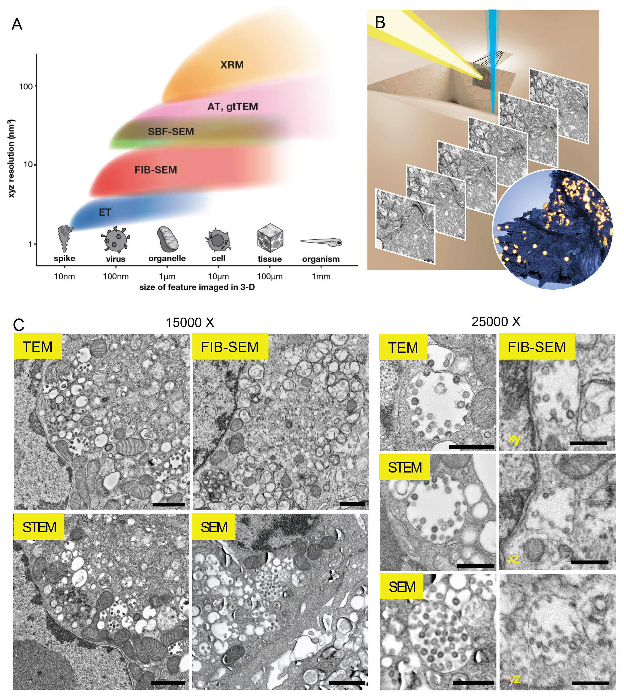
- FIB milling is orthogonal to the top surface, SEM imaging is at a ~54° angle, and the sample is retained at the “coincidence point”, where both beams converge in focus (Figure 1B). In Zeiss instruments with ATLAS (Fibics Inc., Ottawa, ON, Canada), a patterned platinum and carbon pad is deposited over the area of interest by the FIB. Milling the pad (and resin underneath) reveals the patterned notches in cross-section along with the sample. When captured in the image, notch locations allow the SEM to correct for sample drifts in x and y, and, critically, the FIB to correct for drifts in z “on the fly”. This feedback loop allows stable and continuous milling at z slice thicknesses reliably measured up to 3 nm [66], but this geometry causes some attenuation of signal from the deepest regions of the milled trench.
- In another approach (Thermo Fisher/FEI), the stage tilts and rises after each FIB mill for a high-resolution SEM raster, and then lowers and tilts back for the next mill, which again is orthogonal to the top surface and parallel to the “cliff face”. Markers on the top surface and cliff face adjacent to the ROI allow for drift correction. The stage can recover its location and tilt accurately, leading to small additions to total run time, and downstream alignment algorithms can correct for drifts in the imaging plane. Z thicknesses are more difficult to accurately measure, but SEM imaging is less affected by the geometry of the trench. The stage movements may also place some limits on the size and orientations of the sample allowed in the tool.
- An “L” shaped configuration (Hitachi) has an orthogonal orientation between the FIB and SEM columns, but it is the hybrid “Feiss” (FEI Magnum FIB + Zeiss Gemini SEM) custom tool from the Hess group [38] that has shown spectacular use in biology. Here, pre-trimmed resin samples are milled at finely controlled increments and imaged at optimal parameters, and innovative Ga tip regeneration protocols vastly increase volumes that can be imaged continuously. This orientation reveals the entire milled face of the sample for imaging, but ROI location may be limited as sputtering efficiencies at the FIB currents typically used will fall off within tens or a few hundred microns at best from the sample edge.
4.2. Sample Preparation
- There is no opportunity to post-stain samples. While lead citrate or phosphotungstic acid “post-staining” of sections on TEM grids boosts signal and contrast, this is not possible in FIB-SEM experiment. This is because these protocols are executed on accessible sections, while here, the FIB continuously and automatically ablates away material in the vacuum of an SEM chamber during a run. Instead, increased en-bloc heavy metal staining of samples to boost signal is possible, such as the rOTO protocol and its variants [70].
- One cannot perform traditional post-embedding or Tokayasu method-based [71] immunostaining during a FIB-SEM run, for the same reason as above. Modifying these immunostaining protocols so as to complete all steps before final resin-embedding is possible [72], but 5–15 nm gold particles must be enhanced to be reliably discerned, and in our hands, this can result in significant background signals.
- Resin hardness is important for stability under the electron and ion beams. A variety of resins including lower viscosity formulations like LR White have applications in immunocytochemistry. We and others have observed [72] that softer and even some medium hardness resin formulations perform poorly under the abrasive FIB and the constant switching between positively charged Ga ions and negatively charged electrons during a run. Hard formulations are safe and Durcupan is popular especially for tissue work [38], with newer formulations being developed.
- Sample orientation and trimming is critical for FIB-SEM. As milling efficiency typically drops off within 100 µm, it is important to position the ROI at or just below the top surface, and firmly fix this to the stub to prevent sample movements. Adherent cells are relatively easy, as they are typically milled “upside down” following resin embedment, substrate (gridded cover slip for correlative experiments) removal, and ROI location [66]. Targeted ROI imaging in tissue samples requires correlation, which can be done with near infrared branding [73], but this still requires careful trimming before FIB-SEM steps. For deeply embedded features that do not require exact spatial correlation, aggressive semi-thick microtome sectioning and occasional visual check by toluidine blue staining is commonly used to approach or even expose the feature of interest.
4.3. Image Generation and Resolution
- Long dwell times (how long and how many times the electron beam visits each pixel) have diminishing returns. This adds to the imaging overhead, and as useful signal is typically extracted and noise averaged out within several µs, accumulated negative charges imparted to the sample result in “baking” of the resin and image degradation. A dwell time of 3–4 µs is usually sufficient.
- At the parameters typically used (1–2 kV accelerating voltage, 0.5–2 nA current for the electron beam), beam widths are typically on the order of ~5 nm, meaning that artificial increasing of magnification ends up supersampling the ROI (pixel sampling << beam spot size) and blurring the image. Similarly, artificially setting very thin z slices risks including information from deeper within the sample, again causing image blurriness. Usually, either a ~5 nm z slice with faster imaging, or 10–20 nm slice with slower imaging is chosen.
- FIBs have limited “sweep”: the FIB moves back and forth rapidly at the cliff face while inching forward to mill away controlled amounts of resin. A limit of ~ 100 µm width is practical, as milling meaningful depths at these widths necessarily slows down the FIB to the point that sample drift in the SEM chamber rivals the FIB advancement rate. It is possible to ablate away larger volumes, or alternatively fixed volumes faster, with the FIB operated at a higher current, but the user loses fine control with the larger beam profile. Further, unlike in the material sciences, soft and insulating resin polymers are prone to warping and uncontrolled sputtering at high FIB currents. Irrespective, FIB-SEM imaging is not a high-throughput approach: as a rule of thumb, ~10,000 µm3 or a volume of 25 × 20 × 20 µm per day is a reasonable limit without significantly compromising resolutions or signal-to-noise (SNR) ratios.
- A simple empirical test for resolution is that at minimum, both membranes in the nuclear envelope and ER should be resolved and crisp in all planes throughout the run, and ideally, intercristal spaces should be visible in mitochondria. Automated and frequent beam tuning on the fly should prevent focus and stigmation issues, and these are immediately visible to the eye.
4.4. Processing and Correlation
- Registration and inversion: FIB-SEM images are typically acquired as grayscale.tiff files, with some drifts and jitters, and possibly poor SNR. 8-bit depth is sufficient for most applications and helps control file size. Contrast inversion is often an option during acquisition itself. A popular approach to register the newly acquired stack is to generate and apply transforms between successive images to correct for translation, rotation, skew etc. Some acquisition software can perform simple registration. The registration, stitching, and TrakEM2 [84] plugins in Fiji are popular, and some groups use custom solutions [85]. Stitching of FIB-SEM image tiles is unnecessary given small ROIs, but z-spacing correction [86] may be required.
- Binning and Denoising: FIB-SEM imaging uniquely allows high-resolution 3-D reconstructions with isotropic voxels, enabling equal and warp-free sampling. Many groups directly acquire data with isotropic settings (e.g., 8 × 8 nm image pixel and 8 nm FIB step size), but it is also possible to acquire at say 4 × 4 × 8 and then “bin” or average by 2 in xy to yield final 8 nm voxel sizes. Binning averages out noise and reduces file size, but at the expense of pixel size, so this needs to be deployed wisely. A hidden advantage is that coarser z spacing speeds up FIB advancement rates, stabilizing milling and reducing artifacts. Various denoising algorithms may also be applied and most recently, deep learning-based approaches for denoising [87] are showing appreciable results.
- Correlation: Combining FIB-SEM with other techniques, most frequently light or fluorescence microscopy (correlative light electron microscopy, CLEM) allows live, high-throughput screening or specific imaging of targets and 3-D ultrastructural reconstructions of the same regions of interest. Much work has been done recently, including an excellent handbook published on correlative imaging [75]. There are two connotations for CLEM: correlation for identification or relocation, where modalities like live light microscopy (LM) [66], cryo-LM [69], or XRM [88] are used to identify the coordinates to be imaged by FIB-SEM subsequently. The other is correlation for colocalization or registration, which requires high-resolution registration of signals from two modalities [89], which by default requires 3-D reconstructions for LM and FIB-SEM since the two imaging planes are orthogonal to each other. The correlation must take into account the differences in resolution, orientation, and differential warping and movements that occurs during sample processing. Recently developed software modules address these [90] but correlative experiments still need to be designed carefully to be correct and insightful.
4.5. Segmentation and Visualization
5. Observations of SARS-CoV-2-Cell Interactions in 3-D
Data Handling and Sharing
- Raw vs. “clean” image data: Even with acquisition at 8-bit depth, raw image datasets on the order of TB are common these days; cropping, binning down and processing data to the minimum “clean” image volume required to answer the question at hand will greatly ease handling and transfers. One possibility is creating two data streams: one raw data to archive, and more volatile targeted sub-volumes which can be probed as required. Appropriate sub-volumes are sufficient for most correlation and segmentation, although care is needed to maintain spatial fidelity.
- Derived data such as segmentation and quantitation: as the reporting of FIB-SEM data transitions from representative 2-D images to 3-D reconstructions to quantitative analyses from 3-D models, it is critical to track intermediate files and parameters used to ensure continuity of these derived data. Strong lab record practices and naming conventions help, and workflow builders such as KNIME can help teams repeat proven analysis pipelines or construct new ones. Further, as metadata conventions in vEM get established, converge, and finally integrated into imaging workflows, we expect that distinct data types will be easier to combine and connect.
- Data sharing: While the advantages (and encouragement) of sharing data have become clear, we acknowledge a hurdle specific to FIB-SEM or vEM data. Often, only a small fraction of these datasets is mined and published, leading to some reluctance to share these large image volumes. We hope for more concrete incentives for data sharing in the future, but for now make the case for undeniable collaborative benefits (and gratification!) of such datasets being re-used for scientific progress. There are many institutional repositories such as EMBL-EBI is accepting vEM datasets via EMPIAR (https://www.ebi.ac.uk/pdbe/emdb/empiar/, accessed on 31 March 2021), with several large datasets already deposited.
6. Conclusions
Supplementary Materials
Author Contributions
Funding
Institutional Review Board Statement
Informed Consent Statement
Data Availability Statement
Acknowledgments
Conflicts of Interest
References
- Knott, G.; Genoud, C. Is EM dead? J. Cell Sci. 2013, 126 Pt 20, 4545–4552. [Google Scholar] [CrossRef] [PubMed] [Green Version]
- Gentile, M.; Gelderblom, H.R. Electron microscopy in rapid viral diagnosis: An update. New Microbiol. 2014, 37, 403–422. [Google Scholar] [PubMed]
- Hazelton, P.R.; Gelderblom, H.R. Electron Microscopy for Rapid Diagnosis of Emerging Infectious Agents1. Emerg. Infect. Dis. 2003, 9, 294–303. [Google Scholar] [CrossRef]
- Beck, M.; Baumeister, W. Cryo-Electron Tomography: Can it Reveal the Molecular Sociology of Cells in Atomic Detail? Trends Cell Biol. 2016, 26, 825–837. [Google Scholar] [CrossRef]
- Tocheva, E.I.; Li, Z.; Jensen, G.J. Electron cryotomography. Cold Spring Harb Perspect. Biol. 2010, 2, a003442. [Google Scholar] [CrossRef] [Green Version]
- Iwasaki, K.; Omura, T. Electron tomography of the supramolecular structure of virus-infected cells. Curr. Opin. Struct. Biol. 2010, 20, 632–639. [Google Scholar] [CrossRef]
- Peddie, C.J.; Collinson, L.M. Exploring the third dimension: Volume electron microscopy comes of age. Micron 2014, 61, 9–19. [Google Scholar] [CrossRef] [PubMed]
- Lu, R.; Zhao, X.; Li, J.; Niu, P.; Yang, B.; Wu, H.; Wang, W.; Song, H.; Huang, B.; Zhu, N.; et al. Genomic characterisation and epidemiology of 2019 novel coronavirus: Implications for virus origins and receptor binding. Lancet 2020, 395, 565–574. [Google Scholar] [CrossRef] [Green Version]
- Ksiazek, T.G.; Erdman, D.; Goldsmith, C.S.; Zaki, S.R.; Peret, T.; Emery, S.; Tong, S.; Urbani, C.; Comer, J.A.; Lim, W.; et al. A novel coronavirus associated with severe acute respiratory syndrome. N. Engl. J. Med. 2003, 348, 1953–1966. [Google Scholar] [CrossRef] [PubMed]
- Goldsmith, C.S.; Tatti, K.M.; Ksiazek, T.G.; Rollin, P.E.; Comer, J.A.; Lee, W.W.; Rota, P.A.; Bankamp, B.; Bellini, W.J.; Zaki, S.R. Ultrastructural characterization of SARS coronavirus. Emerg. Infect. Dis. 2004, 10, 320–326. [Google Scholar] [CrossRef]
- Goldsmith, C.S.; Miller, S.E. Modern uses of electron microscopy for detection of viruses. Clin. Microbiol. Rev. 2009, 22, 552–563. [Google Scholar] [CrossRef] [Green Version]
- Hamre, D.; Kindig, D.A.; Mann, J. Growth and intracellular development of a new respiratory virus. J. Virol. 1967, 1, 810–816. [Google Scholar] [CrossRef] [PubMed] [Green Version]
- Gan, L.; Jensen, G.J. Electron tomography of cells. Q. Rev. Biophys. 2012, 45, 27–56. [Google Scholar] [CrossRef] [PubMed] [Green Version]
- Lucic, V.; Forster, F.; Baumeister, W. Structural studies by electron tomography: From cells to molecules. Annu. Rev. Biochem. 2005, 74, 833–865. [Google Scholar] [CrossRef] [PubMed]
- Cortese, M.; Lee, J.Y.; Cerikan, B.; Neufeldt, C.J.; Oorschot, V.M.J.; Kohrer, S.; Hennies, J.; Schieber, N.L.; Ronchi, P.; Mizzon, G.; et al. Integrative Imaging Reveals SARS-CoV-2-Induced Reshaping of Subcellular Morphologies. Cell Host Microbe 2020, 28, 853–866.e855. [Google Scholar] [CrossRef] [PubMed]
- Snijder, E.J.; Limpens, R.; de Wilde, A.H.; de Jong, A.W.M.; Zevenhoven-Dobbe, J.C.; Maier, H.J.; Faas, F.; Koster, A.J.; Barcena, M. A unifying structural and functional model of the coronavirus replication organelle: Tracking down RNA synthesis. PLoS Biol. 2020, 18, e3000715. [Google Scholar] [CrossRef]
- Klein, S.; Cortese, M.; Winter, S.L.; Wachsmuth-Melm, M.; Neufeldt, C.J.; Cerikan, B.; Stanifer, M.L.; Boulant, S.; Bartenschlager, R.; Chlanda, P. SARS-CoV-2 structure and replication characterized by in situ cryo-electron tomography. Nat. Commun. 2020, 11, 5885. [Google Scholar] [CrossRef] [PubMed]
- Wolff, G.; Limpens, R.W.A.L.; Zevenhoven-Dobbe, J.C.; Laugks, U.; Zheng, S.; de Jong, A.W.M.; Koning, R.I.; Agard, D.A.; Grünewald, K.; Koster, A.J.; et al. A molecular pore spans the double membrane of the coronavirus replication organelle. Science 2020, 369, 1395–1398. [Google Scholar] [CrossRef] [PubMed]
- Marko, M.; Hsieh, C.; Schalek, R.; Frank, J.; Mannella, C. Focused-ion-beam thinning of frozen-hydrated biological specimens for cryo-electron microscopy. Nat. Methods 2007, 4, 215–217. [Google Scholar] [CrossRef]
- Rigort, A.; Bauerlein, F.J.; Villa, E.; Eibauer, M.; Laugks, T.; Baumeister, W.; Plitzko, J.M. Focused ion beam micromachining of eukaryotic cells for cryoelectron tomography. Proc. Natl. Acad. Sci. USA 2012, 109, 4449–4454. [Google Scholar] [CrossRef] [PubMed] [Green Version]
- Maurer, U.E.; Sodeik, B.; Grunewald, K. Native 3D intermediates of membrane fusion in herpes simplex virus 1 entry. Proc. Natl. Acad. Sci. USA 2008, 105, 10559–10564. [Google Scholar] [CrossRef] [PubMed] [Green Version]
- Peng, L.; Ryazantsev, S.; Sun, R.; Zhou, Z.H. Three-dimensional visualization of gammaherpesvirus life cycle in host cells by electron tomography. Structure 2010, 18, 47–58. [Google Scholar] [CrossRef] [PubMed] [Green Version]
- Sougrat, R.; Bartesaghi, A.; Lifson, J.D.; Bennett, A.E.; Bess, J.W.; Zabransky, D.J.; Subramaniam, S. Electron tomography of the contact between T cells and SIV/HIV-1: Implications for viral entry. PLoS Pathog. 2007, 3, e63. [Google Scholar] [CrossRef] [PubMed] [Green Version]
- Risco, C.; de Castro, I.F.; Sanz-Sanchez, L.; Narayan, K.; Grandinetti, G.; Subramaniam, S. Three-Dimensional Imaging of Viral Infections. Annu. Rev. Virol. 2014, 1, 453–473. [Google Scholar] [CrossRef] [PubMed]
- Romero-Brey, I.; Bartenschlager, R. Viral Infection at High Magnification: 3D Electron Microscopy Methods to Analyze the Architecture of Infected Cells. Viruses 2015, 7, 6316–6345. [Google Scholar] [CrossRef] [PubMed]
- Jiang, Y.F.; Lin, H.L.; Fu, C.Y. 3D Mitochondrial Ultrastructure of Drosophila Indirect Flight Muscle Revealed by Serial-section Electron Tomography. J. Vis. Exp. 2017. [Google Scholar] [CrossRef] [PubMed] [Green Version]
- Sun, S.; Fisher, R.L.; Bowser, S.S.; Pentecost, B.T.; Sui, H. Three-dimensional architecture of epithelial primary cilia. Proc. Natl. Acad. Sci. USA 2019, 116, 9370–9379. [Google Scholar] [CrossRef] [PubMed] [Green Version]
- Marsh, B.J. Reconstructing Mammalian Membrane Architecture by Large Area Cellular Tomography. Methods Cell Biol. 2007, 79, 193–220. [Google Scholar] [CrossRef] [PubMed]
- He, Q.; Hsueh, M.; Zhang, G.; Joy, D.C.; Leapman, R.D. Biological serial block face scanning electron microscopy at improved z-resolution based on Monte Carlo model. Sci. Rep. 2018, 8, 1–13. [Google Scholar] [CrossRef] [PubMed] [Green Version]
- Ryan, K.; Lu, Z.; Meinertzhagen, I.A. The CNS connectome of a tadpole larva of Ciona intestinalis (L.) highlights sidedness in the brain of a chordate sibling. eLife 2016, 5, e16962. [Google Scholar] [CrossRef]
- White, J.G.; Southgate, E.; Thomson, J.N.; Brenner, S. The structure of the nervous system of the nematode Caenorhabditis elegans. Philos Trans. R. Soc. Lond B Biol. Sci. 1986, 314, 1–340. [Google Scholar] [CrossRef] [PubMed]
- Graham, B.J.; Hildebrand, D.G.C.; Kuan, A.T.; Maniates-Selvin, J.T.; Thomas, L.A.; Shanny, B.L.; Lee, W.-C.A. High-throughput transmission electron microscopy with automated serial sectioning. bioRxiv 2019. [Google Scholar] [CrossRef] [Green Version]
- Gholinia, A.; Curd, M.E.; Bousser, E.; Taylor, K.; Hosman, T.; Coyle, S.; Shearer, M.H.; Hunt, J.; Withers, P.J. Coupled Broad Ion Beam–Scanning Electron Microscopy (BIB–SEM) for polishing and three dimensional (3D) serial section tomography (SST). Ultramicroscopy 2020, 214, 112989. [Google Scholar] [CrossRef] [PubMed]
- Burnett, T.L.; Kelley, R.; Winiarski, B.; Contreras, L.; Daly, M.; Gholinia, A.; Burke, M.G.; Withers, P.J. Large volume serial section tomography by Xe Plasma FIB dual beam microscopy. Ultramicroscopy 2016, 161, 119–129. [Google Scholar] [CrossRef] [PubMed]
- Bushong, E.A.; Johnson, D.D.; Kim, K.-Y.; Terada, M.; Hatori, M.; Peltier, S.T.; Panda, S.; Merkle, A.; Ellisman, M.H. X-Ray Microscopy as an Approach to Increasing Accuracy and Efficiency of Serial Block-Face Imaging for Correlated Light and Electron Microscopy of Biological Specimens. Microsc. Microanal. 2015, 21, 231–238. [Google Scholar] [CrossRef] [PubMed] [Green Version]
- Kislinger, G.; Gnagi, H.; Kerschensteiner, M.; Simons, M.; Misgeld, T.; Schifferer, M. Multiscale ATUM-FIB Microscopy Enables Targeted Ultrastructural Analysis at Isotropic Resolution. iScience 2020, 23, 101290. [Google Scholar] [CrossRef] [PubMed]
- Kizilyaprak, C.; Stierhof, Y.D.; Humbel, B.M. Volume microscopy in biology: FIB-SEM tomography. Tissue Cell 2019, 57, 123–128. [Google Scholar] [CrossRef]
- Xu, C.S.; Hayworth, K.J.; Lu, Z.; Grob, P.; Hassan, A.M.; Garcia-Cerdan, J.G.; Niyogi, K.K.; Nogales, E.; Weinberg, R.J.; Hess, H.F. Enhanced FIB-SEM systems for large-volume 3D imaging. eLife 2017, 6, e25916. [Google Scholar] [CrossRef]
- Engel, A. Chapter 9 Scanning Transmission Electron Microscopy; Elsevier: Amsterdam, The Netherlands, 2009. [Google Scholar]
- Höhn, K.; Sailer, M.; Wang, L.; Lorenz, M.; Schneider, M.; Walther, P. Preparation of cryofixed cells for improved 3D ultrastructure with scanning transmission electron tomography. Histochem. Cell Biol. 2011, 135, 1–9. [Google Scholar] [CrossRef] [PubMed]
- Sousa, A.A.; Azari, A.A.; Zhang, G.; Leapman, R.D. Dual-axis electron tomography of biological specimens: Extending the limits of specimen thickness with bright-field STEM imaging. J. Struct. Biol. 2011, 174, 107–114. [Google Scholar] [CrossRef] [PubMed] [Green Version]
- Hohmann-Marriott, M.F.; Sousa, A.A.; Azari, A.A.; Glushakova, S.; Zhang, G.; Zimmerberg, J.; Leapman, R.D. Nanoscale 3D cellular imaging by axial scanning transmission electron tomography. Nat. Methods 2009, 6, 729–731. [Google Scholar] [CrossRef] [PubMed] [Green Version]
- Pokrovskaya, I.D.; Aronova, M.A.; Kamykowski, J.A.; Prince, A.A.; Hoyne, J.D.; Calco, G.N.; Kuo, B.C.; He, Q.; Leapman, R.D.; Storrie, B. STEM tomography reveals that the canalicular system and α-granules remain separate compartments during early secretion stages in blood platelets. J. Thromb. Haemost. 2016, 14, 572–584. [Google Scholar] [CrossRef] [PubMed]
- Read, C.; Schauflinger, M.; Nikolaenko, D.; Walther, P.; Von Einem, J. Regulation of Human Cytomegalovirus Secondary Envelopment by a C-Terminal Tetralysine Motif in pUL71. J. Virol. 2019, 93. [Google Scholar] [CrossRef] [PubMed] [Green Version]
- Hayworth, K.J.; Kasthuri, N.; Schalek, R.; Lichtman, J.W. Automating the Collection of Ultrathin Serial Sections for Large Volume TEM Reconstructions. Microsc. Microanal. 2006, 12, 86–87. [Google Scholar] [CrossRef] [Green Version]
- Baena, V.; Schalek, R.L.; Lichtman, J.W.; Terasaki, M. Serial-section electron microscopy using automated tape-collecting ultramicrotome (ATUM). Methods Cell Biol. 2019, 152, 41–67. [Google Scholar] [CrossRef] [PubMed]
- Kasthuri, N.; Hayworth, K.J.; Berger, D.R.; Schalek, R.L.; Conchello, J.A.; Knowles-Barley, S.; Lee, D.; Vazquez-Reina, A.; Kaynig, V.; Jones, T.R.; et al. Saturated Reconstruction of a Volume of Neocortex. Cell 2015, 162, 648–661. [Google Scholar] [CrossRef] [Green Version]
- Eberle, A.L.; Mikula, S.; Schalek, R.; Lichtman, J.; Tate, M.L.K.; Zeidler, D. High-resolution, high-throughput imaging with a multibeam scanning electron microscope. J. Microsc. 2015, 259, 114–120. [Google Scholar] [CrossRef] [PubMed] [Green Version]
- Norris, R.P.; Terasaki, M. Gap junction internalization and processing in vivo: A 3D immuno-electron microscopy study. J. Cell Sci. 2021, 134. [Google Scholar] [CrossRef] [PubMed]
- Micheva, K.D.; Smith, S.J. Array tomography: A new tool for imaging the molecular architecture and ultrastructure of neural circuits. Neuron 2007, 55, 25–36. [Google Scholar] [CrossRef] [PubMed] [Green Version]
- Denk, W.; Horstmann, H. Serial block-face scanning electron microscopy to reconstruct three-dimensional tissue nanostructure. PLoS Biol. 2004, 2, e329. [Google Scholar] [CrossRef]
- Briggman, K.L.; Helmstaedter, M.; Denk, W. Wiring specificity in the direction-selectivity circuit of the retina. Nature 2011, 471, 183–188. [Google Scholar] [CrossRef] [PubMed]
- Helmstaedter, M.; Briggman, K.L.; Turaga, S.C.; Jain, V.; Seung, H.S.; Denk, W. Connectomic reconstruction of the inner plexiform layer in the mouse retina. Nature 2013, 500, 168–174. [Google Scholar] [CrossRef] [PubMed]
- Bohorquez, D.V.; Samsa, L.A.; Roholt, A.; Medicetty, S.; Chandra, R.; Liddle, R.A. An enteroendocrine cell-enteric glia connection revealed by 3D electron microscopy. PLoS ONE 2014, 9, e89881. [Google Scholar] [CrossRef] [PubMed] [Green Version]
- Ewald, A.J.; Huebner, R.J.; Palsdottir, H.; Lee, J.K.; Perez, M.J.; Jorgens, D.M.; Tauscher, A.N.; Cheung, K.J.; Werb, Z.; Auer, M. Mammary collective cell migration involves transient loss of epithelial features and individual cell migration within the epithelium. J. Cell Sci. 2012, 125 Pt 11, 2638–2654. [Google Scholar] [CrossRef] [PubMed] [Green Version]
- Deerinck, T.J.; Shone, T.M.; Bushong, E.A.; Ramachandra, R.; Peltier, S.T.; Ellisman, M.H. High-performance serial block-face SEM of nonconductive biological samples enabled by focal gas injection-based charge compensation. J. Microsc. 2018, 270, 142–149. [Google Scholar] [CrossRef]
- Heymann, J.A.; Shi, D.; Kim, S.; Bliss, D.; Milne, J.L.; Subramaniam, S. 3D imaging of mammalian cells with ion-abrasion scanning electron microscopy. J. Struct. Biol. 2009, 166, 1–7. [Google Scholar] [CrossRef] [PubMed] [Green Version]
- Narayan, K.; Subramaniam, S. Focused ion beams in biology. Nat. Methods 2015, 12, 1021–1031. [Google Scholar] [CrossRef] [PubMed]
- Aggarwal, A.; Iemma, T.L.; Shih, I.; Newsome, T.P.; McAllery, S.; Cunningham, A.L.; Turville, S.G. Mobilization of HIV spread by diaphanous 2 dependent filopodia in infected dendritic cells. PLoS Pathog. 2012, 8, e1002762. [Google Scholar] [CrossRef] [PubMed] [Green Version]
- Do, T.; Murphy, G.; Earl, L.A.; Del Prete, G.Q.; Grandinetti, G.; Li, G.H.; Estes, J.D.; Rao, P.; Trubey, C.M.; Thomas, J.; et al. Three-dimensional imaging of HIV-1 virological synapses reveals membrane architectures involved in virus transmission. J. Virol. 2014, 88, 10327–10339. [Google Scholar] [CrossRef] [PubMed] [Green Version]
- Sherer, N.M.; Lehmann, M.J.; Jimenez-Soto, L.F.; Horensavitz, C.; Pypaert, M.; Mothes, W. Retroviruses can establish filopodial bridges for efficient cell-to-cell transmission. Nat. Cell Biol. 2007, 9, 310–315. [Google Scholar] [CrossRef] [PubMed] [Green Version]
- Bennett, A.E.; Narayan, K.; Shi, D.; Hartnell, L.M.; Gousset, K.; He, H.; Lowekamp, B.C.; Yoo, T.S.; Bliss, D.; Freed, E.O.; et al. Ion-abrasion scanning electron microscopy reveals surface-connected tubular conduits in HIV-infected macrophages. PLoS Pathog. 2009, 5, e1000591. [Google Scholar] [CrossRef] [PubMed] [Green Version]
- Felts, R.L.; Narayan, K.; Estes, J.D.; Shi, D.; Trubey, C.M.; Fu, J.; Hartnell, L.M.; Ruthel, G.T.; Schneider, D.K.; Nagashima, K.; et al. 3D visualization of HIV transfer at the virological synapse between dendritic cells and T cells. Proc. Natl. Acad. Sci. USA 2010, 107, 13336–13341. [Google Scholar] [CrossRef] [PubMed] [Green Version]
- Ospina Stella, A.; Turville, S. All-Round Manipulation of the Actin Cytoskeleton by HIV. Viruses 2018, 10, 63. [Google Scholar] [CrossRef] [PubMed] [Green Version]
- Villa, E.; Schaffer, M.; Plitzko, J.M.; Baumeister, W. Opening windows into the cell: Focused-ion-beam milling for cryo-electron tomography. Curr. Opin. Struct. Biol. 2013, 23, 771–777. [Google Scholar] [CrossRef] [PubMed]
- Narayan, K.; Danielson, C.M.; Lagarec, K.; Lowekamp, B.C.; Coffman, P.; Laquerre, A.; Phaneuf, M.W.; Hope, T.J.; Subramaniam, S. Multi-resolution correlative focused ion beam scanning electron microscopy: Applications to cell biology. J. Struct. Biol. 2014, 185, 278–284. [Google Scholar] [CrossRef] [PubMed] [Green Version]
- McDonald, K.; Schwarz, H.; Müller-Reichert, T.; Webb, R.; Buser, C.; Morphew, M. “Tips and Tricks” for High-Pressure Freezing of Model Systems. Methods Cell Biol. 2010, 96, 671–693. [Google Scholar]
- Rahman, M.; Chang, I.Y.; Harned, A.; Maheshwari, R.; Amoateng, K.; Narayan, K.; Cohen-Fix, O. C. elegans pronuclei fuse after fertilization through a novel membrane structure. J. Cell Biol. 2020, 219. [Google Scholar] [CrossRef] [PubMed]
- Chang, I.Y.; Rahman, M.; Harned, A.; Cohen-Fix, O.; Narayan, K. Cryo-fluorescence microscopy of high-pressure frozen C. elegans enables correlative FIB-SEM imaging of targeted embryonic stages in the intact worm. Methods Cell Biol. 2020, 162, 223–252. [Google Scholar] [CrossRef] [PubMed]
- Tapia, J.C.; Kasthuri, N.; Hayworth, K.J.; Schalek, R.; Lichtman, J.W.; Smith, S.J.; Buchanan, J. High-contrast en bloc staining of neuronal tissue for field emission scanning electron microscopy. Nat. Protoc. 2012, 7, 193–206. [Google Scholar] [CrossRef] [PubMed]
- Tokuyasu, K.T. A Technique for ultracryotomy of cell suspensions and tissues. J. Cell Biol. 1973, 57, 551–565. [Google Scholar] [CrossRef]
- Murphy, G.E.; Narayan, K.; Lowekamp, B.C.; Hartnell, L.M.; Heymann, J.A.; Fu, J.; Subramaniam, S. Correlative 3D imaging of whole mammalian cells with light and electron microscopy. J. Struct. Biol. 2011, 176, 268–278. [Google Scholar] [CrossRef] [PubMed] [Green Version]
- Bishop, D.; Nikic, I.; Brinkoetter, M.; Knecht, S.; Potz, S.; Kerschensteiner, M.; Misgeld, T. Near-infrared branding efficiently correlates light and electron microscopy. Nat. Methods 2011, 8, 568–570. [Google Scholar] [CrossRef] [PubMed]
- Insinna, C.; Lu, Q.; Teixeira, I.; Harned, A.; Semler, E.M.; Stauffer, J.; Magidson, V.; Tiwari, A.; Kenworthy, A.K.; Narayan, K.; et al. Investigation of F-BAR domain PACSIN proteins uncovers membrane tubulation function in cilia assembly and transport. Nat. Commun. 2019, 10, 1–17. [Google Scholar] [CrossRef] [Green Version]
- Collinson, L.; Verkade, P. The Future of CLEM. In Correlative Imaging; Verkade, P., Collinson, L., Eds.; Wiley: Hoboken, NJ, USA, 2019; pp. 223–226. [Google Scholar] [CrossRef]
- Saalfeld, S. Computational methods for stitching, alignment, and artifact correction of serial section data. Methods Cell Biol. 2019, 152, 261–276. [Google Scholar] [CrossRef] [PubMed]
- Schindelin, J.; Arganda-Carreras, I.; Frise, E.; Kaynig, V.; Longair, M.; Pietzsch, T.; Preibisch, S.; Rueden, C.; Saalfeld, S.; Schmid, B.; et al. Fiji: An open-source platform for biological-image analysis. Nat. Methods 2012, 9, 676–682. [Google Scholar] [CrossRef] [PubMed] [Green Version]
- Schneider, C.A.; Rasband, W.S.; Eliceiri, K.W. NIH Image to ImageJ: 25 years of image analysis. Nat. Methods 2012, 9, 671–675. [Google Scholar] [CrossRef]
- Kikinis, R.; Pieper, S.D.; Vosburgh, K.G. 3D Slicer: A Platform for Subject-Specific Image Analysis, Visualization, and Clinical Support. In Intraoperative Imaging and Image-Guided Therapy; Springer: New York, NY, USA, 2014; pp. 277–289. [Google Scholar] [CrossRef]
- Belevich, I.; Joensuu, M.; Kumar, D.; Vihinen, H.; Jokitalo, E. Microscopy Image Browser: A Platform for Segmentation and Analysis of Multidimensional Datasets. PLoS Biol. 2016, 14, e1002340. [Google Scholar] [CrossRef]
- Kremer, J.R.; Mastronarde, D.N.; McIntosh, J.R. Computer Visualization of Three-Dimensional Image Data Using IMOD. J. Struct. Biol. 1996, 116, 71–76. [Google Scholar] [CrossRef] [PubMed] [Green Version]
- Berg, S.; Kutra, D.; Kroeger, T.; Straehle, C.N.; Kausler, B.X.; Haubold, C.; Schiegg, M.; Ales, J.; Beier, T.; Rudy, M.; et al. ilastik: Interactive machine learning for (bio)image analysis. Nat. Methods 2019, 16, 1226–1232. [Google Scholar] [CrossRef] [PubMed]
- Berger, D.R.; Seung, H.S.; Lichtman, J.W. VAST (Volume Annotation and Segmentation Tool): Efficient Manual and Semi-Automatic Labeling of Large 3D Image Stacks. Front. Neural Circuits 2018, 12, 88. [Google Scholar] [CrossRef] [PubMed] [Green Version]
- Cardona, A.; Saalfeld, S.; Schindelin, J.; Arganda-Carreras, I.; Preibisch, S.; Longair, M.; Tomancak, P.; Hartenstein, V.; Douglas, R.J. TrakEM2 software for neural circuit reconstruction. PLoS ONE 2012, 7, e38011. [Google Scholar] [CrossRef] [Green Version]
- Hennies, J.; Lleti, J.M.S.; Schieber, N.L.; Templin, R.M.; Steyer, A.M.; Schwab, Y. AMST: Alignment to Median Smoothed Template for Focused Ion Beam Scanning Electron Microscopy Image Stacks. Sci. Rep. 2020, 10, 1–10. [Google Scholar] [CrossRef] [PubMed]
- Hanslovsky, P.; Bogovic, J.A.; Saalfeld, S. Post-acquisition image based compensation for thickness variation in microscopy section series. In Proceedings of the 2015 IEEE 12th International Symposium on Biomedical Imaging (ISBI), Brooklyn, NY, USA, 16–19 April 2015. [Google Scholar]
- Roels, J.; Vernaillen, F.; Kremer, A.; Goncalves, A.; Aelterman, J.; Luong, H.Q.; Goossens, B.; Philips, W.; Lippens, S.; Saeys, Y. An interactive ImageJ plugin for semi-automated image denoising in electron microscopy. Nat. Commun. 2020, 11, 771. [Google Scholar] [CrossRef] [PubMed]
- Bayguinov, P.O.; Fisher, M.R.; Fitzpatrick, J.A.J. Assaying three-dimensional cellular architecture using X-ray tomographic and correlated imaging approaches. J. Biol. Chem. 2020, 295, 15782–15793. [Google Scholar] [CrossRef] [PubMed]
- Kukulski, W.; Schorb, M.; Welsch, S.; Picco, A.; Kaksonen, M.; Briggs, J.A. Correlated fluorescence and 3D electron microscopy with high sensitivity and spatial precision. J. Cell Biol. 2011, 192, 111–119. [Google Scholar] [CrossRef] [PubMed]
- Paul-Gilloteaux, P.; Heiligenstein, X.; Belle, M.; Domart, M.C.; Larijani, B.; Collinson, L.; Raposo, G.; Salamero, J. eC-CLEM: Flexible multidimensional registration software for correlative microscopies. Nat. Methods 2017, 14, 102–103. [Google Scholar] [CrossRef] [PubMed]
- Spiers, H.; Songhurst, H.; Nightingale, L.; De Folter, J.; Hutchings, R.; Peddie, C.J.; Weston, A.; Strange, A.; Hindmarsh, S.; Lintott, C.; et al. Citizen Science, Cells and CNNs–Deep Learning for Automatic Segmentation of the Nuclear Envelope in Electron Microscopy Data, Trained with Volunteer Segmentations; Cold Spring Harbor Laboratory: Cold Spring Harbor, NY, USA, 2020. [Google Scholar] [CrossRef]
- Marx, V. Neuroscience waves to the crowd. Nat. Methods 2013, 10, 1069–1074. [Google Scholar] [CrossRef] [PubMed]
- Conrad, R.; Ruth, T.; Löffler, F.; Hadlak, S.; Konrad, S.; Götze, C.; Zugates, C.; McQuilken, M.; Narayan, K. Efficient Skeleton Editing in a VR Environment Facilitates Accurate Modeling of Highly Branched Mitochondria. Microsc. Microanal. 2020, 26, 1158–1161. [Google Scholar] [CrossRef]
- Arganda-Carreras, I.; Kaynig, V.; Rueden, C.; Eliceiri, K.W.; Schindelin, J.; Cardona, A.; Sebastian Seung, H. Trainable Weka Segmentation: A machine learning tool for microscopy pixel classification. Bioinformatics 2017, 33, 2424–2426. [Google Scholar] [CrossRef]
- Haberl, M.G.; Churas, C.; Tindall, L.; Boassa, D.; Phan, S.; Bushong, E.A.; Madany, M.; Akay, R.; Deerinck, T.J.; Peltier, S.T.; et al. CDeep3M-Plug-and-Play cloud-based deep learning for image segmentation. Nat. Methods 2018, 15, 677–680. [Google Scholar] [CrossRef]
- Isensee, F.; Jaeger, P.F.; Kohl, S.A.A.; Petersen, J.; Maier-Hein, K.H. nnU-Net: A self-configuring method for deep learning-based biomedical image segmentation. Nat. Methods 2020, 18, 203–211. [Google Scholar] [CrossRef] [PubMed]
- Conrad, R.; Narayan, K. CEM500K—A large-scale heterogeneous unlabeled cellular electron microscopy image dataset for deep learning. bioRxiv 2020. [Google Scholar] [CrossRef]
- Lehmann, M.J.; Sherer, N.M.; Marks, C.B.; Pypaert, M.; Mothes, W. Actin- and myosin-driven movement of viruses along filopodia precedes their entry into cells. J. Cell Biol. 2005, 170, 317–325. [Google Scholar] [CrossRef] [PubMed] [Green Version]
- Jolly, C.; Sattentau, Q.J. Retroviral spread by induction of virological synapses. Traffic 2004, 5, 643–650. [Google Scholar] [CrossRef]
- Bayliss, R.J.; Piguet, V. Masters of manipulation: Viral modulation of the immunological synapse. Cell Microbiol. 2018, 20, e12944. [Google Scholar] [CrossRef] [PubMed] [Green Version]
- Iwami, S.; Takeuchi, J.S.; Nakaoka, S.; Mammano, F.; Clavel, F.; Inaba, H.; Kobayashi, T.; Misawa, N.; Aihara, K.; Koyanagi, Y.; et al. Cell-to-cell infection by HIV contributes over half of virus infection. eLife 2015, 4, e08150. [Google Scholar] [CrossRef] [PubMed]
- Sattentau, Q. Avoiding the void: Cell-to-cell spread of human viruses. Nat. Rev. Microbiol. 2008, 6, 815–826. [Google Scholar] [CrossRef]
- Yang, Z.Y.; Huang, Y.; Ganesh, L.; Leung, K.; Kong, W.P.; Schwartz, O.; Subbarao, K.; Nabel, G.J. pH-dependent entry of severe acute respiratory syndrome coronavirus is mediated by the spike glycoprotein and enhanced by dendritic cell transfer through DC-SIGN. J. Virol. 2004, 78, 5642–5650. [Google Scholar] [CrossRef] [PubMed] [Green Version]
- Fehr, A.R.; Perlman, S. Coronaviruses: An overview of their replication and pathogenesis. Methods Mol. Biol. 2015, 1282, 1–23. [Google Scholar] [CrossRef] [Green Version]
- Fung, T.S.; Liu, D.X. Human Coronavirus: Host-Pathogen Interaction. Annu. Rev. Microbiol. 2019, 73, 529–557. [Google Scholar] [CrossRef] [PubMed] [Green Version]
- Bracq, L.; Xie, M.; Benichou, S.; Bouchet, J. Mechanisms for Cell-to-Cell Transmission of HIV-1. Front. Immunol. 2018, 9, 260. [Google Scholar] [CrossRef] [PubMed]
- Aggarwal, A.; Stella, A.O.; Henry, C.; Narayan, K.; Turville, S.G. HIV corruption of the Arp2/3-Cdc42-IQGAP1 axis to hijack cortical F-Actin to promote cell-cell viral spread. bioRxiv 2021. [Google Scholar] [CrossRef] [Green Version]
- Cohen-Steiner, D.; Morvan, J.-M. Restricted Delaunay Triangulations and Normal Cycle; ACM Press: Times Square, NY, USA, 2003. [Google Scholar]
- Ouyang, W.; Zimmer, C. The imaging tsunami: Computational opportunities and challenges. Curr. Opin. Syst. Biol. 2017, 4, 105–113. [Google Scholar] [CrossRef] [Green Version]
- Lichtman, J.W.; Pfister, H.; Shavit, N. The big data challenges of connectomics. Nat. Neurosci. 2014, 17, 1448–1454. [Google Scholar] [CrossRef] [PubMed]
- Riesterer, J.L.; López, C.S.; Stempinski, E.S.; Williams, M.; Loftis, K.; Stoltz, K.; Thibault, G.; Lanicault, C.; Williams, T.; Gray, J.W. A Workflow for Visualizing Human Cancer Biopsies Using Large-Format Electron Microscopy; Cold Spring Harbor Laboratory: Cold Spring Harbor, NY, USA, 2019. [Google Scholar]


Publisher’s Note: MDPI stays neutral with regard to jurisdictional claims in published maps and institutional affiliations. |
© 2021 by the authors. Licensee MDPI, Basel, Switzerland. This article is an open access article distributed under the terms and conditions of the Creative Commons Attribution (CC BY) license (https://creativecommons.org/licenses/by/4.0/).
Share and Cite
Baena, V.; Conrad, R.; Friday, P.; Fitzgerald, E.; Kim, T.; Bernbaum, J.; Berensmann, H.; Harned, A.; Nagashima, K.; Narayan, K. FIB-SEM as a Volume Electron Microscopy Approach to Study Cellular Architectures in SARS-CoV-2 and Other Viral Infections: A Practical Primer for a Virologist. Viruses 2021, 13, 611. https://doi.org/10.3390/v13040611
Baena V, Conrad R, Friday P, Fitzgerald E, Kim T, Bernbaum J, Berensmann H, Harned A, Nagashima K, Narayan K. FIB-SEM as a Volume Electron Microscopy Approach to Study Cellular Architectures in SARS-CoV-2 and Other Viral Infections: A Practical Primer for a Virologist. Viruses. 2021; 13(4):611. https://doi.org/10.3390/v13040611
Chicago/Turabian StyleBaena, Valentina, Ryan Conrad, Patrick Friday, Ella Fitzgerald, Taeeun Kim, John Bernbaum, Heather Berensmann, Adam Harned, Kunio Nagashima, and Kedar Narayan. 2021. "FIB-SEM as a Volume Electron Microscopy Approach to Study Cellular Architectures in SARS-CoV-2 and Other Viral Infections: A Practical Primer for a Virologist" Viruses 13, no. 4: 611. https://doi.org/10.3390/v13040611
APA StyleBaena, V., Conrad, R., Friday, P., Fitzgerald, E., Kim, T., Bernbaum, J., Berensmann, H., Harned, A., Nagashima, K., & Narayan, K. (2021). FIB-SEM as a Volume Electron Microscopy Approach to Study Cellular Architectures in SARS-CoV-2 and Other Viral Infections: A Practical Primer for a Virologist. Viruses, 13(4), 611. https://doi.org/10.3390/v13040611