

Apprehending the NAD+–ADPr-Dependent Systems in the Virus World

Abstract
:1. Introduction
2. Material and Methods
2.1. Dataset
2.2. Sequence Analysis
2.3. Structure Analysis
2.4. Comparative Genomics and Phylogenetic Analysis
3. Results and Discussion
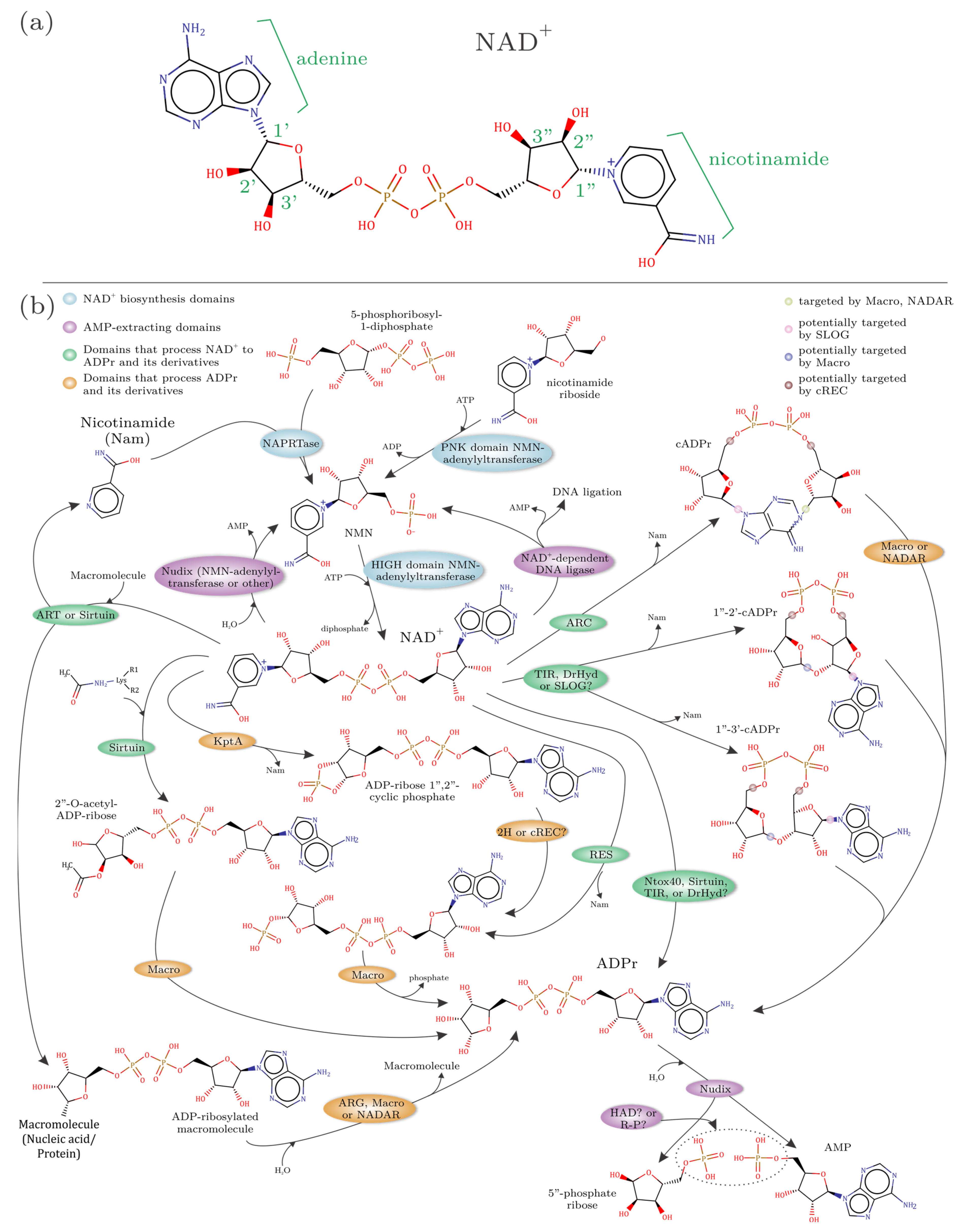
3.1. Defining the Building Blocks of the NAD+–ADPr Network
- (1)
- Domains that use NAD(P)+ as a substrate to release ADPr and its derivatives. This group includes ART, sirtuin, TIR, and DrHyd superfamily domains. The basic reaction catalyzed by these enzymes (best studied in the case of the ART superfamily) is an SN1 reaction, where an oxacarbenium ion intermediate is formed from NAD+ at the 1″ position of ADPr upon nicotinamide leaving [76,77] (Figure 1b). This intermediate is then available for nucleophilic attack. If the attack is by water, then free ADPr is released, i.e., a NADase reaction. Alternatively, it might be attacked by phosphate (e.g., in the case of KptA and RES domains of the ART superfamily) or acetyl (certain sirtuin domains) groups resulting in ADPr derivatives conjugated to these moieties. An attack by the adenine group from within NAD+ results in cyclic ADPr (cADPr; e.g., catalyzed by the ARC clade of the DrHyd superfamily) [58]. Similarly, attacks by the 2′ or 3′ ribose hydroxyls result in the variant cADPrs with 1″-2′ or 1″-3′ linkages (catalyzed by some members of the TIR and likely DrHyd superfamilies) (Figure 1b). If NADP is used as a substrate, an attack by a free nicotinate results in nicotinic acid adenine dinucleotide (NAADP; catalyzed by the ARC clade of the DrHyd superfamily) [78]. Finally, an attack by groups in macromolecules, such as bases in nucleic acids or amino acid sidechains from proteins can result in ADPr being conjugated to them (catalyzed by the ART and sirtuin superfamilies) [14,21,23,33,79,80].
- (2)
- Domains that process ADPr derivatives. This group includes structurally unrelated but catalytically comparable domains from the Macro, ARG, 2H, cREC, NADAR and SLOG superfamilies, which are known or predicted to act on the products generated by the above group (Figure 1b). The Macro superfamily shows considerable versatility in acting on both soluble ADPr derivatives and macromolecule-conjugated versions [81,82,83]. The characterized members of the ARG superfamily act on ADP-ribosylated proteins, but contextual evidence suggests a potentially wider range of substrates such as the Macro domain [83]. The 2H family acts on 1″-2″cyclic ADPr phosphate (ADPr > P, also known as Appr > p) generated by the action of KptA [57], the ART domain involved in the clean-up of cyclic phosphate RNA-termini [53]. While the cREC, NADAR and SLOG domains are strongly predicted to process ADPr derivatives they remain experimentally uncharacterized [12,14,40].
- (3)
- Domains that extract AMP from NAD+ or ADPr. This group includes two rather distinct types of domains. The first is the NAD+-dependent ligases, which are nucleotidyltransferases that use NAD+ as a substrate to adenylate DNA ends during phage DNA repair and replication [10]. The second is the Nudix domain that is involved in the breakdown of NAD+ to AMP and NMN and ADPr to AMP and ribose-3′-phosphate [84] (Figure 1b).
- (4)
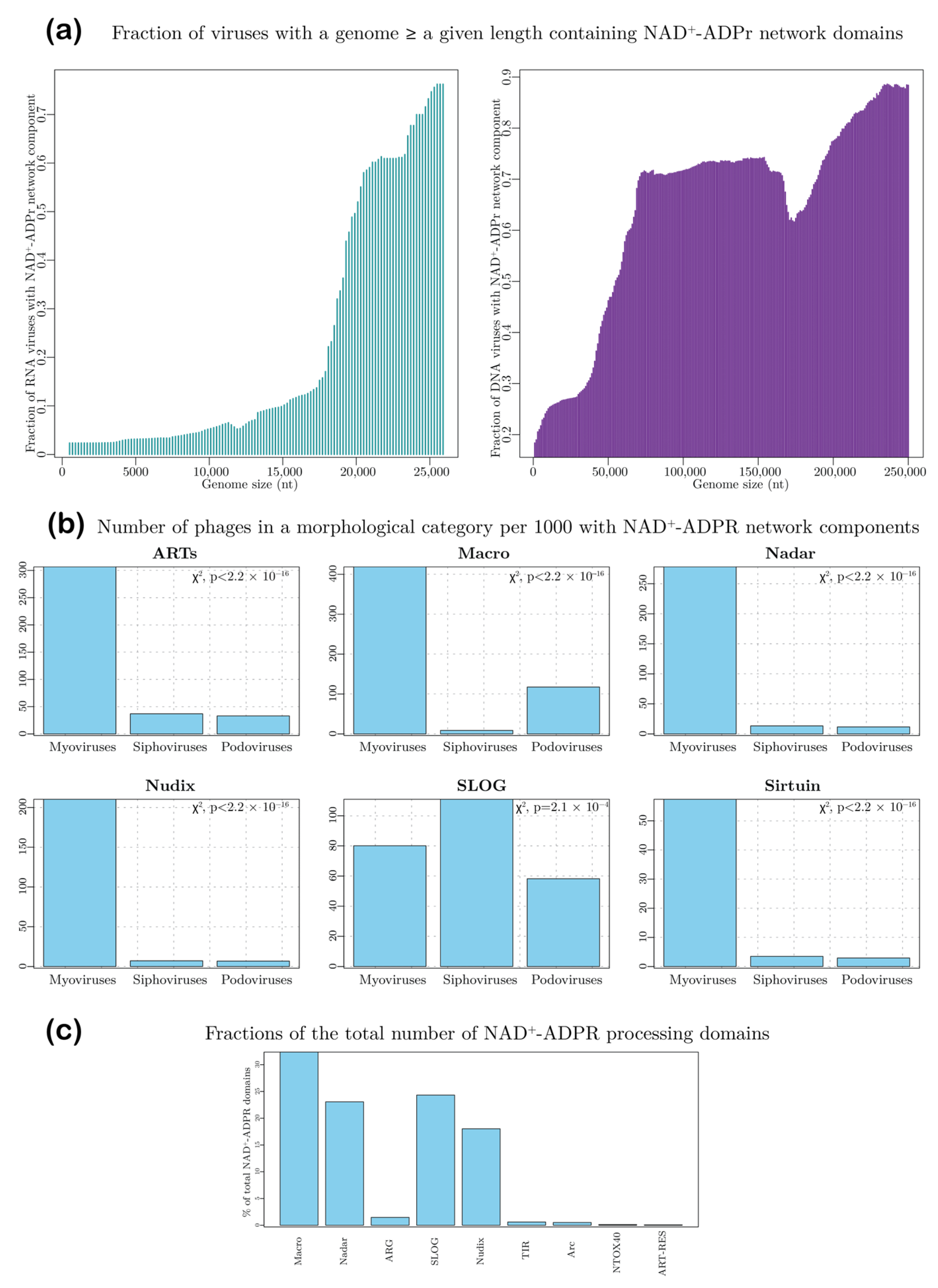
3.2. NAD+–ADPr Network Is a Feature of Both Large RNA and DNA Viruses
3.3. Distribution of NAD+-Utilizing Domains of the NAD+–ADPr Network in Viruses
3.3.1. The ART Superfamily
3.3.2. The Sirtuin Superfamily
3.3.3. The TIR-DrHyd-SLOG Class of Rossmannoid Domains
3.4. Distribution of Viral ADPr Processing Domains
3.4.1. Macro Domains
3.4.2. ADP Ribosyl Glycohydrolases (ARGs)
3.4.3. The NADAR Domain
3.4.4. The SLOG Domains
3.4.5. The 2H and cRec Superfamilies
3.5. Domains Generating AMP from NAD+ or ADPr-Derivatives
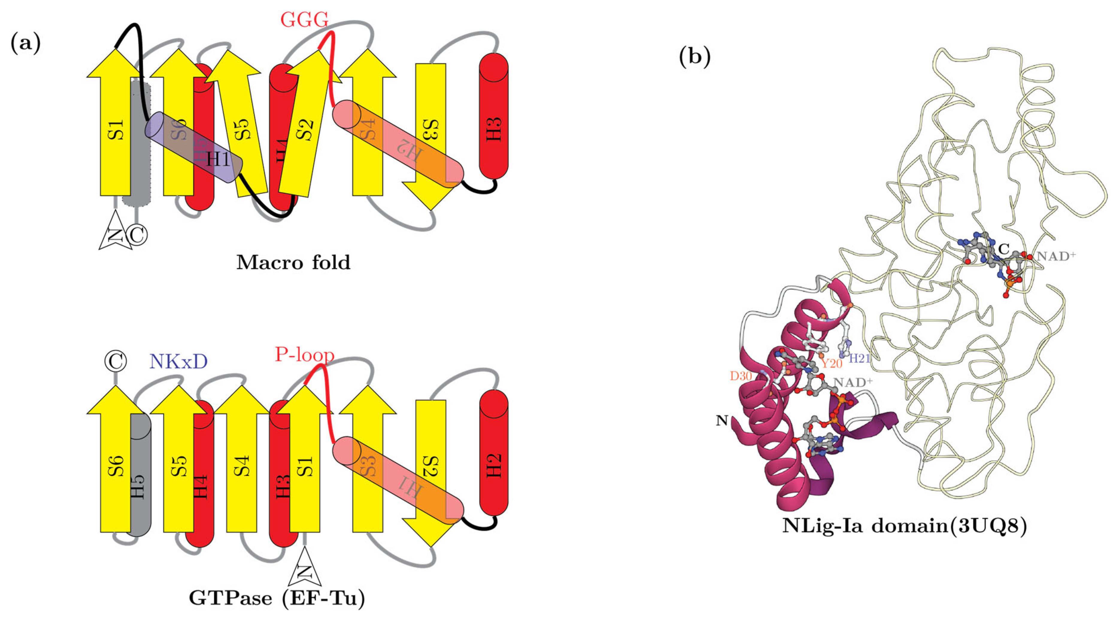
3.5.1. The NAD+-Dependent Ligases and the NAD+-Binding NLig-Ia Domain
3.5.2. Nudix Domains
3.6. Phage-Encoded NAD+-Synthesis Domains
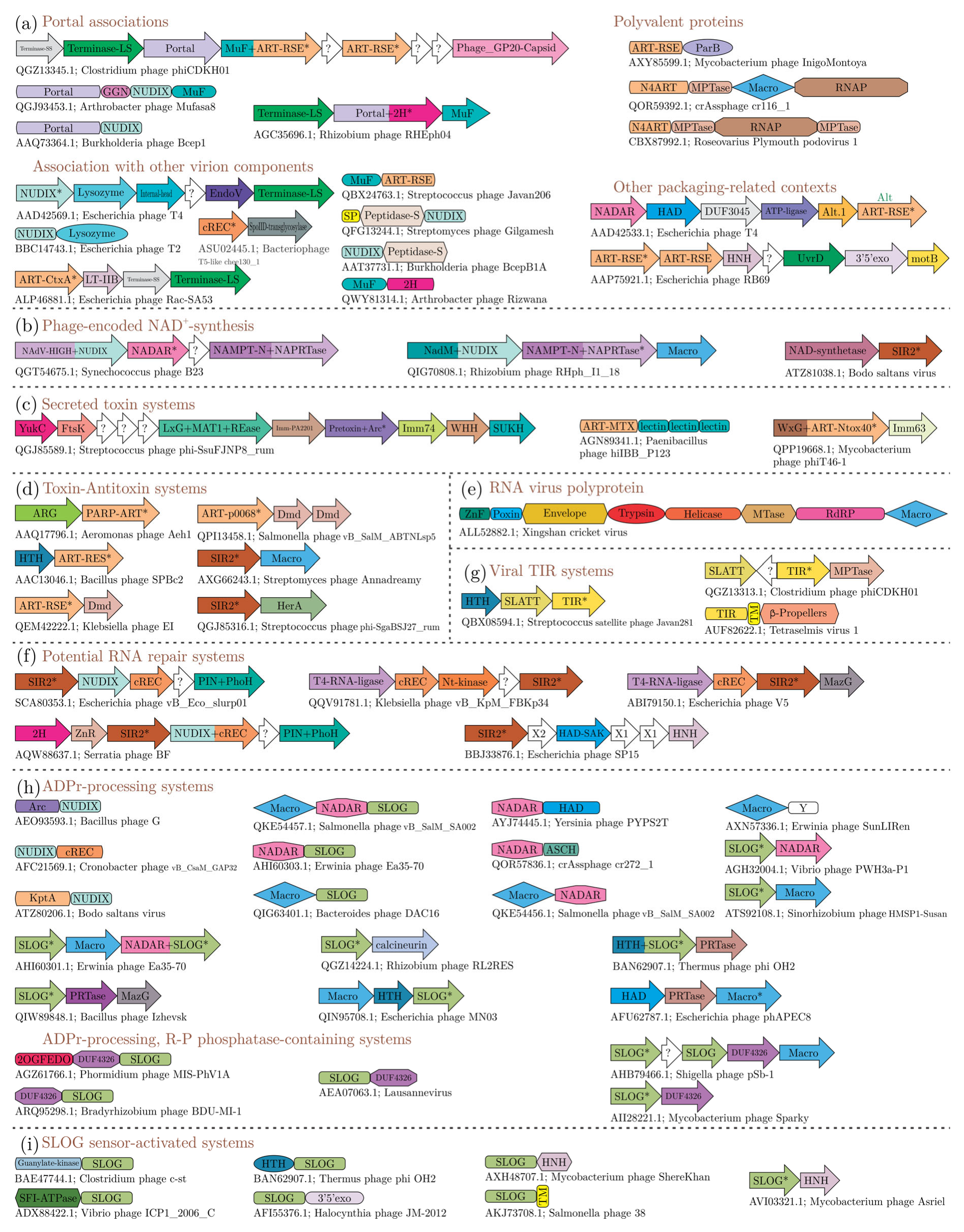
3.7. Reconstructing Viral NAD+–ADPr-Based Systems
3.7.1. Systems Involved in Macromolecular Modifications Are the Largest Group of DNA Viral Domains in the NAD+–ADPr Network
3.7.2. Systems Deconjugating ADPr Adducts to Macromolecules Are Widespread in Animal RNA Viruses
3.7.3. Systems Involved in RNA Repair
3.7.4. Viral Networks Generating and Processing Low-Molecular-Weight NAD+–ADPr-Derived Metabolites and Signaling Messengers
- (1)
- Modification enzymes that confer resistance against small molecule toxins—for example, the ARTs of the rifampin ADP-ribosyltransferase family that are predicted to modify rifamycin-like antibiotics [155]. These are borne by several phages of the actinobacteria Microbacterium and Gordonia, and the Sinorhizobium phage PBC5. A straightforward interpretation of these proteins would be that the virus confers resistance to their host against such antibiotics produced by rival bacteria. However, one cannot rule out the possibility that these ARTs are deployed against diffusible compounds that might participate in immunity against viruses as hinted by the production of such molecules in the recently discovered apoptosis and antiviral systems of actinobacteria [100,156,157].
- (2)
- Some T–A systems borne by phages feature a divergent ART family, RES, coupled to a helix-turn-helix transcription factor antitoxin (Figure 5d) [19]. In the RES family conserved S and E residues typical of the R-S-E clade of ARTs have been substituted by a Y and an N, respectively. These ART domains catalyze the phosphorolysis of NAD+ to generate ADPr-1″P, a toxic metabolite, that might either inhibit superinfecting phages or induce host dormancy to survive adverse environmental conditions [19].
- (3)
- Some mycobacterial phages also code for polymorphic toxin systems that display a Ntox40 (TNT) family ART toxin domain coupled to a gene for its characteristic immunity protein Imm63 [134]. Recent studies have shown that the NADase activity of the Ntox40 domain from mycobacteria is also directed against the macrophages of their animal host, wherein it degrades NAD+ to ADPr and triggers their apoptosis [158,159](Figure 5c). Similarly, we found a Type-VII secretion system-dependent polymorphic toxin with an Arc domain and its cognate immunity protein (Imm74) toxin [134] to be encoded in the Streptococcus phage phi-SsuFJNP8_rum (Figure 5c). Based on the precedence of the experimentally characterized Ntox40 system, here too it is conceivable that the Arc toxin acts in targeting the immune response of the eukaryotic host of the Streptococcus phage. The Arc domains generate cADPr or NAADP from NAD+ and NADP; given that these molecules are also generated by the cell-surface receptor Arcs in animal immune systems (e.g., CD38 and CD157) and have an important role in the antibacterial response [15,58], it is possible that virally encoded Arc domain toxins provide a mechanism for the bacterium to interfere with its host’s immunity. Moreover, polymorphic toxins are a key mechanism for kin-cooperation among bacteria [134]. Thus, in both these cases, the virally encoded ART and Arc polymorphic toxins appear to be part of a multilevel biological conflict. The virus, when in a (pseudo)lysogenic state likely enhances its own survival by both fostering kin-cooperation in its bacterial host and aiding it against the immunity of the bacterium’s eukaryotic host.
- (4)
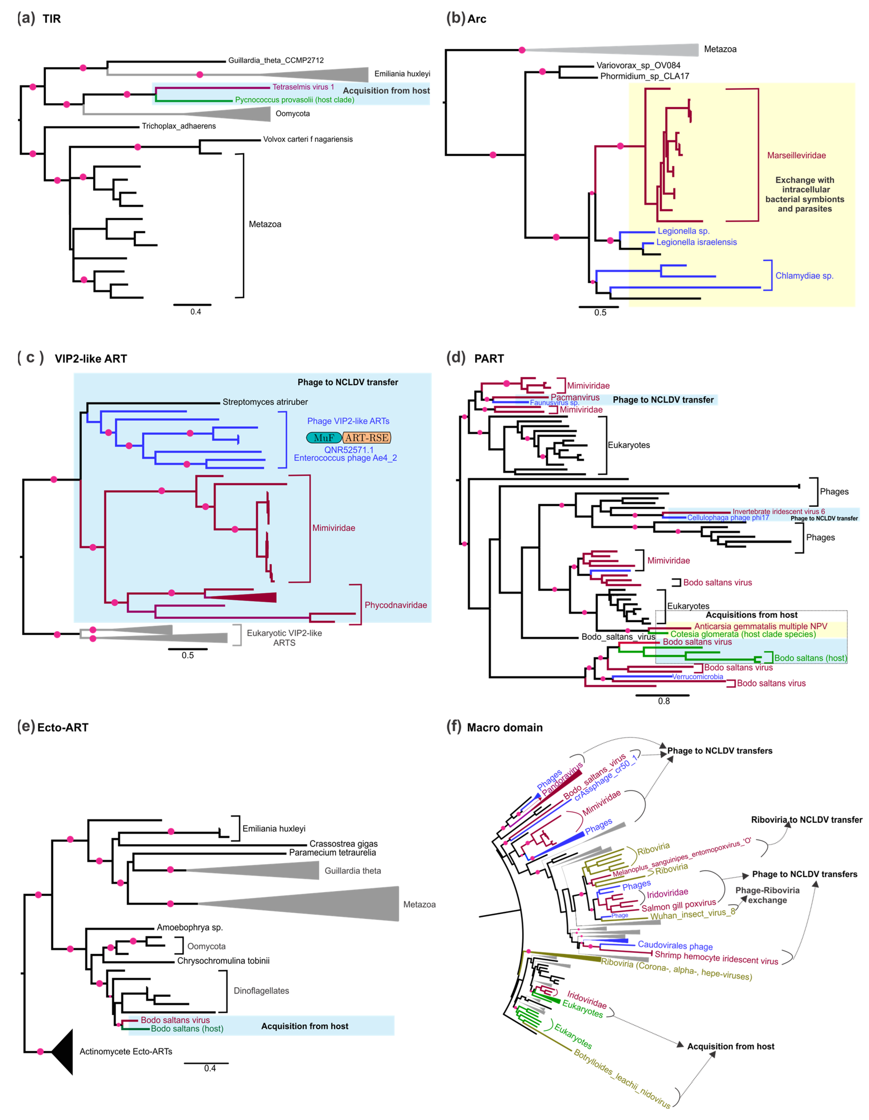
- A relatively small set of prokaryotic and eukaryotic viruses code for TIR and Arc domains (outside of polymorphic toxins). TIR domains are carried by multiple firmicute phages (e.g., the Streptococcus phage Javan281 and the Staphylococcus phage SN10), typically in a conserved gene–neighborhood association with the 2TM SLATT domain (Figure 5g). These gene neighborhoods sometimes also code for a helix-turn-helix (HTH) domain transcription factor or a zincin-like metallopeptidase. The SLATT domain is predicted to function as an ADPr derivative-regulated effector controlling membrane permeability across diverse prokaryotic antiviral systems [12]. The TIR and Arc domains found sporadically in NCLDVs have been respectively acquired from their eukaryotic hosts and bacterial endosymbionts. Given their role in host immune systems, their presence in viruses is rather enigmatic. In the case of the TIR domain, it is conceivable that it limits superinfection via an apoptotic mechanism that relies on kin selection in a host population/multicellular ensemble that has either been lysogenized or carries the dormant virus. The cADPr or NAADP signals generated by the Arc domain could also manipulate host immunity in favor of the virus.
3.8. Prediction of Immune Mechanisms That Counter Viral ADPr Modifications of Host Macromolecules
3.9. Evolutionary Considerations
4. Conclusions
Supplementary Materials
Author Contributions
Funding
Conflicts of Interest
References
- Hayes, W. Max Delbrück and the Birth of Molecular Biology. Soc. Res. 1984, 51, 641–673. [Google Scholar] [CrossRef]
- Kuhn, A.; Thomas, J.A. The Beauty of Bacteriophage T4 Research: Lindsay W. Black and the T4 Head Assembly. Viruses 2022, 14, 700. [Google Scholar] [CrossRef]
- Miller, E.S.; Kutter, E.; Mosig, G.; Arisaka, F.; Kunisawa, T.; Ruger, W. Bacteriophage T4 genome. Microbiol. Mol. Biol. Rev. 2003, 67, 86–156. [Google Scholar] [CrossRef]
- Rao, V.B.; Black, L.W. Structure and assembly of bacteriophage T4 head. Virol. J. 2010, 7, 356. [Google Scholar] [CrossRef]
- Goff, C.G. Chemical structure of a modification of the Escherichia coli ribonucleic acid polymerase alpha polypeptides induced by bacteriophage T4 infection. J. Biol. Chem. 1974, 249, 6181–6190. [Google Scholar] [CrossRef]
- Depping, R.; Lohaus, C.; Meyer, H.E.; Ruger, W. The mono-ADP-ribosyltransferases Alt and ModB of bacteriophage T4: Target proteins identified. Biochem. Biophys. Res. Commun. 2005, 335, 1217–1223. [Google Scholar] [CrossRef]
- Collier, R.J. Diphtheria toxin: Mode of action and structure. Bacteriol. Rev. 1975, 39, 54–85. [Google Scholar] [CrossRef]
- Holmes, R.K. Biology and molecular epidemiology of diphtheria toxin and the tox gene. J. Infect. Dis. 2000, 181 (Suppl. S1), S156–S167. [Google Scholar] [CrossRef]
- Berg, J.M.; Tymoczko, J.L.; Stryer, L.; Stryer, L. Biochemistry; W.H. Freeman: New York, NY, USA, 2002. [Google Scholar]
- Shuman, S. DNA ligases: Progress and prospects. J. Biol. Chem. 2009, 284, 17365–17369. [Google Scholar] [CrossRef] [PubMed]
- Burroughs, A.M.; Aravind, L. Identification of uncharacterized components of prokaryotic immune systems and their diverse eukaryotic reformulations. J. Bacteriol. 2020, 202, e00365-20. [Google Scholar] [CrossRef] [PubMed]
- Burroughs, A.M.; Zhang, D.; Schaffer, D.E.; Iyer, L.M.; Aravind, L. Comparative genomic analyses reveal a vast, novel network of nucleotide-centric systems in biological conflicts, immunity and signaling. Nucleic Acids Res. 2015, 43, 10633–10654. [Google Scholar] [CrossRef]
- Aravind, L.; Iyer, L.M.; Burroughs, A.M. Discovering Biological Conflict Systems through Genome Analysis: Evolutionary Principles and Biochemical Novelty. Annu. Rev. Biomed. Data Sci. 2022, 5, 367–391. [Google Scholar] [CrossRef]
- Iyer, L.M.; Anantharaman, V.; Krishnan, A.; Burroughs, A.M.; Aravind, L. Jumbo Phages: A Comparative Genomic Overview of Core Functions and Adaptions for Biological Conflicts. Viruses 2021, 13, 63. [Google Scholar] [CrossRef]
- Shang, J.; Smith, M.R.; Anmangandla, A.; Lin, H. NAD+-consuming enzymes in immune defense against viral infection. Biochem. J. 2021, 478, 4071–4092. [Google Scholar] [CrossRef]
- Essuman, K.; Summers, D.W.; Sasaki, Y.; Mao, X.; Yim, A.K.Y.; DiAntonio, A.; Milbrandt, J. TIR Domain Proteins Are an Ancient Family of NAD+-Consuming Enzymes. Curr. Biol. 2018, 28, 421–430.e4. [Google Scholar] [CrossRef] [PubMed]
- Ka, D.; Oh, H.; Park, E.; Kim, J.H.; Bae, E. Structural and functional evidence of bacterial antiphage protection by Thoeris defense system via NAD+ degradation. Nat. Commun. 2020, 11, 2816. [Google Scholar] [CrossRef] [PubMed]
- Ofir, G.; Herbst, E.; Baroz, M.; Cohen, D.; Millman, A.; Doron, S.; Tal, N.; Malheiro, D.B.A.; Malitsky, S.; Amitai, G.; et al. Antiviral activity of bacterial TIR domains via immune signalling molecules. Nature 2021, 600, 116–120. [Google Scholar] [CrossRef]
- Skjerning, R.B.; Senissar, M.; Winther, K.S.; Gerdes, K.; Brodersen, D.E. The RES domain toxins of RES-Xre toxin-antitoxin modules induce cell stasis by degrading NAD+. Mol. Microbiol. 2019, 111, 221–236. [Google Scholar] [CrossRef]
- Wan, L.; Essuman, K.; Anderson, R.G.; Sasaki, Y.; Monteiro, F.; Chung, E.H.; Osborne Nishimura, E.; DiAntonio, A.; Milbrandt, J.; Dangl, J.L.; et al. TIR domains of plant immune receptors are NAD+-cleaving enzymes that promote cell death. Science 2019, 365, 799–803. [Google Scholar] [CrossRef] [PubMed]
- Koch-Nolte, F.; Petersen, D.; Balasubramanian, S.; Haag, F.; Kahlke, D.; Willer, T.; Kastelein, R.; Bazan, F.; Thiele, H.G. Mouse T cell membrane proteins Rt6-1 and Rt6-2 are arginine/protein mono(ADPribosyl)transferases and share secondary structure motifs with ADP-ribosylating bacterial toxins. J. Biol. Chem. 1996, 271, 7686–7693. [Google Scholar] [CrossRef] [Green Version]
- Wilkens, K.; Tiemann, B.; Bazan, F.; Ruger, W. ADP-ribosylation and early transcription regulation by bacteriophage T4. Adv. Exp. Med. Biol. 1997, 419, 71–82. [Google Scholar] [PubMed]
- Aravind, L.; Zhang, D.; de Souza, R.F.; Anand, S.; Iyer, L.M. The natural history of ADP-ribosyltransferases and the ADP-ribosylation system. Curr. Top. Microbiol. Immunol. 2015, 384, 3–32. [Google Scholar] [PubMed]
- Daugherty, M.D.; Young, J.M.; Kerns, J.A.; Malik, H.S. Rapid evolution of PARP genes suggests a broad role for ADP-ribosylation in host-virus conflicts. PLoS Genet. 2014, 10, e1004403. [Google Scholar] [CrossRef] [PubMed]
- Mikolčević, P.; Hloušek-Kasun, A.; Ahel, I.; Mikoč, A. ADP-ribosylation systems in bacteria and viruses. Comput. Struct. Biotechnol. J. 2021, 19, 2366–2383. [Google Scholar] [CrossRef]
- Höfer, K.; Schauerte, M.; Grawenhoff, J.; Wulf, A.; Welp, L.M.; Billau, F.A.; Urlaub, H.; Jäschke, A. Viral ADP-ribosyltransferases attach RNA chains to host proteins. bioRxiv 2021. [Google Scholar] [CrossRef]
- Koch, T.; Raudonikiene, A.; Wilkens, K.; Ruger, W. Overexpression, purification, and characterization of the ADP-ribosyltransferase (gpAlt) of bacteriophage T4: ADP-ribosylation of E. coli RNA polymerase modulates T4 “early” transcription. Gene Expr. 1995, 4, 253–264. [Google Scholar] [PubMed]
- Tiemann, B.; Depping, R.; Gineikiene, E.; Kaliniene, L.; Nivinskas, R.; Rüger, W. ModA and ModB, two ADP-ribosyltransferases encoded by bacteriophage T4: Catalytic properties and mutation analysis. J. Bacteriol. 2004, 186, 7262–7272. [Google Scholar] [CrossRef] [PubMed]
- Iyer, L.M.; Burroughs, A.M.; Anand, S.; de Souza, R.F.; Aravind, L. Polyvalent Proteins, a Pervasive Theme in the Intergenomic Biological Conflicts of Bacteriophages and Conjugative Elements. J. Bacteriol 2017, 199, e00245-17. [Google Scholar] [CrossRef]
- LeRoux, M.; Srikant, S.; Teodoro, G.I.C.; Zhang, T.; Littlehale, M.L.; Doron, S.; Badiee, M.; Leung, A.K.L.; Sorek, R.; Laub, M.T. The DarTG toxin-antitoxin system provides phage defence by ADP-ribosylating viral DNA. Nat. Microbiol. 2022, 7, 1028–1040. [Google Scholar] [CrossRef]
- Otto, H.; Reche, P.A.; Bazan, F.; Dittmar, K.; Haag, F.; Koch-Nolte, F. In silico characterization of the family of PARP-like poly(ADP-ribosyl)transferases (pARTs). BMC Genom. 2005, 6, 139. [Google Scholar] [CrossRef] [Green Version]
- Li, C.; Debing, Y.; Jankevicius, G.; Neyts, J.; Ahel, I.; Coutard, B.; Canard, B. Viral Macro Domains Reverse Protein ADP-Ribosylation. J. Virol. 2016, 90, 8478–8486. [Google Scholar] [CrossRef]
- Hawse, W.F.; Wolberger, C. Structure-based mechanism of ADP-ribosylation by sirtuins. J. Biol. Chem. 2009, 284, 33654–33661. [Google Scholar] [CrossRef] [PubMed]
- Beauharnois, J.M.; Bolivar, B.E.; Welch, J.T. Sirtuin 6: A review of biological effects and potential therapeutic properties. Mol. Biosyst. 2013, 9, 1789–1806. [Google Scholar] [CrossRef] [PubMed]
- Whiteley, A.T.; Eaglesham, J.B.; de Oliveira Mann, C.C.; Morehouse, B.R.; Lowey, B.; Nieminen, E.A.; Danilchanka, O.; King, D.S.; Lee, A.S.Y.; Mekalanos, J.J.; et al. Bacterial cGAS-like enzymes synthesize diverse nucleotide signals. Nature 2019, 567, 194–199. [Google Scholar] [CrossRef] [PubMed]
- Eaglesham, J.B.; Pan, Y.; Kupper, T.S.; Kranzusch, P.J. Viral and metazoan poxins are cGAMP-specific nucleases that restrict cGAS-STING signalling. Nature 2019, 566, 259–263. [Google Scholar] [CrossRef] [PubMed]
- Morehouse, B.R.; Govande, A.A.; Millman, A.; Keszei, A.F.A.; Lowey, B.; Ofir, G.; Shao, S.; Sorek, R.; Kranzusch, P.J. STING cyclic dinucleotide sensing originated in bacteria. Nature 2020, 586, 429–433. [Google Scholar] [CrossRef] [PubMed]
- Essuman, K.; Summers, D.W.; Sasaki, Y.; Mao, X.; DiAntonio, A.; Milbrandt, J. The SARM1 Toll/Interleukin-1 Receptor Domain Possesses Intrinsic NAD+ Cleavage Activity that Promotes Pathological Axonal Degeneration. Neuron 2017, 93, 1334–1343.e5. [Google Scholar] [CrossRef] [PubMed]
- Samanovic, M.I.; Tu, S.; Novak, O.; Iyer, L.M.; McAllister, F.E.; Aravind, L.; Gygi, S.P.; Hubbard, S.R.; Strnad, M.; Darwin, K.H. Proteasomal control of cytokinin synthesis protects Mycobacterium tuberculosis against nitric oxide. Mol. Cell 2015, 57, 984–994. [Google Scholar] [CrossRef]
- de Souza, R.F.; Aravind, L. Identification of novel components of NAD-utilizing metabolic pathways and prediction of their biochemical functions. Mol. Biosyst. 2012, 8, 1661–1677. [Google Scholar] [CrossRef]
- Manik, M.K.; Shi, Y.; Li, S.; Zaydman, M.A.; Damaraju, N.; Eastman, S.; Smith, T.G.; Gu, W.; Masic, V.; Mosaiab, T.; et al. Chemical structures of cyclic ADP ribose (cADPR) isomers and the molecular basis of their production and signaling. bioRxiv 2022. [Google Scholar] [CrossRef]
- Rack, J.G.; Perina, D.; Ahel, I. Macrodomains: Structure, Function, Evolution, and Catalytic Activities. Annu. Rev. Biochem. 2016, 85, 431–454. [Google Scholar] [CrossRef]
- Schuller, M.; Ahel, I. Beyond protein modification: The rise of non-canonical ADP-ribosylation. Biochem. J. 2022, 479, 463–477. [Google Scholar] [CrossRef] [PubMed]
- Lee, J.Y.; Li, Z.; Miller, E.S. Vibrio phage KVP40 encodes a functional NAD+ salvage pathway. J. Bacteriol. 2017, 199, e00855-16. [Google Scholar] [CrossRef]
- Mazumder, R.; Iyer, L.M.; Vasudevan, S.; Aravind, L. Detection of novel members, structure-function analysis and evolutionary classification of the 2H phosphoesterase superfamily. Nucleic Acids Res. 2002, 30, 5229–5243. [Google Scholar] [CrossRef]
- Zhang, R.; Jha, B.K.; Ogden, K.M.; Dong, B.; Zhao, L.; Elliott, R.; Patton, J.T.; Silverman, R.H.; Weiss, S.R. Homologous 2′,5′-phosphodiesterases from disparate RNA viruses antagonize antiviral innate immunity. Proc. Natl. Acad. Sci. USA 2013, 110, 13114–13119. [Google Scholar] [CrossRef] [PubMed]
- Parrish, S.; Resch, W.; Moss, B. Vaccinia virus D10 protein has mRNA decapping activity, providing a mechanism for control of host and viral gene expression. Proc. Natl. Acad. Sci. USA 2007, 104, 2139–2144. [Google Scholar] [CrossRef] [PubMed]
- Ge, J.; Wei, Z.; Huang, Y.; Yin, J.; Zhou, Z.; Zhong, J. AcMNPV ORF38 protein has the activity of ADP-ribose pyrophosphatase and is important for virus replication. Virology 2007, 361, 204–211. [Google Scholar] [CrossRef]
- Xu, W.; Gauss, P.; Shen, J.; Dunn, C.A.; Bessman, M.J. The gene e.1 (nudE.1) of T4 bacteriophage designates a new member of the Nudix hydrolase superfamily active on flavin adenine dinucleotide, adenosine 5′-triphospho-5′-adenosine, and ADP-ribose. J. Biol. Chem. 2002, 277, 23181–23185. [Google Scholar] [CrossRef] [PubMed]
- Kaufmann, G. Anticodon nucleases. Trends Biochem. Sci. 2000, 25, 70–74. [Google Scholar] [CrossRef]
- Phizicky, E.M.; Hopper, A.K. tRNA processing, modification, and subcellular dynamics: Past, present, and future. RNA 2015, 21, 483–485. [Google Scholar] [CrossRef] [Green Version]
- Makarova, K.S.; Anantharaman, V.; Aravind, L.; Koonin, E.V. Live virus-free or die: Coupling of antivirus immunity and programmed suicide or dormancy in prokaryotes. Biol. Direct 2012, 7, 40. [Google Scholar] [CrossRef] [PubMed]
- Burroughs, A.M.; Aravind, L. RNA damage in biological conflicts and the diversity of responding RNA repair systems. Nucleic Acids Res. 2016, 44, 8525–8555. [Google Scholar] [CrossRef] [PubMed]
- Spinelli, S.L.; Kierzek, R.; Turner, D.H.; Phizicky, E.M. Transient ADP-ribosylation of a 2′-phosphate implicated in its removal from ligated tRNA during splicing in yeast. J. Biol. Chem. 1999, 274, 2637–2644. [Google Scholar] [CrossRef]
- Culver, G.M.; Consaul, S.A.; Tycowski, K.T.; Filipowicz, W.; Phizicky, E.M. tRNA splicing in yeast and wheat germ. A cyclic phosphodiesterase implicated in the metabolism of ADP-ribose 1″,2″-cyclic phosphate. J. Biol. Chem. 1994, 269, 24928–24934. [Google Scholar] [CrossRef]
- Shull, N.P.; Spinelli, S.L.; Phizicky, E.M. A highly specific phosphatase that acts on ADP-ribose 1″-phosphate, a metabolite of tRNA splicing in Saccharomyces cerevisiae. Nucleic Acids Res. 2005, 33, 650–660. [Google Scholar] [CrossRef] [PubMed]
- Steiger, M.A.; Jackman, J.E.; Phizicky, E.M. Analysis of 2′-phosphotransferase (Tpt1p) from Saccharomyces cerevisiae: Evidence for a conserved two-step reaction mechanism. RNA 2005, 11, 99–106. [Google Scholar] [CrossRef]
- Hogan, K.A.; Chini, C.C.S.; Chini, E.N. The Multi-faceted Ecto-enzyme CD38: Roles in Immunomodulation, Cancer, Aging, and Metabolic Diseases. Front. Immunol. 2019, 10, 1187. [Google Scholar] [CrossRef]
- Younker, I.T.; Duffy, C. Jumbo phages. In Reference Module in Life Sciences; Elsevier: Amsterdam, The Netherlands, 2020. [Google Scholar]
- Tan, Y.; Schneider, T.; Leong, M.; Aravind, L.; Zhang, D. Novel Immunoglobulin Domain Proteins Provide Insights into Evolution and Pathogenesis of SARS-CoV-2-Related Viruses. mBio 2020, 11, e00760-20. [Google Scholar] [CrossRef] [PubMed]
- Altschul, S.F.; Madden, T.L.; Schaffer, A.A.; Zhang, J.; Zhang, Z.; Miller, W.; Lipman, D.J. Gapped BLAST and PSI-BLAST: A new generation of protein database search programs. Nucleic Acids Res. 1997, 25, 3389–3402. [Google Scholar] [CrossRef] [PubMed]
- Eddy, S.R. A new generation of homology search tools based on probabilistic inference. Genome Inform. 2009, 23, 205–211. [Google Scholar] [PubMed]
- Mirdita, M.; Steinegger, M.; Soding, J. MMseqs2 desktop and local web server app for fast, interactive sequence searches. Bioinformatics 2019, 35, 2856–2858. [Google Scholar] [CrossRef]
- El-Gebali, S.; Mistry, J.; Bateman, A.; Eddy, S.R.; Luciani, A.; Potter, S.C.; Qureshi, M.; Richardson, L.J.; Salazar, G.A.; Smart, A.; et al. The Pfam protein families database in 2019. Nucleic Acids Res. 2019, 47, D427–D432. [Google Scholar] [CrossRef] [PubMed]
- Lassmann, T.; Frings, O.; Sonnhammer, E.L. Kalign2: High-performance multiple alignment of protein and nucleotide sequences allowing external features. Nucleic Acids Res. 2009, 37, 858–865. [Google Scholar] [CrossRef] [PubMed]
- Rozewicki, J.; Li, S.; Amada, K.M.; Standley, D.M.; Katoh, K. MAFFT-DASH: Integrated protein sequence and structural alignment. Nucleic Acids Res. 2019, 47, W5–W10. [Google Scholar] [CrossRef]
- Soding, J.; Biegert, A.; Lupas, A.N. The HHpred interactive server for protein homology detection and structure prediction. Nucleic Acids Res. 2005, 33, W244–W248. [Google Scholar] [CrossRef]
- Cole, C.; Barber, J.D.; Barton, G.J. The Jpred 3 secondary structure prediction server. Nucleic Acids Res. 2008, 36, W197–W201. [Google Scholar] [CrossRef]
- Holm, L. Benchmarking fold detection by DaliLite v.5. Bioinformatics 2019, 35, 5326–5327. [Google Scholar] [CrossRef]
- Sehnal, D.; Bittrich, S.; Deshpande, M.; Svobodova, R.; Berka, K.; Bazgier, V.; Velankar, S.; Burley, S.K.; Koca, J.; Rose, A.S. Mol* Viewer: Modern web app for 3D visualization and analysis of large biomolecular structures. Nucleic Acids Res. 2021, 49, W431–W437. [Google Scholar] [CrossRef]
- The PyMOL Molecular Graphics System, version 1.8; Schrodinger, LLC.: New York, NY, USA, 2015.
- Baek, M.; DiMaio, F.; Anishchenko, I.; Dauparas, J.; Ovchinnikov, S.; Lee, G.R.; Wang, J.; Cong, Q.; Kinch, L.N.; Schaeffer, R.D.; et al. Accurate prediction of protein structures and interactions using a three-track neural network. Science 2021, 373, 871–876. [Google Scholar] [CrossRef] [PubMed]
- Jumper, J.; Evans, R.; Pritzel, A.; Green, T.; Figurnov, M.; Ronneberger, O.; Tunyasuvunakool, K.; Bates, R.; Zidek, A.; Potapenko, A.; et al. Highly accurate protein structure prediction with AlphaFold. Nature 2021, 596, 583–589. [Google Scholar] [CrossRef] [PubMed]
- Price, M.N.; Dehal, P.S.; Arkin, A.P. FastTree 2—Approximately maximum-likelihood trees for large alignments. PLoS ONE 2010, 5, e9490. [Google Scholar] [CrossRef] [PubMed]
- Minh, B.Q.; Schmidt, H.A.; Chernomor, O.; Schrempf, D.; Woodhams, M.D.; von Haeseler, A.; Lanfear, R. IQ-TREE 2: New Models and Efficient Methods for Phylogenetic Inference in the Genomic Era. Mol. Biol. Evol. 2020, 37, 1530–1534. [Google Scholar] [CrossRef] [PubMed]
- Jorgensen, R.; Wang, Y.; Visschedyk, D.; Merrill, A.R. The nature and character of the transition state for the ADP-ribosyltransferase reaction. EMBO Rep. 2008, 9, 802–809. [Google Scholar] [CrossRef] [PubMed]
- Avalos, J.L.; Boeke, J.D.; Wolberger, C. Structural basis for the mechanism and regulation of Sir2 enzymes. Mol. Cell 2004, 13, 639–648. [Google Scholar] [CrossRef]
- Guse, A.H.; Lee, H.C. NAADP: A universal Ca2+ trigger. Sci. Signal. 2008, 1, re10. [Google Scholar] [CrossRef]
- Nakano, T.; Takahashi-Nakaguchi, A.; Yamamoto, M.; Watanabe, M. Pierisins and CARP-1: ADP-ribosylation of DNA by ARTCs in butterflies and shellfish. Curr. Top. Microbiol. Immunol. 2015, 384, 127–149. [Google Scholar]
- Rack, J.G.; Morra, R.; Barkauskaite, E.; Kraehenbuehl, R.; Ariza, A.; Qu, Y.; Ortmayer, M.; Leidecker, O.; Cameron, D.R.; Matic, I.; et al. Identification of a Class of Protein ADP-Ribosylating Sirtuins in Microbial Pathogens. Mol. Cell 2015, 59, 309–320. [Google Scholar] [CrossRef]
- Rack, J.G.M.; Zorzini, V.; Zhu, Z.; Schuller, M.; Ahel, D.; Ahel, I. Viral macrodomains: A structural and evolutionary assessment of the pharmacological potential. Open Biol. 2020, 10, 200237. [Google Scholar] [CrossRef] [PubMed]
- Han, W.; Li, X.; Fu, X. The macro domain protein family: Structure, functions, and their potential therapeutic implications. Mutat. Res. 2011, 727, 86–103. [Google Scholar] [CrossRef] [PubMed]
- Slade, D.; Dunstan, M.S.; Barkauskaite, E.; Weston, R.; Lafite, P.; Dixon, N.; Ahel, M.; Leys, D.; Ahel, I. The structure and catalytic mechanism of a poly(ADP-ribose) glycohydrolase. Nature 2011, 477, 616–620. [Google Scholar] [CrossRef]
- Mildvan, A.S.; Xia, Z.; Azurmendi, H.F.; Saraswat, V.; Legler, P.M.; Massiah, M.A.; Gabelli, S.B.; Bianchet, M.A.; Kang, L.W.; Amzel, L.M. Structures and mechanisms of Nudix hydrolases. Arch. Biochem. Biophys. 2005, 433, 129–143. [Google Scholar] [CrossRef] [PubMed]
- Dulyaninova, N.G.; Podlepa, E.M.; Toulokhonova, L.V.; Bykhovsky, V.Y. Salvage pathway for NAD biosynthesis in Brevibacterium ammoniagenes: Regulatory properties of triphosphate-dependent nicotinate phosphoribosyltransferase. Biochim. Biophys. Acta 2000, 1478, 211–220. [Google Scholar] [CrossRef]
- Charles, J.; Tangudu, C.S.; Hurt, S.L.; Tumescheit, C.; Firth, A.E.; Garcia-Rejon, J.E.; Machain-Williams, C.; Blitvich, B.J. Discovery of a novel Tymoviridae-like virus in mosquitoes from Mexico. Arch. Virol. 2019, 164, 649–652. [Google Scholar] [CrossRef]
- Alhammad, Y.M.O.; Kashipathy, M.M.; Roy, A.; Gagne, J.P.; McDonald, P.; Gao, P.; Nonfoux, L.; Battaile, K.P.; Johnson, D.K.; Holmstrom, E.D.; et al. The SARS-CoV-2 Conserved Macrodomain Is a Mono-ADP-Ribosylhydrolase. J. Virol. 2021, 95, e01969-20. [Google Scholar] [CrossRef] [PubMed]
- Yuan, Y.; Gao, M. Jumbo bacteriophages: An overview. Front. Microbiol. 2017, 8, 403. [Google Scholar] [CrossRef] [PubMed]
- Iyer, L.M.; Aravind, L.; Koonin, E.V. Common origin of four diverse families of large eukaryotic DNA viruses. J. Virol. 2001, 75, 11720–11734. [Google Scholar] [CrossRef]
- Koonin, E.V.; Yutin, N. Evolution of the Large Nucleocytoplasmic DNA Viruses of Eukaryotes and Convergent Origins of Viral Gigantism. Adv. Virus Res. 2019, 103, 167–202. [Google Scholar]
- Iyer, L.M.; Balaji, S.; Koonin, E.V.; Aravind, L. Evolutionary genomics of nucleo-cytoplasmic large DNA viruses. Virus Res. 2006, 117, 156–184. [Google Scholar] [CrossRef]
- Smith, J.M. Evolutionary Genetics; Oxford University Press: Oxford, UK, 1989. [Google Scholar]
- Waldor, M.K.; Mekalanos, J.J. Lysogenic conversion by a filamentous phage encoding cholera toxin. Science 1996, 272, 1910–1914. [Google Scholar] [CrossRef] [PubMed]
- Blander, G.; Guarente, L. The Sir2 family of protein deacetylases. Annu. Rev. Biochem. 2004, 73, 417–435. [Google Scholar] [CrossRef] [PubMed]
- Garb, J.; Lopatina, A.; Bernheim, A.; Zaremba, M.; Siksnys, V.; Melamed, S.; Leavitt, A.; Millman, A.; Amitai, G.; Sorek, R. Multiple phage resistance systems inhibit infection via SIR2-dependent NAD+ depletion. bioRxiv 2021. [Google Scholar] [CrossRef]
- Tsang, A.W.; Escalante-Semerena, J.C. CobB, a new member of the SIR2 family of eucaryotic regulatory proteins, is required to compensate for the lack of nicotinate mononucleotide: 5,6-dimethylbenzimidazole phosphoribosyltransferase activity in cobT mutants during cobalamin biosynthesis in Salmonella typhimurium LT2. J. Biol. Chem. 1998, 273, 31788–31794. [Google Scholar] [PubMed] [Green Version]
- Li, R.; Gu, J.; Chen, Y.Y.; Xiao, C.L.; Wang, L.W.; Zhang, Z.P.; Bi, L.J.; Wei, H.P.; Wang, X.D.; Deng, J.Y.; et al. CobB regulates Escherichia coli chemotaxis by deacetylating the response regulator CheY. Mol. Microbiol. 2010, 76, 1162–1174. [Google Scholar] [CrossRef] [PubMed]
- Iyer, L.M.; Makarova, K.S.; Koonin, E.V.; Aravind, L. Comparative genomics of the FtsK-HerA superfamily of pumping ATPases: Implications for the origins of chromosome segregation, cell division and viral capsid packaging. Nucleic Acids Res. 2004, 32, 5260–5279. [Google Scholar] [CrossRef]
- Burroughs, A.M.; Iyer, L.M.; Aravind, L. Two novel PIWI families: Roles in inter-genomic conflicts in bacteria and Mediator-dependent modulation of transcription in eukaryotes. Biol. Direct 2013, 8, 13. [Google Scholar] [CrossRef]
- Kaur, G.; Iyer, L.M.; Burroughs, A.M.; Aravind, L. Bacterial death and TRADD-N domains help define novel apoptosis and immunity mechanisms shared by prokaryotes and metazoans. Elife 2021, 10, e70394. [Google Scholar] [CrossRef]
- Burroughs, A.M.; Zhang, D.; Aravind, L. The eukaryotic translation initiation regulator CDC123 defines a divergent clade of ATP-grasp enzymes with a predicted role in novel protein modifications. Biol. Direct 2015, 10, 21. [Google Scholar] [CrossRef]
- Aravind, L.; Anantharaman, V.; Zhang, D.; de Souza, R.F.; Iyer, L.M. Gene flow and biological conflict systems in the origin and evolution of eukaryotes. Front. Cell. Infect. Microbiol. 2012, 2, 89. [Google Scholar] [CrossRef] [PubMed]
- Leipe, D.D.; Wolf, Y.I.; Koonin, E.V.; Aravind, L. Classification and evolution of P-loop GTPases and related ATPases. J. Mol. Biol. 2002, 317, 41–72. [Google Scholar] [CrossRef]
- Moure, V.R.; Costa, F.F.; Cruz, L.M.; Pedrosa, F.O.; Souza, E.M.; Li, X.D.; Winkler, F.; Huergo, L.F. Regulation of nitrogenase by reversible mono-ADP-ribosylation. Curr. Top. Microbiol. Immunol. 2015, 384, 89–106. [Google Scholar]
- Ono, T.; Kasamatsu, A.; Oka, S.; Moss, J. The 39-kDa poly(ADP-ribose) glycohydrolase ARH3 hydrolyzes O-acetyl-ADP-ribose, a product of the Sir2 family of acetyl-histone deacetylases. Proc. Natl. Acad. Sci. USA 2006, 103, 16687–16691. [Google Scholar] [CrossRef]
- Mueller-Dieckmann, C.; Kernstock, S.; Lisurek, M.; von Kries, J.P.; Haag, F.; Weiss, M.S.; Koch-Nolte, F. The structure of human ADP-ribosylhydrolase 3 (ARH3) provides insights into the reversibility of protein ADP-ribosylation. Proc. Natl. Acad. Sci. USA 2006, 103, 15026–15031. [Google Scholar] [CrossRef] [PubMed]
- Frelin, O.; Huang, L.; Hasnain, G.; Jeffryes, J.G.; Ziemak, M.J.; Rocca, J.R.; Wang, B.; Rice, J.; Roje, S.; Yurgel, S.N.; et al. A directed-overflow and damage-control N-glycosidase in riboflavin biosynthesis. Biochem. J. 2015, 466, 137–145. [Google Scholar] [CrossRef] [PubMed] [Green Version]
- Brzozowski, R.S.; Huber, M.; Burroughs, A.M.; Graham, G.; Walker, M.; Alva, S.S.; Aravind, L.; Eswara, P.J. Deciphering the Role of a SLOG Superfamily Protein YpsA in Gram-Positive Bacteria. Front. Microbiol. 2019, 10, 623. [Google Scholar] [CrossRef] [PubMed]
- Makarova, K.S.; Anantharaman, V.; Grishin, N.V.; Koonin, E.V.; Aravind, L. CARF and WYL domains: Ligand-binding regulators of prokaryotic defense systems. Front. Genet. 2014, 5, 102. [Google Scholar] [CrossRef] [PubMed]
- Blankenchip, C.L.; Nguyen, J.V.; Lau, R.K.; Ye, Q.; Gu, Y.; Corbett, K.D. Control of bacterial immune signaling by a WYL domain transcription factor. Nucleic Acids Res. 2022, 50, 5239–5250. [Google Scholar] [CrossRef]
- Asthana, A.; Gaughan, C.; Dong, B.; Weiss, S.R.; Silverman, R.H. Specificity and Mechanism of Coronavirus, Rotavirus, and Mammalian Two-Histidine Phosphoesterases That Antagonize Antiviral Innate Immunity. mBio 2021, 12, e0178121. [Google Scholar] [CrossRef]
- Renzi, F.; Caffarelli, E.; Laneve, P.; Bozzoni, I.; Brunori, M.; Vallone, B. The structure of the endoribonuclease XendoU: From small nucleolar RNA processing to severe acute respiratory syndrome coronavirus replication. Proc. Natl. Acad. Sci. USA 2006, 103, 12365–12370. [Google Scholar] [CrossRef] [PubMed]
- Ricagno, S.; Egloff, M.P.; Ulferts, R.; Coutard, B.; Nurizzo, D.; Campanacci, V.; Cambillau, C.; Ziebuhr, J.; Canard, B. Crystal structure and mechanistic determinants of SARS coronavirus nonstructural protein 15 define an endoribonuclease family. Proc. Natl. Acad. Sci. USA 2006, 103, 11892–11897. [Google Scholar] [CrossRef]
- Ivanov, K.A.; Hertzig, T.; Rozanov, M.; Bayer, S.; Thiel, V.; Gorbalenya, A.E.; Ziebuhr, J. Major genetic marker of nidoviruses encodes a replicative endoribonuclease. Proc. Natl. Acad. Sci. USA 2004, 101, 12694–12699. [Google Scholar] [CrossRef] [PubMed]
- Burroughs, A.M.; Iyer, L.M.; Aravind, L. Comparative genomics and evolutionary trajectories of viral ATP dependent DNA-packaging systems. Genome Dyn. 2007, 3, 48–65. [Google Scholar] [PubMed]
- Aravind, L.; Koonin, E.V. Gleaning non-trivial structural, functional and evolutionary information about proteins by iterative database searches. J. Mol. Biol. 1999, 287, 1023–1040. [Google Scholar] [CrossRef]
- Shuman, S.; Schwer, B. RNA capping enzyme and DNA ligase: A superfamily of covalent nucleotidyl transferases. Mol. Microbiol. 1995, 17, 405–410. [Google Scholar] [CrossRef]
- Balaji, S.; Aravind, L. The RAGNYA fold: A novel fold with multiple topological variants found in functionally diverse nucleic acid, nucleotide and peptide-binding proteins. Nucleic Acids Res. 2007, 35, 5658–5671. [Google Scholar] [CrossRef] [PubMed]
- Sriskanda, V.; Shuman, S. Conserved residues in domain Ia are required for the reaction of Escherichia coli DNA ligase with NAD+. J. Biol. Chem. 2002, 277, 9695–9700. [Google Scholar] [CrossRef]
- Lahiri, S.D.; Gu, R.F.; Gao, N.; Karantzeni, I.; Walkup, G.K.; Mills, S.D. Structure guided understanding of NAD+ recognition in bacterial DNA ligases. ACS Chem. Biol. 2012, 7, 571–580. [Google Scholar] [CrossRef]
- Burroughs, A.M.; Balaji, S.; Iyer, L.M.; Aravind, L. Small but versatile: The extraordinary functional and structural diversity of the beta-grasp fold. Biol. Direct 2007, 2, 18. [Google Scholar] [CrossRef]
- Sorci, L.; Kurnasov, O.; Rodionov, D.A.; Osterman, A.L.L. Genomics and enzymology of NAD biosynthesis. In Comprehensive Natural Products II; Elsevier: Amsterdam, The Netherlands, 2010; Volume 7, pp. 213–257. [Google Scholar]
- Huang, N.; Sorci, L.; Zhang, X.; Brautigam, C.A.; Li, X.; Raffaelli, N.; Magni, G.; Grishin, N.V.; Osterman, A.L.; Zhang, H. Bifunctional NMN adenylyltransferase/ADP-ribose pyrophosphatase: Structure and function in bacterial NAD metabolism. Structure 2008, 16, 196–209. [Google Scholar] [CrossRef] [PubMed]
- Kago, G.; Parrish, S. The Mimivirus L375 Nudix enzyme hydrolyzes the 5′ mRNA cap. PLoS ONE 2021, 16, e0245820. [Google Scholar] [CrossRef] [PubMed]
- Parrish, S.; Hurchalla, M.; Liu, S.W.; Moss, B. The African swine fever virus g5R protein possesses mRNA decapping activity. Virology 2009, 393, 177–182. [Google Scholar] [CrossRef]
- Freire, D.M.; Gutierrez, C.; Garza-Garcia, A.; Grabowska, A.D.; Sala, A.J.; Ariyachaokun, K.; Panikova, T.; Beckham, K.S.H.; Colom, A.; Pogenberg, V.; et al. An NAD+ Phosphorylase Toxin Triggers Mycobacterium tuberculosis Cell Death. Mol. Cell 2019, 73, 1282–1291.e8. [Google Scholar] [CrossRef]
- Vinga, I.; Droge, A.; Stiege, A.C.; Lurz, R.; Santos, M.A.; Daugelavicius, R.; Tavares, P. The minor capsid protein gp7 of bacteriophage SPP1 is required for efficient infection of Bacillus subtilis. Mol. Microbiol. 2006, 61, 1609–1621. [Google Scholar] [CrossRef] [PubMed]
- Morgan, G.J.; Hatfull, G.F.; Casjens, S.; Hendrix, R.W. Bacteriophage Mu genome sequence: Analysis and comparison with Mu-like prophages in Haemophilus, Neisseria and Deinococcus11Edited by M. Gottesman. J. Mol. Biol. 2002, 317, 337–359. [Google Scholar] [CrossRef] [PubMed]
- Carpusca, I.; Jank, T.; Aktories, K. Bacillus sphaericus mosquitocidal toxin (MTX) and pierisin: The enigmatic offspring from the family of ADP-ribosyltransferases. Mol. Microbiol. 2006, 62, 621–630. [Google Scholar] [CrossRef]
- Lyons, B.; Ravulapalli, R.; Lanoue, J.; Lugo, M.R.; Dutta, D.; Carlin, S.; Merrill, A.R. Scabin, a Novel DNA-acting ADP-ribosyltransferase from Streptomyces scabies. J. Biol. Chem. 2016, 291, 11198–11215. [Google Scholar] [CrossRef] [PubMed]
- Han, S.; Craig, J.A.; Putnam, C.D.; Carozzi, N.B.; Tainer, J.A. Evolution and mechanism from structures of an ADP-ribosylating toxin and NAD complex. Nat. Struct. Biol. 1999, 6, 932–936. [Google Scholar] [PubMed]
- Treiber, N.; Reinert, D.J.; Carpusca, I.; Aktories, K.; Schulz, G.E. Structure and mode of action of a mosquitocidal holotoxin. J. Mol. Biol. 2008, 381, 150–159. [Google Scholar] [CrossRef]
- Song, N.; Chen, L.; Zhou, Z.; Ren, X.; Liu, B.; Zhou, S.; Wang, C.; Wu, Y.; Waterfield, N.R.; Yang, J.; et al. Genome-wide dissection reveals diverse pathogenic roles of bacterial Tc toxins. PLoS Pathog. 2021, 17, e1009102. [Google Scholar] [CrossRef]
- Zhang, D.; de Souza, R.F.; Anantharaman, V.; Iyer, L.M.; Aravind, L. Polymorphic toxin systems: Comprehensive characterization of trafficking modes, processing, mechanisms of action, immunity and ecology using comparative genomics. Biol. Direct 2012, 7, 18. [Google Scholar] [CrossRef]
- Koga, M.; Otsuka, Y.; Lemire, S.; Yonesaki, T. Escherichia coli rnlA and rnlB compose a novel toxin-antitoxin system. Genetics 2011, 187, 123–130. [Google Scholar] [CrossRef]
- Short, F.L.; Blower, T.R.; Salmond, G.P. A promiscuous antitoxin of bacteriophage T4 ensures successful viral replication. Mol. Microbiol. 2012, 83, 665–668. [Google Scholar] [CrossRef] [PubMed]
- Uzan, M. RNA processing and decay in bacteriophage T4. Prog. Mol. Biol. Transl. Sci. 2009, 85, 43–89. [Google Scholar] [PubMed]
- Tong, L.; Denu, J.M. Function and metabolism of sirtuin metabolite O-acetyl-ADP-ribose. Biochim. Biophys. Acta 2010, 1804, 1617–1625. [Google Scholar] [CrossRef] [Green Version]
- Sun, C.; Liu, Y.; Hu, Y.; Fan, Q.; Li, W.; Yu, X.; Mao, H.; Hu, C. Gig1 and Gig2 homologs (CiGig1 and CiGig2) from grass carp (Ctenopharyngodon idella) display good antiviral activities in an IFN-independent pathway. Dev. Comp. Immunol. 2013, 41, 477–483. [Google Scholar] [CrossRef] [PubMed]
- Krasnov, A.; Timmerhaus, G.; Schiotz, B.L.; Torgersen, J.; Afanasyev, S.; Iliev, D.; Jorgensen, J.; Takle, H.; Jorgensen, S.M. Genomic survey of early responses to viruses in Atlantic salmon, Salmo salar L. Mol. Immunol. 2011, 49, 163–174. [Google Scholar] [CrossRef]
- Burroughs, A.M.; Aravind, L. The Origin and Evolution of Release Factors: Implications for Translation Termination, Ribosome Rescue, and Quality Control Pathways. Int. J. Mol. Sci. 2019, 20, 1981. [Google Scholar] [CrossRef]
- Bailly-Bechet, M.; Vergassola, M.; Rocha, E. Causes for the intriguing presence of tRNAs in phages. Genome Res. 2007, 17, 1486–1495. [Google Scholar] [CrossRef] [PubMed]
- Al-Shayeb, B.; Sachdeva, R.; Chen, L.-X.; Ward, F.; Munk, P.; Devoto, A.; Castelle, C.J.; Olm, M.R.; Bouma-Gregson, K.; Amano, Y.; et al. Clades of huge phages from across Earth’s ecosystems. Nature 2020, 578, 425–431. [Google Scholar] [CrossRef] [PubMed]
- Delesalle, V.A.; Tanke, N.T.; Vill, A.C.; Krukonis, G.P. Testing hypotheses for the presence of tRNA genes in mycobacteriophage genomes. Bacteriophage 2016, 6, e1219441. [Google Scholar] [CrossRef]
- Ho, C.K.; Shuman, S. Bacteriophage T4 RNA ligase 2 (gp24.1) exemplifies a family of RNA ligases found in all phylogenetic domains. Proc. Natl. Acad. Sci. USA 2002, 99, 12709–12714. [Google Scholar] [CrossRef]
- Gu, W.; Jackman, J.E.; Lohan, A.J.; Gray, M.W.; Phizicky, E.M. tRNAHis maturation: An essential yeast protein catalyzes addition of a guanine nucleotide to the 5′ end of tRNAHis. Genes Dev. 2003, 17, 2889–2901. [Google Scholar] [CrossRef] [PubMed]
- Chakravarty, A.K.; Shuman, S. RNA 3′-phosphate cyclase (RtcA) catalyzes ligase-like adenylylation of DNA and RNA 5′-monophosphate ends. J. Biol. Chem. 2011, 286, 4117–4122. [Google Scholar] [CrossRef]
- Tanaka, N.; Shuman, S. RtcB is the RNA ligase component of an Escherichia coli RNA repair operon. J. Biol. Chem. 2011, 286, 7727–7731. [Google Scholar] [CrossRef] [Green Version]
- Sztuba-Solinska, J.; Stollar, V.; Bujarski, J.J. Subgenomic messenger RNAs: Mastering regulation of (+)-strand RNA virus life cycle. Virology 2011, 412, 245–255. [Google Scholar] [CrossRef] [PubMed]
- Markiewicz, L.; Drazkowska, K.; Sikorski, P.J. Tricks and threats of RNA viruses—Towards understanding the fate of viral RNA. RNA Biol. 2021, 18, 669–687. [Google Scholar] [CrossRef] [PubMed]
- Anantharaman, V.; Makarova, K.S.; Burroughs, A.M.; Koonin, E.V.; Aravind, L. Comprehensive analysis of the HEPN superfamily: Identification of novel roles in intra-genomic conflicts, defense, pathogenesis and RNA processing. Biol. Direct 2013, 8, 15. [Google Scholar] [CrossRef]
- Chakrabarti, A.; Jha, B.K.; Silverman, R.H. New insights into the role of RNase L in innate immunity. J. Interferon Cytokine Res. 2011, 31, 49–57. [Google Scholar] [CrossRef]
- Li, J.; Boix, E. Host Defence RNases as Antiviral Agents against Enveloped Single Stranded RNA Viruses. Virulence 2021, 12, 444–469. [Google Scholar] [CrossRef] [PubMed]
- Moroz, O.V.; Murzin, A.G.; Makarova, K.S.; Koonin, E.V.; Wilson, K.S.; Galperin, M.Y. Dimeric dUTPases, HisE, and MazG belong to a new superfamily of all-alpha NTP pyrophosphohydrolases with potential “house-cleaning” functions. J. Mol. Biol. 2005, 347, 243–255. [Google Scholar] [CrossRef]
- Baysarowich, J.; Koteva, K.; Hughes, D.W.; Ejim, L.; Griffiths, E.; Zhang, K.; Junop, M.; Wright, G.D. Rifamycin antibiotic resistance by ADP-ribosylation: Structure and diversity of Arr. Proc. Natl. Acad. Sci. USA 2008, 105, 4886–4891. [Google Scholar] [CrossRef]
- Tenconi, E.; Traxler, M.F.; Hoebreck, C.; van Wezel, G.P.; Rigali, S. Production of Prodiginines Is Part of a Programmed Cell Death Process in Streptomyces coelicolor. Front. Microbiol. 2018, 9, 1742. [Google Scholar] [CrossRef] [PubMed]
- Kaur, G.; Burroughs, A.M.; Iyer, L.M.; Aravind, L. Highly regulated, diversifying NTP-dependent biological conflict systems with implications for the emergence of multicellularity. Elife 2020, 9, e52696. [Google Scholar] [CrossRef]
- Sun, J.; Siroy, A.; Lokareddy, R.K.; Speer, A.; Doornbos, K.S.; Cingolani, G.; Niederweis, M. The tuberculosis necrotizing toxin kills macrophages by hydrolyzing NAD. Nat. Struct. Mol. Biol. 2015, 22, 672–678. [Google Scholar] [CrossRef] [Green Version]
- Tak, U.; Vlach, J.; Garza-Garcia, A.; William, D.; Danilchanka, O.; de Carvalho, L.P.S.; Saad, J.S.; Niederweis, M. The tuberculosis necrotizing toxin is an NAD+ and NADP+ glycohydrolase with distinct enzymatic properties. J. Biol. Chem. 2019, 294, 3024–3036. [Google Scholar] [CrossRef] [PubMed]
- Gabelli, S.B.; Bianchet, M.A.; Bessman, M.J.; Amzel, L.M. The structure of ADP-ribose pyrophosphatase reveals the structural basis for the versatility of the Nudix family. Nat. Struct. Biol. 2001, 8, 467–472. [Google Scholar] [CrossRef]
- Gao, R.; Wei, W.; Hassan, B.H.; Li, J.; Deng, J.; Feng, Y. A single regulator NrtR controls bacterial NAD+ homeostasis via its acetylation. Elife 2019, 8, e51603. [Google Scholar] [CrossRef] [PubMed]
- Chang, C.; Tesar, C.; Li, X.; Kim, Y.; Rodionov, D.A.; Joachimiak, A. A novel transcriptional regulator of L-arabinose utilization in human gut bacteria. Nucleic Acids Res. 2015, 43, 10546–10559. [Google Scholar] [CrossRef]
- Huang, N.; De Ingeniis, J.; Galeazzi, L.; Mancini, C.; Korostelev, Y.D.; Rakhmaninova, A.B.; Gelfand, M.S.; Rodionov, D.A.; Raffaelli, N.; Zhang, H. Structure and function of an ADP-ribose-dependent transcriptional regulator of NAD metabolism. Structure 2009, 17, 939–951. [Google Scholar] [CrossRef]
- Rodionov, D.A.; De Ingeniis, J.; Mancini, C.; Cimadamore, F.; Zhang, H.; Osterman, A.L.; Raffaelli, N. Transcriptional regulation of NAD metabolism in bacteria: NrtR family of Nudix-related regulators. Nucleic Acids Res. 2008, 36, 2047–2059. [Google Scholar] [CrossRef]
- Hughes, K.J.; Chen, X.; Burroughs, A.M.; Aravind, L.; Wolin, S.L. An RNA Repair Operon Regulated by Damaged tRNAs. Cell Rep. 2020, 33, 108527. [Google Scholar] [CrossRef]
- Hofmann, K.; Bucher, P.; Kajava, A.V. A model of Cdc25 phosphatase catalytic domain and Cdk-interaction surface based on the presence of a rhodanese homology domain. J. Mol. Biol. 1998, 282, 195–208. [Google Scholar] [CrossRef]
- Hobiger, K.; Friedrich, T. Voltage sensitive phosphatases: Emerging kinship to protein tyrosine phosphatases from structure-function research. Front. Pharmacol. 2015, 6, 20. [Google Scholar] [CrossRef] [PubMed]
- Denu, J.M.; Dixon, J.E. Protein tyrosine phosphatases: Mechanisms of catalysis and regulation. Curr. Opin. Chem. Biol. 1998, 2, 633–641. [Google Scholar] [CrossRef]
- Langton, M.; Sun, S.; Ueda, C.; Markey, M.; Chen, J.; Paddy, I.; Jiang, P.; Chin, N.; Milne, A.; Pandelia, M.E. The HD-Domain Metalloprotein Superfamily: An Apparent Common Protein Scaffold with Diverse Chemistries. Catalysts 2020, 10, 1191. [Google Scholar] [CrossRef]
- Aravind, L.; Koonin, E.V. Phosphoesterase domains associated with DNA polymerases of diverse origins. Nucleic Acids Res. 1998, 26, 3746–3752. [Google Scholar] [CrossRef] [PubMed]
- Aravind, L.; Koonin, E.V. The HD domain defines a new superfamily of metal-dependent phosphohydrolases. Trends Biochem. Sci. 1998, 23, 469–472. [Google Scholar] [CrossRef]
- Burroughs, A.M.; Allen, K.N.; Dunaway-Mariano, D.; Aravind, L. Evolutionary genomics of the HAD superfamily: Understanding the structural adaptations and catalytic diversity in a superfamily of phosphoesterases and allied enzymes. J. Mol. Biol. 2006, 361, 1003–1034. [Google Scholar] [CrossRef] [PubMed]
- Ruiz-Albert, J.; Mundy, R.; Yu, X.J.; Beuzon, C.R.; Holden, D.W. SseA is a chaperone for the SseB and SseD translocon components of the Salmonella pathogenicity-island-2-encoded type III secretion system. Microbiology 2003, 149 Pt 5, 1103–1111. [Google Scholar] [CrossRef] [PubMed]
- Zurawski, D.V.; Stein, M.A. SseA acts as the chaperone for the SseB component of the Salmonella Pathogenicity Island 2 translocon. Mol. Microbiol. 2003, 47, 1341–1351. [Google Scholar] [CrossRef]
- Tripp, J.; Hahn, A.; Koenig, P.; Flinner, N.; Bublak, D.; Brouwer, E.M.; Ertel, F.; Mirus, O.; Sinning, I.; Tews, I.; et al. Structure and conservation of the periplasmic targeting factor Tic22 protein from plants and cyanobacteria. J. Biol. Chem. 2012, 287, 24164–24173. [Google Scholar] [CrossRef]
- Bartho, J.D.; Bellini, D.; Wuerges, J.; Demitri, N.; Toccafondi, M.; Schmitt, A.O.; Zhao, Y.; Walsh, M.A.; Benini, S. The crystal structure of Erwinia amylovora AmyR, a member of the YbjN protein family, shows similarity to type III secretion chaperones but suggests different cellular functions. PLoS ONE 2017, 12, e0176049. [Google Scholar] [CrossRef]
- Norais, C.; Servant, P.; Bouthier-de-la-Tour, C.; Coureux, P.D.; Ithurbide, S.; Vannier, F.; Guerin, P.P.; Dulberger, C.L.; Satyshur, K.A.; Keck, J.L.; et al. The Deinococcus radiodurans DR1245 protein, a DdrB partner homologous to YbjN proteins and reminiscent of type III secretion system chaperones. PLoS ONE 2013, 8, e56558. [Google Scholar] [CrossRef]
- Chen, X.; Shen, D.; Zhou, B. Analysis of the temperature-sensitive mutation of Escherichia coli pantothenate kinase reveals YbjN as a possible protein stabilizer. Biochem. Biophys. Res. Commun. 2006, 345, 834–842. [Google Scholar] [CrossRef] [PubMed]
- Masai, H. TT-pocket/HIRAN: Binding to 3′-terminus of DNA for recognition and processing of stalled replication forks. J. Biochem. 2022, 172, 57–60. [Google Scholar] [CrossRef] [PubMed]
- Iyer, L.M.; Babu, M.M.; Aravind, L. The HIRAN domain and recruitment of chromatin remodeling and repair activities to damaged DNA. Cell Cycle 2006, 5, 775–782. [Google Scholar] [CrossRef]
- Aravind, L.; Anand, S.; Iyer, L.M. Novel autoproteolytic and DNA-damage sensing components in the bacterial SOS response and oxidized methylcytosine-induced eukaryotic DNA demethylation systems. Biol. Direct 2013, 8, 20. [Google Scholar] [CrossRef] [PubMed]
- Halabelian, L.; Ravichandran, M.; Li, Y.; Zeng, H.; Rao, A.; Aravind, L.; Arrowsmith, C.H. Structural basis of HMCES interactions with abasic DNA and multivalent substrate recognition. Nat. Struct. Mol. Biol. 2019, 26, 607–612. [Google Scholar] [CrossRef]
- Harden, A.; Young, W.J.; Martin, C.J. The alcoholic ferment of yeast-juice. Part II—The coferment of yeast-juice. Proc. R. Soc. Lond. Ser. B 1906, 78, 369–375. [Google Scholar]
- Bonkowski, M.S.; Sinclair, D.A. Slowing ageing by design: The rise of NAD+ and sirtuin-activating compounds. Nat. Rev. Mol. Cell Biol. 2016, 17, 679–690. [Google Scholar] [CrossRef]
- Hatfull, G.F.; Hendrix, R.W. Bacteriophages and their genomes. Curr. Opin. Virol. 2011, 1, 298–303. [Google Scholar] [CrossRef] [PubMed] [Green Version]


Publisher’s Note: MDPI stays neutral with regard to jurisdictional claims in published maps and institutional affiliations. |
© 2022 by the authors. Licensee MDPI, Basel, Switzerland. This article is an open access article distributed under the terms and conditions of the Creative Commons Attribution (CC BY) license (https://creativecommons.org/licenses/by/4.0/).
Share and Cite
Iyer, L.M.; Burroughs, A.M.; Anantharaman, V.; Aravind, L. Apprehending the NAD+–ADPr-Dependent Systems in the Virus World. Viruses 2022, 14, 1977. https://doi.org/10.3390/v14091977
Iyer LM, Burroughs AM, Anantharaman V, Aravind L. Apprehending the NAD+–ADPr-Dependent Systems in the Virus World. Viruses. 2022; 14(9):1977. https://doi.org/10.3390/v14091977
Chicago/Turabian StyleIyer, Lakshminarayan M., A. Maxwell Burroughs, Vivek Anantharaman, and L. Aravind. 2022. "Apprehending the NAD+–ADPr-Dependent Systems in the Virus World" Viruses 14, no. 9: 1977. https://doi.org/10.3390/v14091977
APA StyleIyer, L. M., Burroughs, A. M., Anantharaman, V., & Aravind, L. (2022). Apprehending the NAD+–ADPr-Dependent Systems in the Virus World. Viruses, 14(9), 1977. https://doi.org/10.3390/v14091977