Article

Deciphering the Anti-Aflatoxinogenic Properties of Eugenol Using a Large-Scale q-PCR Approach

1
Toxalim, Université de Toulouse, INRA, ENVT, INP Purpan, UPS, Toulouse, France
2
Laboratoire de Mycologie et Sécurité des Aliments (LMSA), Département de Biochimie, Faculté des Sciences, Université Saint-Joseph, P.O. Box 11-514, Beirut 1107 2050, Lebanon
3
Applied Mycology Group, School of Energy, Environment and AgriFood, Cranfield University, Cranfield MK43 0AL, Bedfordshire, UK
4
Laboratory of Microbiology, Department of Natural Sciences and Earth, Faculty of Sciences I, Lebanese University, Hadath Campus, P.O. Box 11-8281, Beirut, Lebanon
*
Author to whom correspondence should be addressed.
These authors contributed equally to this work.
Toxins 2016, 8(5), 123; https://doi.org/10.3390/toxins8050123
Submission received: 31 March 2016 / Revised: 13 April 2016 / Accepted: 18 April 2016 / Published: 26 April 2016
(This article belongs to the Collection Aflatoxins)

Abstract

:
Produced by several species of Aspergillus, Aflatoxin B1 (AFB1) is a carcinogenic mycotoxin contaminating many crops worldwide. The utilization of fungicides is currently one of the most common methods; nevertheless, their use is not environmentally or economically sound. Thus, the use of natural compounds able to block aflatoxinogenesis could represent an alternative strategy to limit food and feed contamination. For instance, eugenol, a 4-allyl-2-methoxyphenol present in many essential oils, has been identified as an anti-aflatoxin molecule. However, its precise mechanism of action has yet to be clarified. The production of AFB1 is associated with the expression of a 70 kB cluster, and not less than 21 enzymatic reactions are necessary for its production. Based on former empirical data, a molecular tool composed of 60 genes targeting 27 genes of aflatoxin B1 cluster and 33 genes encoding the main regulatory factors potentially involved in its production, was developed. We showed that AFB1 inhibition in Aspergillus flavus following eugenol addition at 0.5 mM in a Malt Extract Agar (MEA) medium resulted in a complete inhibition of the expression of all but one gene of the AFB1 biosynthesis cluster. This transcriptomic effect followed a down-regulation of the complex composed by the two internal regulatory factors, AflR and AflS. This phenomenon was also influenced by an over-expression of veA and mtfA, two genes that are directly linked to AFB1 cluster regulation.

Graphical Abstract

1. Introduction

Aflatoxin B1 (AFB1) is a mycotoxin produced by many fungal species belonging to the Flavi section of the Aspergillus genus, A. flavus being the most preoccupying species [1]. AFB1 is the most potent naturally occurring carcinogen [2], responsible for hepatocarcinoma; it is also an immunosuppressive agent and has been linked to growth impairment in children [3,4].
AFB1 is problematic in countries with tropical and sub-tropical climates where temperature and humidity conditions are optimal for fungal growth and toxin production [5]. However, this danger has spread beyond its predicted geographical borders and has reached countries previously considered as safe. In fact, in recent years, several surveys demonstrated the contamination of European crops by this toxin [6,7]. Fungal infection can occur at a pre- or post-harvest stage of cereal production, especially corn, but also oilseeds, nuts, spices and dried fruit [8].
Many strategies have been developed to reduce AFB1 contamination, either by preventing the fungal development or by blocking the toxin’s production after infection [9]. Natural extracts are considered as a possible alternative antimicrobial agent [10]. Indeed, organic and aqueous extracts from plants and spices, as well as essential oils, have demonstrated fungicidal and/or anti-toxinogenic properties [11,12,13]. For example, eugenol (4-allyl-2-methoxyphenol), the active compound of many anti-toxinogenic essential oils [14,15], blocks AFB1 production in aflatoxigenic fungi [16,17]. However, little is known about the molecular mechanism of this inhibition and the fungal pathways affected by eugenol remain to be determined.
AFB1’s biosynthetic pathway is well characterized and consists in A. flavus, of a cluster of 27 genes whose expression is governed by two internal regulators (AflR and AflS). AFB1’s biosynthesis is also interconnected with developmental genes that play a role in morphology, conidiation, or sclerotia formation [18,19], as well as genes encoding the velvet regulating proteins that coordinate primary and secondary metabolism (SM) [20]. Transcription factors (TFs) such as mtfA [21] and fcr3 (A. nidulans rsmA orthologous) [22] and other TFs influenced by environmental factors like pH, nitrogen and carbon, can also interfere with aflatoxin production [23]. In the same way, genes related to oxidative stress regulation [24] as well as genes encoding cellular signal mediators such as rasA, [25] G-protein receptors [26] and oxylipins’ biosynthetic genes [27] influence toxin synthesis. This demonstrates the complexity of environmental signals and cellular pathways involved or interfering with mycotoxin production.
In the present study, a molecular tool was developed including genes directly implicated in aflatoxin production as well as genes involved in the upstream regulation of this toxin. This tool was used to determine the molecular mechanism of AFB1’s inhibition by eugenol. Hence, we demonstrate that this later compound acts at the transcriptomic level to inhibit AFB1 production by restricting the expression of its biosynthetic cluster. Moreover, we also reveal that this inhibition is mainly governed by the modification of mtfA and veA’s expression levels.

2. Results

2.1. Effect of Eugenol on Fungal Growth and Aflatoxin B1 Production

Five different concentrations of eugenol were tested for their effect on both fungal development and AFB1 biosynthesis (Figure 1). Fungal growth was only slightly affected and colony diameter was reduced by 11.4% for 0.5 mM and 34.5% for 1 mM eugenol. By contrast, eugenol decreased AFB1 production in a dose-dependent manner with inhibitions of 19.8, 30.9, 70.2 and 100% at eugenol concentrations of 0.01, 0.05, 0.1 and 0.5–1 mM, respectively.
For subsequent assays, a concentration of 0.5 mM of eugenol was used allowing complete inhibition of AFB1 production with a limited impact on fungal growth. Since AFB1 production can be modulated by pH, this parameter was measured in the fungal cultures. Before incubation, pH values were 5.30 ± 0.06 and 5.23 ± 0.08 in control and treated cultures, respectively. After four days at 27 °C, both cultures displayed a mild but statistically significant (p-value = 0.049) acidification with pH means ± Standard Error of Mean (SEM) of 4.7 ± 0.03 for control and 4.4 ± 0.2 for eugenol treated cultures.

2.2. Effect of Eugenol on Aflatoxin Biosynthetic Pathway

In A. flavus, AFB1’s biosynthetic pathway consists of 27 genes regrouped in a cluster where aflR and aflS are the two internal regulators. Following the addition of 0.5 mM eugenol, AFB1 was completely inhibited and all cluster genes with the exception of aflT (p-value = 0.8667) were strongly down-regulated. In fact, the expression of 19 out of 27 genes was almost completely inhibited, whereas five others had 10- to 20-fold reductions in expression levels compared to control conditions (Figure 2). Therefore, the extent of down-regulation was mildly different according to the chronological intervention level of the encoded enzyme in the biosynthetic pathway.
As an illustration, fundamental genes involved in the first steps of AFB1’s enzymatic cascade, such as aflC, encoding the polyketide synthase A, and two of fatty acid synthase genes aflA (fas-2) and aflB (fas-1), appeared less affected by eugenol compared to further intermediate genes such as aflO (omtB), aflP (omtA) and aflQ (ordA), the latter being an enzyme in charge of the final transformation of AFB1. For those genes, expression was almost completely inhibited by eugenol (p-values < 0.0001).
The reduced expression of cluster genes went with a decreased expression of the internal regulators. Concerning aflS, it saw a 3.9-fold down-regulation (p-value = 0.0030) whereas aflR’s expression level was not significantly affected by eugenol addition (p-value = 0.0522), although a diminution averaging at half was observed.

2.3. Effect of Eugenol on Regulatory Factors Linked to AFB1 Production

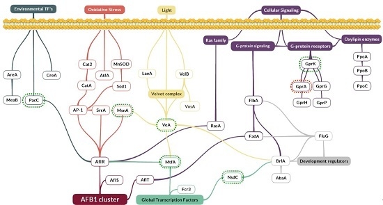
Since AFB1 biosynthesis is strongly interconnected with several other fungal biosynthetic pathways, a large number of genes considered as regulatory factors were analyzed. They included the velvet complex, genes involved in oxidative stress response, environmental and global transcription factors, genes involved in cellular signaling (oxylipins, Ras family and G-protein signaling and receptors) and developmental regulators (Figure 3).
As shown in Figure 4, among the 33 tested genes, only seven presented significant modifications of their level of expression upon eugenol exposure.
These genes were:
  • The global regulator gene veA, belonging to the velvet complex. It was over-expressed with a 3.8-fold change compared to the control (p-value = 0.002);
  • mtfA, a putative C2H2 zinc finger transcription factor. It presented the same up-regulated pattern, increasing its expression by 2.2 times (p-value = 0.0297);
  • nsdC, of the global transcription factors, whose expression was increased by 1.7 times (p-value = 0.0100);
  • gprK, which was the most affected gene among the five G-protein coupled receptors analyzed here. This gene was over-expressed by 4.5 times (p-value = 0.0009). By contrast, gprA was down-regulated by 0.45 times (p-value = 0.0177);
  • The msnA gene was increased by 1.9 times (p-value < 0.0001), whereas no significant changes were observed for other genes implicated in the oxidative stress response such as superoxidase dismutases, catalases or oxylipins;
  • Finally, pacC’s expression, a zinc finger transcription factor related to pH, was increased by 2.3 times (p-value = 0.0098).

3. Discussion

A number of natural extracts were identified as being able to down-modulate the synthesis of the carcinogenic mycotoxin AFB1 in A. flavus [39]. The use of such inhibiting compounds could therefore represent an alternative strategy to the use of pesticides to control crop contamination. However, to date, the precise molecular mechanism responsible for this effect is only poorly documented. Indeed, mycotoxin production is a complex phenomenon based on the presence of biosynthetic clusters in toxigenic fungi whose regulation is governed by many environmental and physiological processes.
In order to better understand the inhibition of AFB1 production by such natural extracts, we developed a molecular tool allowing the simultaneous analysis of the expression of both the AFB1 cluster and a large number of global regulatory genes involved in different cellular pathways. We used it to characterize the mechanism of action of eugenol, a compound present in many essential oils and that has been previously identified as an AFB1 inhibitor. Eugenol has been extensively studied for its many biological effects including antimicrobial, anti-inflammatory, anti-oxidant and anticancer activities. However, to date, no precise cellular target was identified even though the interaction with cell membrane may represent a key point in the biological effects of this molecule [40].

3.1. Eugenol Inhibits the Expression of Aflatoxin Cluster Genes in A. flavus

The inhibition of AFB1 production by a toxigenic strain of A. flavus following eugenol addition is accompanied by the down-regulation of all but one aflatoxin cluster genes.
We demonstrated that the expression of 19 out of 27 genes of the cluster was almost no more detectable after eugenol exposure, whereas the others saw 10- to 20-fold reductions in their expression levels. These results are coherent with a very recent work of Jahanshiri et al. [16] showing that eugenol decreased the expression of some of Aflatoxins (AF)’s cluster genes in A. parasiticus. In that previous study, only five genes of the pathway were analyzed. Our present work extended this finding to the whole genes involved in the biosynthesis of AFB1.
AFB1’s cluster is internally regulated by two genes, aflR and aflS, whose activation is governed, independently one from another, by external regulators. An interaction between AflR and AflS is reportedly required for aflatoxinogenesis [41]. Kong et al. [42] reported that the activation by these two proteins led to the formation of a functional activation complex in the proper ratio of four AflS to one AflR. In our study, the expression of aflR was not significantly decreased in cultures exposed to eugenol, although a major down-regulation tendency was observed. Such a finding was already described in several studies where even low-level changes of aflR’s expression levels were accompanied by a severe decrease of structural genes [25,43,44]. However, levels of aflS transcripts were significantly reduced, which might have led to an alteration of the ratio between AflR and AflS and thus the formation of a limited number of active complexes. Consequently, AflR-binding sites were not attained and the transcription of the cluster was not activated (Figure 2). All of the cluster genes regulated by aflR had their expression levels severely decreased by eugenol’s addition. The genes intervening in the later stages of AFB1’s enzymatic cascade (aflM, aflN, aflX, aflO, aflP, aflQ and hypB) were more impacted than those involved in the beginning stages (aflA, aflB, aflC) leading to the polyketide structure [45]. The limited AflR/AflS complexes formed might have been promptly used up at the beginning of AFB1’s synthesis and were no longer sufficiently available for the proper activation of the rest of the cluster genes.
For aflT, the expression levels were not significantly different between control and eugenol-treated cultures. In fact, aflT, a Major Facilitator Superfamily (MFS) transporter encoding gene, is regulated neither by AflR nor by its co-activator AflS, due to the absence of an AflR binding-site on its promoter, but rather by the FadA-dependent G-protein signaling pathway [46]. Moreover, the expression of none of the genes belonging or affected by the latter pathway, notably fadA, flbA, fluG and brlA [34], has been altered by eugenol’s addition (Table S1).

3.2. Eugenol Alters the Expression of Global Regulation Factors

Eugenol‘s transcriptomic effect goes upstream of AF’s cluster genes, affecting genes encoding general transcriptional regulating factors.

3.2.1. The Pivotal Role of MtfA, VeA and MsnA in Eugenols Molecular Mechanism

MtfA is a global transcription factor, regulating sterigmatocystin/aflatoxin biosynthesis as well as other secondary metabolites clusters [31]. The deletion as well as the over-expression of mtfA has been shown to inhibit the expression of aflR and subsequent sterigmatocystin production in A. nidulans [21]. Also, in the recent work of Zhuang et al. [47] on peanut seeds infected with ΔmtfA and over-expressed (OE) mtfA gene on A. flavus strains, it was observed that the decrease in AFB1 production was greater when there was a 2.75-fold mtfA over-expression compared to the wide-type strain, than deleted, and went with a decreased expression of aflR. Although the mtfA gene is over-expressed at a similar level as in the above-quoted study [47], this does not mean that MtfA is the direct molecular target of eugenol. A kinetic study of gene expression, using our molecular tool coupled to AF production analysis, could be used in order to determine the time course evolution of modulated genes.
Furthermore, mtfAs expression is highly dependent on that of veA. As discussed in the study by Lind et al. [31], MtfA interacts with VeA in A. nidulans and the expression of mtfA (AN8741.2) was decreased by 5 times in an A. nidulans ΔveA. All of the above findings are in favor of the notion that the over-expression of mtfA in A. flavus is a result of the increased expression of veA upon eugenol addition. The interaction of these two regulators could then be responsible for aflRs down-regulation and the succeeding inhibition of AFs biosynthetic pathway in A. flavus. Moreover, VeA is by itself essential for the transcription of both aflR and aflS and, consequently, the production of aflatoxins [48]. This global transcription regulator plays a key role in secondary metabolite production [49]. Depending on its abundance in the cell, it may act as a repressor or as an activator [50]. For instance, the expression of fumagillin and fumitremorgin G gene clusters was inhibited in a overexpressing veA (OE:veA) strain of A. fumigatus [51]. Therefore, an over-expression of veA (3.8-fold) observed here is compatible with the down-regulation of aflR and aflS and the subsequent inhibition of aflatoxin production in eugenol-treated cultures. Furthermore, as in many filamentous fungi, SM, and aflatoxin production in particular are often induced as a response to Reactive Oxygen Species (ROS) formation [52]. It has been also demonstrated that VeA plays a critical role in protecting A. flavus from oxidative stress. VeA positively regulates the expression of oxidative stress tolerance genes such as msnA in A. flavus [38] and also in A. nidulans [31]. Conversely, levels of ROS as well as AFB1 increased in ΔmsnA A. flavus and A. parasiticus strains [53]. Furthermore, it has been demonstrated that aflatoxin biosynthesis is itself a source of intracellular ROS, and the over-expression of msnA could then also be the outcome of AFB1s inhibition [54].
Accordingly, an oxidative-stress alleviating condition, such as the addition of eugenol, could lead to an increased expression of veA and consequently of msnA. In our study, msnA transcripts increased by 1.9 times along with AFB1s inhibition in eugenol-treated cultures, following veA’s over-expression. This effect is possibly linked to a decrease of intracellular ROS.

3.2.2. The Putative Implication of Other Regulatory Factors and Signaling Proteins

Eugenol addition also alters the expression of other transcription-regulating factors such as (i) nsdC and (ii) pacC encoding a pH-dependent transcription factor, as well as (iii) the grpA and grpK genes involved in the G-protein signaling pathway. Except for grpK, these genes have been shown to be closely related to the expression of AF cluster genes and AFB1 production [19,26,55,56]. However, to date, the exact mechanism of this interaction has to be clarified.

4. Conclusions

In this study, we bring out the transcriptomic inhibition behind eugenols AFB1-repressing action. This is the first time, to our knowledge, that the expression of all the 27 genes involved in AFB1 synthesis has been studied on an inhibitor molecule. In the presence of eugenol, AFB1 cluster genes were strongly down-modulated following decreased expression of its regulating complex AflR/AflS. We also demonstrated that this went with a modulation of seven regulatory factors. We then highlighted the involvement of mtfA, veA and msnA in this inhibition.

5. Materials and Methods

5.1. Chemicals and Reagents

Aflatoxin B1 and eugenol standards were purchased from Sigma-Aldrich (Saint-Quentin-Fallavier, France) and dissolved in methanol and ethanol, respectively. Stock solutions were stored at 4 °C until use. All analytical grade solvents were purchased from Thermo Fisher Scientific (Illkirch, France).

5.2. Fungal Strain and Culture Conditions

The Aspergillus flavus strain NRRL 62477 used in this study [57] was maintened in the dark on a Malt Extract Agar (MEA) medium (Biokar Diagnostics, Allone, France) at 27 °C.
For experiments, 10 μL of a calibrated spore suspension (106 spores/mL) prepared from a seven-day culture was used to centrally inoculate the MEA medium covered with sterile cellophane layers (Hutchinson, Chalette-sur-Loing, France) as described by Leite et al. [58]. Firstly, five different concentrations of eugenol (0.1 to 1 mM) were tested to determine the concentration able to inhibit AFB1 with a limited impact on fungal development, as measured by colony diameter. The concentration of 0.5 mM was selected for further experiments. Eugenol dilutions were prepared to add only 20 μL of ethanol in the culture medium, this concentration having been identified as a no-effect dose on both fungal growth and AFB1 production. Control cultures were performed by adding only 20 μL of ethanol in the medium. Six replicates of each group were prepared and incubated for four days at 27 °C in the dark. At least three replications of the experiment were performed. The pH of all media was taken before and after incubation using a food pH-meter H199161 (Hanna Instruments, Tanneries, France).

5.3. Aflatoxin B1 Extraction and Determination by HPLC

For AFB1 extraction, culture media were mixed with 25 mL of chloroform. Samples were agitated for 2 h on a horizontal shaking table at 160 rpm at room temperature. Chloroform extract was filtered through a Whatman 1PS phase separator (GE Healthcare Life Sciences, Vélizy-Villacoublay, France), evaporated at 60 °C until dry and dissolved in 500 μL of a water-acetonitrile-methanol (65:17.5:17.5; v/v/v) mixture. To eliminate possible impurities, all samples were filtered through a 0.45 μm disk filters (Thermo Scientific Fisher, Villebon-Sur-Yvette, France). The analysis of samples was done with a Dionex Ultimate 3000 UHPLC system (Thermo Scientific, Illkirch, France) using a liquid chromatography column, Luna® C18 (125 × 2 mm, 5 μm, 100 Å) (Phenomenex, Torrance, CA, USA) at 30 °C. Separation conditions were adapted from Fu et al. [59] with mild modifications. A 20 min isocratic mode was delivered at 82.5% of eluent A: acidified water (0.2% of acetic acid) and acetonitrile (79:21 v/v); and 17.5% of eluent B: pure methanol. A flow rate of 0.2 mL/min was used and 10 μL of extract was injected. AFB1 was detected by a fluorescent detector at 365/430 nm excitation/emission wavelengths. Peak identity was confirmed by analyzing absorption spectrum with a diode array detector coupled to the system. Production levels of AFB1 on media were calculated based on a standard calibration curve.

5.4. Isolation of Fungal RNA and Reverse Transcriptase-Polymerase Chain Reaction (RT-PCR)

At the end of incubation, mycelia were separated from the medium and ground up under liquid nitrogen. The RNA was purified as recommended by the manufacturer from 100 mg of mycelium using a Qiagen RNeasy PlusMinikit (Qiagen, Hilden, Germany) and including a gDNA eliminator column. The quality of RNA was verified by gel electrophoresis (1.2% agarose) and concentrations were measured using a NanoDrop ND1000 (Labtech, Palaiseau, France). The A260/A280 ratio was measured [60], and each sample was adjusted to a final RNA concentration of 300 ng/μL.
First-strand cDNA synthesis was carried out by RT-PCR. Reverse transcription took place in a 20 μL reaction mixture containing 10 μL of RNA, 200 U of RevertAid Reverse transcriptase, 4 μL of 5× Reaction Buffer, 20 U of RNase inhibitor (Thermo Scientific, Illkirch, France), 2 μL of 10 mM dNTP (Euromedex, Souffelweyersheim, France), 1 μL of sterile water, and 1 μL of oligo (dT) Bys 3' Primer: (5'-GCTGTCAACGATACGCTATAACGGCATGACAGTGTTTTTTTTTTTTTTT-3').
A first denaturation was done at 70 °C for 5 min and reverse transcription was performed as follows: 5 min at 37 °C; 60 min at 42 °C and 15 min at 85 °C.

5.5. Design and Validation of q-PCR Primers

All primer sets were designed based on the genomic data of the Aspergillus flavus strain NRRL3357 (GenBank accession number EQ963478A). A total of 62 genes primer pairs were designed including all 27 AFB1 cluster genes. It must be noted that aflF and aflU were not followed in this study, as the promoter regions of both genes are missing in A. flavus species, thus their inability to produce type-G aflatoxins. Other regulatory factors directly or indirectly related to AFB1’s production (Figure 3) were included, along with two housekeeping genes, b-tubulin and gpdA. All primer pairs were designed to amplify a 50–150 bp fragment based solely on the coding sequence of the corresponding genes, with at least one of the primers extending on an exon/exon junction in order to avoid undesirable genomic DNA amplification. Primer-dimer or self-complementarities were evaluated using the PrimerExpress 2.0 software (Applied Biosystems, Courtaboeuf, France). All primers were synthesized by Sigma Aldrich (Saint-Quentin Fallavier, France) and tested with four different concentrations (300/300; 300/900; 900/300 and 900/900 nM) after reception to determine their optimal concentrations in the mix. Primer validation was carried out with the best amplification curve and dissociation curves were used to confirm the good amplification of each gene. At least three biological replicates of A. flavus NRRL62477 were used to validate the amplification specificity. Negative controls in which no reverse transcriptase enzyme was added and a no template control were included to control reagents contamination. Primer sequences and their concentrations are listed in Table S2.

5.6. Analysis of the Expression of the Genes Linked to Aflatoxin B1 Biosynthesis

Experiments were carried out using a ViiA7 Real-Time PCR System (Applied Biosystems, Forster City, CA, USA). The 384 well-plates were prepared by an Agilent Bravo Automated Liquid Handling Platform (Agilent Technologies, Santa Clara, CA, USA). Each well contained a final volume of a 5 μL mix: 2.5 μL of Power SYBR® Green PCR Master Mix (Applied Biosystems, Warrington, UK) used as a fluorescent dye, 1.5 μL of each primer set and 1 μL of cDNA material. Three-step quantitative PCRs were performed as follows: a first one-hold stage at 95 °C for 10 min followed by 45 cycles (95 °C for 15 s and 60 °C for 30 s), and a final extending step (95 °C for 15 s, 60 °C for 1 min and 95 °C for 15 s) for melt curve analysis. The results were analyzed with a Quant-Studio Real time PCR software v1.1 (Applied Biosystems, Courtaboeuf, France). Housekeeping genes were analyzed with Normfinder algorithm [61] and the more stable was used as a reference for normalization in the 2−ΔΔCt analysis method [62]. Five distinct experiments were done, each including at least three biological replicates of each condition.

5.7. Statistics

Students t-test was used to analyze the differences between control and treated samples. The differences were considered to be statistically significant when the p-value was lower than 0.05.

Supplementary Materials

The following are available online at www.mdpi.com/2072-6651/8/5/123/s1, Table S1: Gene expression values of regulatory factors that were not impacted upon Eugenol addition. Values are expressed in fold change and compared to a control fixed at 1; Table S2: Primer sequences of the 62 designed genes.

Acknowledgments

The authors would like to thank the Agence Nationale de la Recherche (ANR) and the Research Council of Saint-Joseph University (Lebanon), the two organizations who funded the doctoral studies of Rhoda El Khoury, as well as the Consejo Nacional de Ciencia y Tecnología (CONACYT) México, funding Isaura Caceres. This work was financed by the projects Aflafree (ANR-11-ALID-0003), Aflared (joint project, 001-2012 STDF-AIRD), and ToxinFree (Campus France PHC cèdre 32763). The authors would also like to thank Soraya Tadrist and Annabelle Muller for their contribution to the RNA extraction protocol.

Author Contributions

Isabelle P. Oswald, Olivier Puel, Jean-Denis Bailly, Ali Atoui, André El Khoury conceived, supervised and designed the experiments. Isaura Caceres and Rhoda El Khoury performed the experiments, contributed to experiment design. Isaura Caceres, Rhoda El Khoury, Olivier Puel and Jean-Denis Bailly wrote the paper. Ángel Medina contributed to the RNA extraction protocol and culture condition techniques; Yannick Lippi and Claire Naylies contributed to q-PCR performance and data analysis.

Conflicts of Interest

The authors declare no conflict of interest.

Abbreviations

A. flavusAspergillus flavus
AFAflatoxins
AFB1Aflatoxin B1
GPCRsG-protein coupled receptors
HPLCHigh Performance Liquid Chromatography
MEAMalt Extract Agar
MFSMajor Facilitator Superfamily
mMMillimolar
ndNot detectable
nsNo significant changes
OEOver-expresssed
ROSReactive oxygen species
SEMStandard Error of Mean
SMSecondary metabolism
TFsTranscription factors

References

  1. Amaike, S.; Keller, N.P. Aspergillus flavus. Annu. Rev. Phytopathol. 2011, 49, 107–133. [Google Scholar] [CrossRef] [PubMed]
  2. I.A.R.C. Some naturally occurring substances: Food items and constituents, heterocyclic aromatic amines and mycotoxins. IARC Monogr. Eval. Carcinog. Risks Humans. 1993, 56, 245–395. [Google Scholar]
  3. Bbosa, G.; Kitya, D.; Lubega, A. Review of the biological and health effects of aflatoxins on body organs and body systems. In Aflatoxin—Recent Advances and Future Prospects; INTECH: Hampshire, UK, 2013. [Google Scholar]
  4. Meissonnier, G.M.; Pinton, P.; Laffitte, J.; Cossalter, A.-M.; Gong, Y.Y.; Wild, C.P.; Bertin, G.; Galtier, P.; Oswald, I.P. Immunotoxicity of aflatoxin B1: Impairment of the cell-mediated response to vaccine antigen and modulation of cytokine expression. Toxicol. Appl. Pharmacol. 2008, 231, 142–149. [Google Scholar] [CrossRef] [PubMed]
  5. Groopman, J.D.; Kensler, T.W.; Wild, C.P. Protective interventions to prevent aflatoxin-induced carcinogenesis in developing countries. Annu. Rev. Public Health 2008, 29, 187–203. [Google Scholar] [CrossRef] [PubMed]
  6. Streit, E.; Schatzmayr, G.; Tassis, P.; Tzika, E.; Marin, D.; Taranu, I.; Tabuc, C.; Nicolau, A.; Aprodu, I.; Puel, O.; et al. Current situation of mycotoxin contamination and co-occurrence in animal feed—Focus on Europe. Toxins 2012, 4, 788–809. [Google Scholar] [CrossRef] [PubMed]
  7. Tabuc, C.; Marin, D.; Guerre, P.; Sesan, T.; Bailly, J.D. Molds and mycotoxin content of cereals in southeastern Romania. J. Food Prot. 2009, 72, 662–665. [Google Scholar] [PubMed]
  8. Marin, S.; Ramos, A.J.; Cano-Sancho, G.; Sanchis, V. Mycotoxins: Occurrence, toxicology, and exposure assessment. Food Chem. Toxicol. 2013, 60, 218–237. [Google Scholar] [CrossRef] [PubMed]
  9. Holmes, R.A.; Boston, R.S.; Payne, G.A. Diverse inhibitors of aflatoxin biosynthesis. Appl. Microbiol. Biotechnol. 2008, 78, 559–572. [Google Scholar] [CrossRef] [PubMed]
  10. Ehrlich, K.C. Non-aflatoxigenic Aspergillus flavus to prevent aflatoxin contamination in crops: Advantages and limitations. Front. Microbiol. 2014, 5. [Google Scholar] [CrossRef] [PubMed]
  11. Bluma, R.; Amaiden, M.R.; Daghero, J.; Etcheverry, M. Control of Aspergillus section Flavi growth and aflatoxin accumulation by plant essential oils. J. Appl. Microbiol. 2008, 105, 203–214. [Google Scholar] [CrossRef] [PubMed]
  12. Kohiyama, C.Y.; Yamamoto Ribeiro, M.M.; Mossini, S.A.G.; Bando, E.; Bomfim, N.D.S.; Nerilo, S.B.; Rocha, G.H.O.; Grespan, R.; Mikcha, J.M.G.; Machinski, M. Antifungal properties and inhibitory effects upon aflatoxin production of Thymus vulgaris L. by Aspergillus flavus Link. Food Chem. 2015, 173, 1006–1010. [Google Scholar] [CrossRef] [PubMed]
  13. Kumar, A.; Shukla, R.; Singh, P.; Dubey, N.K. Chemical composition, antifungal and antiaflatoxigenic activities of Ocimum sanctum L. essential oil and its safety assessment as plant based antimicrobial. Food Chem. Toxicol. 2010, 48, 539–543. [Google Scholar] [CrossRef] [PubMed]
  14. Ferreira, F.D.; Kemmelmeier, C.; Arrotéia, C.C.; da Costa, C.L.; Mallmann, C.A.; Janeiro, V.; Ferreira, F.M.D.; Mossini, S.A.G.; Silva, E.L.; Machinski, M. Inhibitory effect of the essential oil of Curcuma longa L. and curcumin on aflatoxin production by Aspergillus flavus Link. Food Chem. 2013, 136, 789–793. [Google Scholar] [CrossRef] [PubMed]
  15. Cheng, S.-S.; Liu, J.-Y.; Hsui, Y.-R.; Chang, S.-T. Chemical polymorphism and antifungal activity of essential oils from leaves of different provenances of indigenous cinnamon (Cinnamomum osmophloeum). Bioresour. Technol. 2006, 97, 306–312. [Google Scholar] [CrossRef] [PubMed]
  16. Jahanshiri, Z.; Shams-Ghahfarokhi, M.; Allameh, A.; Razzaghi-Abyaneh, M. Inhibitory effect of eugenol on aflatoxin B1 production in Aspergillus parasiticus by downregulating the expression of major genes in the toxin biosynthetic pathway. World J. Microbiol. Biotechnol. 2015, 31, 1071–1078. [Google Scholar] [CrossRef] [PubMed]
  17. Jayashree, T.; Subramanyam, C. Antiaflatoxigenic activity of eugenol is due to inhibition of lipid peroxidation. Lett. Appl. Microbiol. 1999, 28, 179–183. [Google Scholar] [CrossRef] [PubMed]
  18. Brodhagen, M.; Keller, N.P. Signalling pathways connecting mycotoxin production and sporulation. Mol. Plant. Pathol. 2006, 7, 285–301. [Google Scholar] [CrossRef] [PubMed]
  19. Cary, J.W.; Harris-Coward, P.Y.; Ehrlich, K.C.; Mack, B.M.; Kale, S.P.; Larey, C.; Calvo, A.M. NsdC and NsdD affect Aspergillus flavus morphogenesis and aflatoxin production. Eukaryot. Cell 2012, 11, 1104–1111. [Google Scholar] [CrossRef] [PubMed]
  20. Bayram, O.; Braus, G.H. Coordination of secondary metabolism and development in fungi: The velvet family of regulatory proteins. FEMS Microbiol. Rev. 2012, 36, 1–24. [Google Scholar] [CrossRef] [PubMed]
  21. Ramamoorthy, V.; Dhingra, S.; Kincaid, A.; Shantappa, S.; Feng, X.; Calvo, A.M. The putative C2H2 transcription factor MtfA is a novel regulator of secondary metabolism and morphogenesis in Aspergillus nidulans. PLoS ONE 2013, 8. [Google Scholar] [CrossRef]
  22. Shaaban, M.I.; Bok, J.W.; Lauer, C.; Keller, N.P. Suppressor mutagenesis identifies a velvet complex remediator of Aspergillus nidulans secondary metabolism. Eukaryot. Cell 2010, 9, 1816–1824. [Google Scholar] [CrossRef] [PubMed]
  23. Yu, J.; Keller, N. Regulation of secondary metabolism in filamentous fungi. Annu. Rev. Phytopathol. 2005, 43, 437–458. [Google Scholar] [CrossRef] [PubMed]
  24. Montibus, M.; Pinson-Gadais, L.; Richard-Forget, F.; Barreau, C.; Ponts, N. Coupling of transcriptional response to oxidative stress and secondary metabolism regulation in filamentous fungi. Crit. Rev. Microbiol. 2013, 7828, 1–14. [Google Scholar] [CrossRef] [PubMed]
  25. Georgianna, D.R.; Payne, G.A. Genetic regulation of aflatoxin biosynthesis: From gene to genome. Fungal Genet. Biol. 2009, 46, 113–125. [Google Scholar] [CrossRef] [PubMed]
  26. Affeldt, K.; Carrig, J.; Amare, M.; Keller, N.P. Global survey of canonical Aspergillus flavus G protein-coupled receptors. MBio 2014, 5, 1501–1514. [Google Scholar] [CrossRef] [PubMed]
  27. Tsitsigiannis, D.I.; Keller, N.P. Oxylipins as developmental and host-fungal communication signals. Trends Microbiol. 2007, 15, 109–118. [Google Scholar] [CrossRef] [PubMed]
  28. Yin, W.; Keller, N.P. Transcriptional regulatory elements in fungal secondary metabolism. J. Microbiol. 2011, 49, 329–339. [Google Scholar] [CrossRef] [PubMed]
  29. Ehrlich, K.C.; Cary, J.W.; Montalbano, B.G. Characterization of the promoter for the gene encoding the aflatoxin biosynthetic pathway regulatory protein AFLR. Biochim. Biophys. Acta 1999, 1444, 412–417. [Google Scholar] [CrossRef]
  30. Hong, S.-Y.; Roze, L.; Linz, J. Oxidative Stress-Related Transcription Factors in the Regulation of Secondary Metabolism. Toxins (Basel) 2013, 5, 683–702. [Google Scholar] [CrossRef] [PubMed]
  31. Lind, A.L.; Wisecaver, J.H.; Smith, T.D.; Feng, X.; Calvo, A.M.; Rokas, A. Examining the evolution of the regulatory circuit controlling secondary metabolism and development in the fungal genus Aspergillus. PLoS Genet. 2015, 11. [Google Scholar] [CrossRef] [PubMed]
  32. Kato, N.; Brooks, W.; Calvo, A. The expression of sterigmatocystin and penicillin genes in Aspergillus nidulans is controlled by veA, a gene required for sexual development. Eukaryot. Cell 2003, 2, 1178–1186. [Google Scholar] [CrossRef] [PubMed]
  33. Shimizu, K.; Hicks, J.K.; Huang, T.P.; Keller, N.P. Pka, Ras and RGS protein interactions regulate activity of AflR, a Zn(II)2Cys6 transcription factor in Aspergillus nidulans. Genetics 2003, 165, 1095–1104. [Google Scholar] [PubMed]
  34. Hicks, J.K.; Yu, J.H.; Keller, N.P.; Adams, T.H. Aspergillus sporulation and mycotoxin production both require inactivation of the FadA Gα protein-dependent signaling pathway. EMBO J. 1997, 16, 4916–4923. [Google Scholar] [CrossRef] [PubMed]
  35. Affeldt, K.J.; Brodhagen, M.; Keller, N.P. Aspergillus oxylipin signaling and quorum sensing pathways depend on G protein-coupled receptors. Toxins (Basel) 2012, 4, 695–717. [Google Scholar] [CrossRef] [PubMed]
  36. Adams, T.H.; Hide, W.A.; Yager, L.N.; Lee, B.N. Isolation of a gene required for programmed initiation of development by Aspergillus nidulans. Mol. Cell. Biol. 1992, 12, 3827–3833. [Google Scholar] [CrossRef] [PubMed]
  37. Gilbert, M.K.; Mack, B.M.; Wei, Q.; Bland, J.M.; Bhatnagar, D.; Cary, J.W. RNA sequencing of an nsdC mutant reveals global regulation of secondary metabolic gene clusters in Aspergillus flavus. Microbiol. Res. 2015, 182, 150–161. [Google Scholar] [CrossRef] [PubMed]
  38. Baidya, S.; Duran, R.M.; Lohmar, J.M.; Harris-Coward, P.Y.; Cary, J.W.; Hong, S.-Y.; Roze, L.V; Linz, J.E.; Calvo, A.M. VeA is associated with the response to oxidative stress in the aflatoxin producer Aspergillus flavus. Eukaryot. Cell 2014, 13, 1095–1103. [Google Scholar] [CrossRef] [PubMed]
  39. Sakuda, S.; Yoshinari, T.; Furukawa, T.; Jermnak, U.; Takagi, K.; Iimura, K.; Yamamoto, T.; Suzuki, M. Search for aflatoxin and trichothecene production inhibitors and analysis of their modes of action. Biosci. Biotechnol. Biochem. 2016, 80, 43–54. [Google Scholar] [CrossRef] [PubMed]
  40. Kamatou, G.P.; Vermaak, I.; Viljoen, A.M. Eugenol—From the remote Maluku Islands to the international market place: A review of a remarkable and versatile molecule. Molecules 2012, 17, 6953–6981. [Google Scholar] [CrossRef] [PubMed]
  41. Chang, P. The Aspergillus parasiticus protein AFLJ interacts with the aflatoxin pathway-specific regulator AFLR. Mol. Genet. Genom. 2003, 268, 711–719. [Google Scholar]
  42. Kong, Q.; Chi, C.; Yu, J.; Shan, S.; Li, Q.; Li, Q.; Guan, B.; Nierman, W.C.; Bennett, J.W. The inhibitory effect of Bacillus megaterium on aflatoxin and cyclopiazonic acid biosynthetic pathway gene expression in Aspergillus flavus. Appl. Microbiol. Biotechnol. 2014, 98, 5161–5172. [Google Scholar] [CrossRef] [PubMed]
  43. Kim, J.H.; Campbell, B.C.; Molyneux, R.; Mahoney, N.; Chan, K.L.; Yu, J.; Wilkinson, J.; Cary, J.; Bhatnagar, D.; Cleveland, T.E. Gene targets for fungal and mycotoxin control. Mycotoxin Res. 2006, 22, 3–8. [Google Scholar] [CrossRef] [PubMed]
  44. Yoshinari, T.; Akiyama, T.; Nakamura, K.; Kondo, T.; Takahashi, Y.; Muraoka, Y.; Nonomura, Y.; Nagasawa, H.; Sakuda, S. Dioctatin A is a strong inhibitor of aflatoxin production by Aspergillus parasiticus. Microbiology 2007, 153, 2774–2780. [Google Scholar] [CrossRef] [PubMed]
  45. Ehrlich, K.C. Predicted roles of the uncharacterized clustered genes in aflatoxin biosynthesis. Toxins (Basel) 2009, 1, 37–58. [Google Scholar] [CrossRef] [PubMed]
  46. Chang, P.-K.; Yu, J.; Yu, J.-H. aflT, a MFS transporter-encoding gene located in the aflatoxin gene cluster, does not have a significant role in aflatoxin secretion. Fungal Genet. Biol. 2004, 41, 911–920. [Google Scholar] [CrossRef] [PubMed]
  47. Zhuang, Z.; Lohmar, J.M.; Satterlee, T.; Cary, J.W.; Calvo, A.M. The master transcription factor mtfA governs aflatoxin production, morphological development and pathogenicity in the fungus Aspergillus flavus. Toxins (Basel) 2016, 8, 1–16. [Google Scholar] [CrossRef] [PubMed]
  48. Calvo, A.M. The VeA regulatory system and its role in morphological and chemical development in fungi. Fungal Genet. Biol. 2008, 45, 1053–1061. [Google Scholar] [CrossRef] [PubMed]
  49. Calvo, A.M.; Bok, J. veA is required for toxin and sclerotial production in Aspergillus parasiticus. Appl. Environ. Microbiol. 2004, 70, 4733–4739. [Google Scholar] [CrossRef] [PubMed]
  50. Sprote, P.; Brakhage, A.A. The light-dependent regulator velvet A of Aspergillus nidulans acts as a repressor of the penicillin biosynthesis. Arch. Microbiol. 2007, 188, 69–79. [Google Scholar] [CrossRef] [PubMed]
  51. Dhingra, S.; Lind, A.L.; Lin, H.-C.; Tang, Y.; Rokas, A.; Calvo, A.M. The fumagillin gene cluster, an example of hundreds of genes under veA control in Aspergillus fumigatus. PLoS ONE 2013, 8. [Google Scholar] [CrossRef] [PubMed]
  52. Reverberi, M.; Zjalic, S.; Ricelli, A.; Punelli, F.; Camera, E.; Fabbri, C.; Picardo, M.; Fanelli, C.; Fabbri, A.A. Modulation of antioxidant defense in Aspergillus parasiticus is involved in aflatoxin biosynthesis: A role for the ApyapA gene. Eukaryot. Cell 2008, 7, 988–1000. [Google Scholar] [CrossRef] [PubMed]
  53. Chang, P.; Scharfenstein, L.; Luo, M. Loss of msnA, a putative stress regulatory gene, in Aspergillus parasiticus and Aspergillus flavus increased production of conidia, aflatoxins and kojic acid. Toxins (Basel) 2011, 3, 82–104. [Google Scholar] [CrossRef] [PubMed]
  54. Roze, L.; Laivenieks, M.; Hong, S.-Y.; Wee, J.; Wong, S.-S.; Vanos, B.; Awad, D.; Ehrlich, K.; Linz, J. Aflatoxin biosynthesis is a novel source of reactive oxygen species—A potential redox signal to initiate resistance to oxidative stress? Toxins (Basel) 2015, 7, 1411–1430. [Google Scholar] [CrossRef] [PubMed]
  55. Keller, N.P.; Nesbitt, C.; Sarr, B.; Phillips, T.D.; Burow, G.B. pH regulation of sterigmatocystin and aflatoxin biosynthesis in Aspergillus spp. Phytopathology 1997, 87, 643–648. [Google Scholar] [CrossRef] [PubMed]
  56. Ehrlich, K.C.; Montalbano, B.G.; Cary, J.W.; Cotty, P.J. Promoter elements in the aflatoxin pathway polyketide synthase gene. Biochim. Biophys. Acta 2002, 1576, 171–175. [Google Scholar] [CrossRef]
  57. El Mahgubi, A.; Puel, O.; Bailly, S.; Tadrist, S.; Querin, A.; Ouadia, A.; Oswald, I.P.; Bailly, J.D. Distribution and toxigenicity of Aspergillus section Flavi in spices marketed in Morocco. Food Control 2013, 32, 143–148. [Google Scholar] [CrossRef]
  58. Leite, G.M.; Magan, N.; Medina, Á. Comparison of different bead-beating RNA extraction strategies: An optimized method for filamentous fungi. J. Microbiol. Methods 2012, 88, 413–418. [Google Scholar] [CrossRef] [PubMed] [Green Version]
  59. Fu, Z.; Huang, X.; Min, S. Rapid determination of aflatoxins in corn and peanuts. J. Chromatogr. A 2008, 1209, 271–274. [Google Scholar] [CrossRef] [PubMed]
  60. Zeng, Y.; Yang, T. RNA isolation from highly viscous samples rich in polyphenols and polysaccharides. Plant Mol. Biol. Rep. 2002, 20. [Google Scholar] [CrossRef]
  61. Andersen, C.L.; Jensen, J.L.; Ørntoft, T.F. Normalization of real-time quantitative reverse transcription-PCR data: A model-based variance estimation approach to identify genes suited for normalization, applied to bladder and colon cancer data sets. Cancer Res. 2004, 64, 5245–5250. [Google Scholar] [CrossRef] [PubMed]
  62. Livak, K.J.; Schmittgen, T.D. Analysis of relative gene expression data using real-time quantitative PCR and the 2(-Delta Delta C(T)) Method. Methods 2001, 25, 402–408. [Google Scholar] [CrossRef] [PubMed]
Figure 1. Effect of eugenol on Aflatoxin B1 (AFB1) production and fungal growth in A. flavus NRRL 62477. Results are expressed as percentage of the control value. AFB1 was estimated by High Performance Liquid Chromatography (HPLC) and fungal growth by colony diameter. Both measures were taken on day 4 on six biological replicates. ns = no significant changes; nd = not detectable; * p-value < 0.05; *** p-value < 0.001.
Toxins 08 00123 g001
Figure 2. Fold change expression of genes belonging to the cluster responsible for aflatoxin biosynthesis in response to eugenol at 0.5 mM. Red line represents control expression level. Gene cluster organization was adapted from Amaike and Keller [1]; ns = no significant changes; * p-value < 0.05; ** p-value < 0.01; *** p-value < 0.001.
Toxins 08 00123 g002
Figure 3. Schematic representation of the 33 selected regulatory factors linked to AFB1 cluster in A. flavus. This hypothetical schema represents a simplified version of the different interactions between the regulatory factors and Aflatoxin‘s cluster. Schema was constructed based on gene interaction data described by the following works: [10,19,20,21,22,23,24,26,28,29,30,31,32,33,34,35,36,37,38]. Up- or down-regulation of genes upon eugenol addition is represented by green an red dotted lines, respectively.
Toxins 08 00123 g003
Figure 4. Fold change levels of the seven affected regulatory factors in presence of eugenol at 0.5 mM. Baseline represents control expression level; * p-value < 0.05; ** p-value < 0.01; *** p-value < 0.001.
Toxins 08 00123 g004