3.1. Beads Fabrication & Coloration Replication
To tune the beads size, several parameters have been optimized; polymer viscosities, voltages, flow-rates and distances between the needle and the substrate.
Figure 4 illustrates the effect of those four parameters on the average size of the beads and on the size distribution given by their average value and standard deviation (error bar) respectively. To elucidate the effect of these parameters and to discern underlying trends, measurements at the extreme of each parameter setting were carried out. With trends established, a fine-tuning of those four parameters was performed in order to reach the 100 to 500 nm beads size as found with the butterfly. According to these experiments, an increase of polymer viscosity tends to increase the beads size but also increase the bead size distribution. Moreover, our experiments confirmed that only low viscosity polymer solutions produce beads as previously shown by Sharma
et al. [
14]. As a consequence this parameter does not present a lot of flexibility for beads size tuning. An increase of the syringe pump flow-rate increases beads size but also increases its distribution rapidly and above 0.6 mL/h beads are not clearly separated from each other anymore. Additionally, we observed that changing the distance between the needle and the substrate has only a minor effect on the bead size as this setting influences simultaneously two parameters that have an opposite effect on the bead size: the electric field and the time of flight. Finally, an increase of the applied voltage decreases the bead size and we found this parameter to be the most efficient for bead size tuning and established that it is possible to produce polymer beads with a size ranging between 100 and 500 nm as found on the
Pieris rapae butterfly using SU-8 2002, a voltage of 12 kV, a distance of 8 cm and a flow-rate of 0.25 mL/h.
Figure 4.
Evolution of the beads average size and distribution (standard deviation of the bead size shown in the error bar) according to the change of the electrospraying parameters such as the polymer viscosity, syringe pump flow-rate, voltage and distance between the needle and the substrate. (a) Variation of the viscosity between SU-8 2002 and SU-8 2005. Other parameters were a voltage of 12 kV, a distance of 8 cm, a gauge 27 needle and a flow-rate of 0.08 mL/h; (b) variations in the flow-rate from 0.3 to 0.9 mL/h. Other parameters were a voltage of 20 kV, a distance of 10 cm, a gauge 23 needle and SU-8 2005 was used; (c) variations in the voltage between 12 and 18 kV Other parameters were a distance of 8 cm, a flow-rate of 0.08 mL/h, a gauge 27 needle and SU-8 2002 was used; (d) variations in the distance needle-substrate between 8 and 12 cm. Other parameters were a voltage of 12 kV, a flow-rate of 0.08 mL/h, a gauge 27 needle and the SU-8 2002 was used. It is shown that the voltage is an efficient parameter to tailor the bead size while parameters such as distance between electrodes tend to show weaker effect.
![Micromachines 05 00216 g004]()
In order to replicate the coloration pattern of the
Pieris rapae butterfly, shadow masks were used to selectively deposit scatterers according to the butterfly color pattern (see
Figure 5). Two different masks and different spraying times were used to generate the two different gray tones patterns replicating the butterfly color pattern as faithful as possible (see
Figure 6). The spraying time for the darkest gray tone was 2 min 50 s and the cumulative spraying time for the lightest area was 5 min 10 s.
Figure 5.
(a) Layer diagram of the butterfly wings replication showing dielectric (SU-8) beads sprayed through a shadow mask onto a silicon wafer previously coated with black SU-8 (I-37, Gersteltec, Pully, Switzerland) that was pyrolyzed to facilitate the bead deposition using electrospraying; (b) SEM picture of dielectric (SU-8) beads in the range of 100 to 500 nm produced by electrospraying to mimic Mie scatterers found on the Pieris rapae butterfly.
Thereby, dielectric beads of a diameter in the range of 100 to 500 nm as found in the
Pieris rapae’s wings have been successfully reproduced using electrospraying of SU-8.
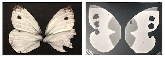
Figure 6 shows the result of the sprayed beads on a black SU-8 absorbing layer. We observe that the white coloration of the
Pieris rapae butterfly has been reproduced using artificial produced SU-8 beads. Grey tones were also obtained at the edges of the wings by varying the beads density using a two-steps spraying process through different shadow masks as described above.
Figure 6.
(a) Actual Pieris rapae butterfly wings showing white coloration with black spots; (b) result of Pieris rapae replication: the white surface is produced by SU-8 beads with diameters in the range of 100 to 500 nm. Black dots are the result of the black pyrolyzed layer without beads.
Figure 6.
(a) Actual Pieris rapae butterfly wings showing white coloration with black spots; (b) result of Pieris rapae replication: the white surface is produced by SU-8 beads with diameters in the range of 100 to 500 nm. Black dots are the result of the black pyrolyzed layer without beads.
3.2. Proof of the Structural nature of the Color Obtained
To demonstrate that we are dealing with structural white color and not a dye we performed reflectance measurement with the produced nanostructured surface. As demonstrated in
Figure 7, the wetting of the replicated surface with DI water decreased the reflectance dramatically something that one does not expect when covering a surface coated with a dye. In the case of the dry surface, an almost flat curve is observed over the whole visible wavelength range as expected from its white appearance and similarly to the real butterfly reflectance spectrum showing a plateau between 450 and 700 nm [
9]. In the case of the DI water wetted surface, the reflected light is decreased as the refractive index of the medium surrounding the beads is shifted now from 1.0 to 1.333 edging toward the SU-8 refractive index of 1.67. These results are in agreement with the Mie scattering model [
12] and give evidences for the structural based white coloration.
Figure 7.
(a) Reflectance spectrum of the Pieris rapae butterfly replica in the case of a dry and wetted surface with DI water; (b) picture of the surface with a dry and a wet circular area qualitatively showing the same effect of light scattering decrease due to the wet condition.
Figure 7.
(a) Reflectance spectrum of the Pieris rapae butterfly replica in the case of a dry and wetted surface with DI water; (b) picture of the surface with a dry and a wet circular area qualitatively showing the same effect of light scattering decrease due to the wet condition.
3.3. Sucrose Sensing Application
The previous experiment demonstrated that the refractive index surrounding the beads determines the intensity of the scattered light. Hence, it suggests the possibility to use such a structured surface for measuring the refractive index of an unknown medium through the reflected light coming from such a structure. The black surface patterned with polymeric beads presented above was thus tested as a possible sucrose sensor.
The refractive index of sucrose solutions is known to depend on the sucrose content and has been measured to vary between 1.333 for pure water to 1.450 for solution containing 700 g of sucrose per liter [
15]. According to van de Hulst [
12], the Mie scattering obtained in the case of spheres depends on the refractive index of the surrounding medium. The closer the indexes of the medium and of the spheres, the smaller the scattering.
Figure 8 illustrates the light reflectance from the artificial butterfly wing structure wetted with different sucrose concentrations. As the sucrose concentration increases from 0 to 250 g/L its refractive index also increases from 1.333 to 1.36 and the scattering of light is reduced. This graph shows that the sucrose concentration can be directly measured by evaluating the light scattering intensity on the artificial butterfly wing structure.
Figure 8.
Evolution of the reflectance of the butterfly wing replica when covered by a sucrose solution of increasing concentration. The reflectance was measured at 600 nm.
Figure 8.
Evolution of the reflectance of the butterfly wing replica when covered by a sucrose solution of increasing concentration. The reflectance was measured at 600 nm.
3.4. Discussion
Although the
Pieris rapae butterfly wing and our replica exhibit a similar white appearance, the artificial butterfly micro structure does differ substantially from the actual wing structure. Importantly, on the actual
Pieris rapae butterfly wing, the beads are attached to a periodic rectangular structure as shown in
Figure 1b. Also, the pterin beads in the actual butterfly wing are not touching the surface but are suspended on those rectangular frames. The rectangular bead suspending frames could have a photonic crystal effect and consequently some specific wavelengths could be attenuated. Those bead suspending rectangular structures were not reproduced in the current study. Making an identical
Pieris rapae structure with electrospraying alone would be hard but could be manufactured using a combination of photolithography and electrospraying. Given the reflectance results obtained in this study without the rectangular structures and with beads directly touching the substate, we believe that effects related to photonic bad gaps or Bragg filter effects are small compared to the effect of the diffusive bead structures. In other words, the dominant effect is the Mie scattering of the randomly organized SU-8 beads. Resonances for specific frequencies given by the bead size and refractive index are cancelled out by the dispersion in bead size, which explains the white spectra obtained both in the replica and the actual butterfly. The effect of the suspending rectangular structures in the butterfly wing could be to minimize the interaction between the substrate and the beads to possibly strengthen the light scattering. This might explain the brighter coloration of the butterfly’s wings compared to that of our replica. In order to mimic such structures, other beads fabrication methods were presented in the literature with an example of Scholz
et al. [
16] synthesizing ZnS nanospheres by homogeneous precipitation. This alternative method has the particularity to produce monodispersed spheres; hence scattering exhibits resonance at specific wavelength.
The replicated beaded structure was also used to make a sucrose sensor. In this case, scattered light from the surface is modulated by the refractive index of the medium surrounding the beads. Hence, increasing sucrose concentration leads to a measurable decrease in scattered light. This refractive index sensor is very easily produced and could be made very inexpensively but its sensitivity is at least one order of magnitude lower than that of a plasmonic-based refractive index sensor. However, our technique seems appropriate to sense sucrose in the range starting from 1 g/L up to the limit of sucrose dissolution in water. This range coincides with the amount of sucrose present in pre-fermented wine must or beer wort. Indeed our technique offers a resolution close to the one of the winemaker portable refractometer (~1 g/L) while presenting the same drawback, which is its lack of specificity. Therefore, this technique could be an alternative to the refractometer used in that industry, which is expensive due to optical components used, such as prisms, and fragile due to optical coating required.


