Article

The Influence of The Light-Activated Titania P25 on Human Breast Cancer Cells

by
Agata Markowska-Szczupak
1,*,
Zhishun Wei
2 and
Ewa Kowalska
3,*
1
Department of Chemical and Process Engineering, West Pomeranian University of Technology in Szczecin, 70-322 Szczecin, Poland
2
Hubei Provincial Key Laboratory of Green Materials for Light Industry, Hubei University of Technology, Wuhan 430068, China
3
Institute for Catalysis, Hokkaido University, Sapporo 001-0021, Japan
*
Authors to whom correspondence should be addressed.
Catalysts 2020, 10(2), 238; https://doi.org/10.3390/catal10020238
Submission received: 24 January 2020 / Revised: 10 February 2020 / Accepted: 14 February 2020 / Published: 17 February 2020
(This article belongs to the Special Issue Commemorative Issue in Honor of Professor Hugo de Lasa)

Abstract

:
Cosmetics and other daily care products contain titanium(IV) oxide (titania). Since multiple risk factors can increase the chance of developing cancer, an evaluation of titania safety has become a matter of concern in recent times. However, it should be pointed out that titania as an efficient photocatalyst has been also applied for inactivation of various pathogens, environmental purification and energy conversion, which might result in significant improvement of human life. Therefore, it is worth considering titania not only as a possible cancer initiator, but also as an efficient solution against cancer cells. Accordingly, in this study, the effect of commercial titania photocatalyst P25 (Degussa/Evonik) on breast adenocarcinoma MCF7 cells (ATCC® HTB-22™, breast adenocarcinoma cell line from human) has been investigated. The cells were treated with titania at doses of 10, 30, and 50 µg/mL under UVA/vis irradiation and in the dark. The significant morphological alterations in living cells were observed for larger doses of titania, such as changes in the shape and the size of cells, the deviation from the normal structure, and an increase in cells’ mortality. Moreover, the effect was significantly higher under irradiation than in the dark confirming strong photocatalytic activity of titania P25. In contrast, the lowest dose of titania (10 µg/mL) did not exhibit a significant impact on MCF7 cells, similarly to the nontreated cells. Accordingly, it has been proposed that locally applied titania might be considered for a cancer therapy after necessary in vivo tests to estimate any possibilities of side effects.

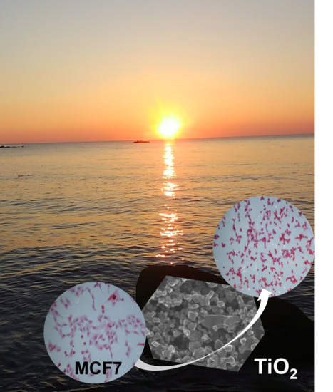
Graphical Abstract

1. Introduction

The concerns on biosafety of nanomaterials, in particular metal-based nanostructures, such as nanogold, nanosilver, and metal oxides (e.g., cerium (IV) oxide, zinc (II) oxide, titanium (IV) oxide), have been raised in the last few years, as evident in the ever-growing number of studies and scientific papers [1,2,3]. However, the last listed above—titania, widespread used as a white pigment of paints and plastics, and in many food applications (as food additive E171) and cosmetics, toothpastes and pills (as CI77891), has been considered as an inert and safe material. Although, some studies have already suggested the toxicity of titania, resulting from nanostructural morphology (nanomaterial toxicity) and photo-reactivity (generation of reactive oxygen species (ROS), including singlet oxygen even in sunscreen creams) [4,5,6], the common and commercial applications of titania for daily products have not been limited. However, the French Agency for Food, Environmental and Occupational Health and Safety (ANSES) classified titania as a category 1b carcinogen in 2017. In April 2019, France banned using E171 as a food additive from 2020 [7]. Furthermore, according to the Committee for Risk Assessment (RAC) of the European Chemical Agency (ECHA) under REACH Regulation, titania could be considered as a class 2 carcinogen, but only through inhalation (H350i). In contrast, the Titanium Dioxide Manufacturers Association (TDMA) has already presented the opposite opinion demonstrating an unclearness and uncertainty of toxicity classification. It is noteworthy that any proposed classification of titania toxicity has not been formerly accepted yet, since no new substantial information that might challenge RAC’s scientific opinion has been put forward, i.e., it has been concluded that titania does not justify the draft commission regulation amending (October 2019) [8,9,10].
The most intriguing question is “what factors of toxicity should be considered?” Firstly, the size of particles differs significantly for various titania products, i.e., nanoparticulate (1 to 100 nm) and microparticulate (>100 nm) titania materials are completely dissimilar, manufactured in different processes and used for various applications. Chloride and sulfate processes are commercially used to produce mainly micron-sized particles (particle sizes from 250 to 350 nm), which are applied to impart a whiteness of papers, paints, coatings, plastics, rubbers and leathers or provide high refractive index for the glass. The inertia of macroscale titania (in rutile form) allows also for its application as additives to food products, cosmetics (e.g., soaps and creams) and pharmaceuticals. Micron-sized titania has been considered as the main titania product, and primarily used as a pigment being two-thirds of all pigments. The world production has exceeded nine million metric tons, and a market has grown at a Compound Annual Growth Rate (CAGR) of 4.2% in terms of value and might reach USD 28.3 billion in 2026 [11]. In contrast, nanoparticulate titania has a much larger specific surface area and consists of mainly anatase phase, which results in enhanced photocatalytic activity (under UV) and high chemical reactivity. For these reasons, high demands for novel technologies of nanoparticulate titania production have been observed. For example, the sol-gel synthesis has been most commonly used for nano-TiO2 production. Moreover, titania nanoparticles (NPs) could be obtained in reaction of titanium chloride with ammonia or oxygen, chemical vapor deposition (CVD), physical vapor deposition (PVD), hydrothermal methods, gas-phase reactions, etc. [12,13]. Ultrafine titania is mainly found in sunscreen creams, cosmetics, self-cleaning coatings, air filtration devices, odor absorbers, textile fibers (also waterproof membranes), wood preservatives and (photo)catalysts, including various consumer products, such as household cleaning products, household self-cleaning coatings, household air filtration devices, computer keyboards and mice, hair styling devices, and various novel applications, e.g., solar cells. Commercially available manufacturers offer nano-TiO2 as pure anatase, pure rutile, and mixed-phase products, which are varied by particle size, specific surface area, purity/modifications (some are doped and/or surface modified), surface characteristics, chemical reactivity, etc. Due to high costs of production, the global production of ultrafine titania is lower than 0.25% of the global production of all titania products. Among the sixty largest titania manufactures, only six in Japan (Sakai Chemical, Ishihara Sangyo Kaisha, Denka, Tayca, Titan Kogyo, Nippon Aerosil (part of Evonik)) and three in Germany (EMD Chemicals Performance Materials, Evonik/Degussa, Sachtleben) boast production facilities for nano-TiO2 [14].
Secondly, due to the widespread use of macro- and nano-TiO2, the overall exposure (as an average individual exposure to titania) is difficult for assessment [15]. Although a large number of toxicology data exist, there are still open questions regarding how different forms of titania, characterized by phase composition, size, specific surface area, surface chemistry, crystallinity, morphology (including shape), solubility, and agglomeration might act in an environment and in living organisms, including human bodies. For this point of view, additional research, dedicated to commercially available products, should pursue the goal of enhancing the knowledge by filling existing gaps in safety/toxicity of titania products.
The aim of the present study is to estimate an influence of commercial titania P25 (Evonik, Germany) on breast adenocarcinoma cells MCF7. Titania P25, commercially known as AEROXIDE® TiO2, AEROPERL®, AEROXIDE® Evonik P25 or Degussa P25, is a fine-particulate, pure titania with high specific surface area and agglomerate structure. The product has a unique combination of an anatase content of 80–90% by weight with a small portion of rutile and amorphous titania [16,17]. Therefore, the P25 structure makes it suitable for use as an effective UV filter in body cosmetics with SPF (sun protection factor). It should be pointed that a sunscreen is a specific body care product, which is directly applied as a thick layer over large areas of the body with high frequency. Accordingly, the influence of titania (as sun creams’ component and other daily care products) on human cells should be analyzed in detail.
Breast cancer is known to be the second most common cancer, and a leading cause of cancer death in women worldwide. Despite more than 100 years of research on breast cancer (mostly over the last 50 years), resulting in the identification of numerous factors that influence the cancer risk, it has been impossible to reduce its appearance so far [18]. Therefore, further and more intensive studies on: (i) Defining factors that contribute to breast cancer development, and (ii) finding methods/materials that might kill/inactivate cancer cells, are highly necessary. Accordingly, the impact of titania, commonly used in various daily products (including both internal and external consumption), on breast cancer cells has been investigated in this study. It should be pointed out that breast cancer cells have been selected for this study because they are considered as: (i) Perfect cells for vitro models, and (ii) self-replicating material, i.e., growing in almost infinite quantities with a high degree of homogeneity (much larger than healthy cells’ line) [19]. Moreover, breast cancer cells have been well described, are easy to handle, and might be obtained fast from frozen stocks. Additionally, the MCF7 cells have been regarded as a model for evaluating response to novel drug therapies.

2. Results

2.1. Titania P25 Photocatalyst

At first, the most adequate (i.e., considering the broad and common application, accessibility, and high activity) titania photocatalyst has been selected for this study. Degussa/Evonik P25 is probably the most “famous” titania photocatalyst, because of extremely high photocatalytic activity for various reactions, and thus commonly used, and available in almost every laboratory focusing on heterogeneous photocatalysis in the world. Based on the literature reports [16,20] and the preliminary photocatalytic experiments for oxidative decomposition of acetic acid (as a model compound), P25 has been chosen for the present study. For those preliminary experiments, commercial titania photocatalysts with different properties (phase composition, crystallite size, specific surface area, etc.) have been taken, and the resultant reaction rates have been compared with that by P25.
Indeed, it has been confirmed that P25 shows the highest activity independently on the surface properties (not the smallest crystallite/particle sizes and not the largest specific surface area), as shown in Figure 1. Generally, anatase has been considered as a more active phase than rutile [21,22,23], which has been also confirmed in this study (solid vs. empty circles in Figure 1). Although P25 (red triangle in Figure 1) also consists of the anatase phase (76–80%), its high photocatalytic activity could not be explained either by anatase presence (other samples contain almost 100% anatase) or surface properties. For example, the photocatalytic activity of ST01 anatase sample with much better surface properties (specific surface area of ca. five times larger (ca. 300 m2 g-1) than that of P25) have been significantly (more than twice) lower than that by P25. Similarly, various literature reports have shown both extremely high and property-unreasonable activity of P25 titania. Therefore, it has been proposed that charge carriers’ transfer between two titania polymorphs (anatase/rutile) could be responsible for the high activity of P25 [24,25,26,27], as well for other mixed-phase titania samples, e.g., anatase/brookite [27,28]. The co-existence of two phases with high intrinsic activities in different reaction systems could also be responsible for high activity of P25, i.e., anatase in photo-oxidation and rutile in photo-reduction reactions, as suggested for single-phase samples (pure anatase and pure rutile) isolated from the original P25 photocatalyst [16,29].
Accordingly, P25 has been widely used for various photocatalytic studies, mainly because of high activity, but also as a “standard” [27] during measurements of photocatalytic activities of various samples in different laboratories (it should be noticed that reliable comparison of photocatalysts is still challenging, even with apparent (not true) quantum yield estimations [30,31,32]). However, it should be pointed that the composition of P25 might slightly differ between samples, and even samples taken from the same container could show divergence, e.g., the content of anatase, rutile and noncrystal part varies from 76% to 80%, 13% to 15% and 6% to 11%, respectively [29,33]. Despite some differences in composition, it should be noted that P25 is mainly composed of the anatase phase (Figure 2d), which has been considered as more active for oxidation reactions [29,34] (also confirmed in the present study (Figure 1)). The crystallite sizes of titania varied from ca. 25–30 nm for anatase to ca. 34–40 nm for rutile [29,35], whereas particles sizes are much larger, due to crystallites’ aggregation, as shown in Figure 2a,b. P25 as all other titania photocatalysts could absorb only UV irradiation (λ < 400 nm), due to its wide bandgap, as shown in Figure 2c.

2.2. P25 Effect on Human Carcinoma Cells

The data of cells’ viability, estimated after their treatment with titania, are shown in Figure 3. It was found that titania significantly affected the viability of MCF7 cells causing their death. About 5.0% cell viability was lost for 10 µg/mL of titania and 7.5% when titania in the same dose was activated by 10-min irradiation. However, the number of dead cells was almost unchanged in control experiments independently on used conditions (dark or irradiation). Interestingly, it was found that P25 exhibited dose-dependent cytotoxicity against breast carcinoma cells both under UVA/vis irradiation and in the dark. About 18% adenocarcinoma breast cells were killed after 10-min treatment with 50 μg/mL of photoexcited titania, which is twice higher than that in the dark. The difference between samples irradiated with UVA/vis light and in dark conditions was noticed as statistically significant in all experiments with titania, i.e., for 10, 30, and 50 μg/mL of P25 (p < 0.05).
As shown in Figure 4a, the untreated MCF7 cells (cultured in media without titania in darkness) maintained their original morphology and kept close contact to each other. The difference in mortality rate among cells irradiated under UVA/vis irradiation and cultivated in darkness in the absence of the photocatalyst was not statistically significant (Figure 3, Figure 4a,e). In contrast, the MCF7 cells lost their original shape after 10-min treatment in the presence of P25 even at a very low dose of 10 μg/mL (Figure 4b). It was observed that in these conditions, cells started to differ forming long edgings. The 30 and 50 µg/mL doses of P25 provided the formation of morphological changes in living cells, but these differences were observed in a greater extent (Figure 4c,d). The typical apoptotic morphological changes, such as cell contraction, condensation of cell content and loss of polygonal or trigonal shape, were observed in the dark only for the highest dose of P25 (50 µg/mL), as shown in Figure 4d. Obviously, UV/vis irradiation intensified the effect, and more suspended cells (dead cells) were observed (Figure 4h).

3. Discussion

The nanostructural titania P25 is characterized by fine particles of 10 to 40 nm; in contrast to the particle size of the majority of titania pigments ranging from 0.2 to 0.5 µm. However, according to the producer, a supplied product does not occur in this form, but as tightly bonded aggregates formed during production [16], as also observed in this study (Figure 1a,b). P25 is a light-sensitive semiconductor of excellent photocatalytic activity [16,17,27,29,33]. When titania absorbs light with an energy equal or larger than its bandgap, i.e., ca. 3.05 eV for rutile and 3.29 eV for anatase, corresponding to an absorption edge of 415 and 385 nm, respectively, it generates pairs of charge carriers, i.e., electrons (e) and holes (h+). The charge carriers migrate to the surface (or recombine with each other being the main shortcoming of all semiconductor photocatalysts) and take part in redox reactions. For example, hydroxyl radicals OH are generated from water molecules adsorbed onto the titania surface, whereas reduction of oxygen results in superoxide ions formation, such as O2−• and O22−. Both direct (by charge carriers) and indirect (via formed reactive oxygen species (ROS)) redox reactions might initiate decomposition of inorganic and organic compounds (leading to complete oxidation, i.e., mineralization (Figure 1)), organic synthesis, solar energy conversion, etc. Therefore, P25 NPs have been intensively examined for many applications, including the capacity of eliminating prions, viruses, and many species of Gram-positive and Gram-negative bacteria from water and air [36,37].
Fungi, yeast and human cells contain a nucleus and organelles enclosed by a plasma membrane, which makes them more complex than bacteria. Based on their structural morphology, it has been hypothesized that the destruction caused by titania photocatalysis would not be effective. However, from our previous studies, it has been known that although titania could not cause the complete fungal growth inhibition, it could cause the reduction of spores’ number and mycotoxin generation [33,35,38]. The highest sensitivity of fungi to titania, activated under irradiation, was observed for young cells just after germination (on day 1).
Unexpectedly, breast adenocarcinoma cells MCF7 have turned out very sensitive to P25 treatment even with only 10 min of UVA/vis irradiation. However, a similar effect has been also observed in the dark. It should be pointed that for the highest dose of photocatalyst, the average number of dead cells is three times higher than that in the absence of titania, proving that photocatalytic mechanism is the most effective against MCF7 cells. In the future, it would be important to expand research to a longer time scale, because from initiation of apoptosis to actual cell death the 10-min incubation might be not enough. It has been reported that the entire process from the initial trigger to the destruction of the cell could take hours or even days [39]. However, it should be pointed out that this study has already shown significant changes in cells caused by titania. Therefore, it is proposed that P25 might attach easily and stick to the cellular membranes. Moreover, it has been found that breast adenocarcinoma cells, treated with photoexcited titania (50 μg/mL of P25), have been effectively damaged (cells are contracted and fragmented), as observed by an optical microscopy (Figure 4). Interestingly, similar results have been reported by Wang et al. [39], where the anatase form of titania has inhibited proliferation and caused DNA damage of human lung cancer A549 cells. On the other hand, Mohammadalipour et al. have demonstrated that titania NPs change significantly the morphology of A375 melanoma cells at much higher concentration of titania (100 µg/mL) [40]. Therefore, it seems to be most important to propose an adjustment of titania dose to the type of cell line. Moreover, the selection of cultivation conditions, e.g., media and pH value, would be equally important. As shown by Yamaguchi et al., all mentioned factors might influence the dispersibility and aggregation ability of titania in aqueous media [41]. It has been demonstrated that a high antitumor activity has been observed for well dispersed titania NPs in water. In contrast, the suppression of photocatalytic effect has been expected for highly aggregable titania (such as P25). However, our results do not confirm this hypothesis (here: An increase in activity with an increase in titania dose), but it should be emphasized that very low doses of titania have been used in this study (to avoid titania sedimentation in well plates, which could disturb cell cultivation). The effect of UV light on breast cancer cells should also be considered. As described by Barough et. al., UV radiation induced apoptosis of breast cancer cells after 24–48 h of irradiation [42]. Interestingly, tumor repair of DNA was weaker than that in healthy cells [43]. Therefore, this phenomenon could play a crucial role in enhancing the effectiveness of anti-cancer therapies based on the photocatalytic effect of titania [43].
The application of nanotechnology has already been proposed for advanced medicine, specifically in cancer therapy. For example, Cai et al. showed for the first time that the in vitro cultured HeLa cells could be effectively killed in the presence of 50 µg/mL titania under 10-min irradiation with a strong 500-W mercury lamp [44]. In the present study, the idea of cancer treatment has been proven for P25 titania under UVA/vis irradiation using simple and cheap gel lamps. Under these conditions, this method could be adapted to an anticancer treatment with locally applied NPs, followed by focused light irradiation on the tumor. Fortunately, UVA, which has been used as an external stimulation in this study, has low penetration capability. Therefore, the treatment using the photocatalytic process might be only effective for surface tumors, i.e., it is necessary to study the effect of titania on epidermoid carcinomas and skin cancer [45]. The exact mechanism of free radical generation, caused by titania, has not been determined yet, but it has been proposed that anatase crystals might aggregate in mitochondria causing some defect in the electron chain, perturbing its function, and even leading to cell death. Moreover, it has been suggested that ROS, produced by irradiated titania, might also agglomerate in cancer cells [46]. According to the described mechanism, a very high number of dead MCF7 cells in comparison to the control experiment (without P25, in the dark) could be caused by the mechanism of photoexcited titania through oxidized chain reactions. Lotfian and Nematiter have demonstrated significant effect of P25 on MCF7 cells at much higher photocatalyst doses (140, 170 and 200 μg/mL) after 48–72 h incubation in dark conditions [47]. Unfortunately, they have not found satisfactory explanation for that observation. Our results showing also some effect of P25 NPs in the dark could be caused by adsorption, similarly to results described for filamentous fungi [33]. It has been known that some proteins and saccharides might adsorb on the surface of rutile and anatase, as well as on brookite [48,49]. The strong interaction between proteins and titania might lead to a major loss of the protein secondary structure and activity [50]. Further studies on the mechanism are necessary to clarify the key-factors of cells’ damage, i.e., which mechanism (photocatalytic (under irradiation) or adsorption (dark conditions)) is more crucial for cancer therapy.
Accordingly, it is believed that the results obtained in this study would raise concerns about the safety of titania applications in the products dedicated for direct contact with human skin. It is important because according to Skocaj et al. the average sizes of titania NPs, applied for sunscreens, range between 10 and 100 nm, but some products contain very fine NPs of sizes even smaller than 5 nm [15]. Our findings have shown that titania NPs under UVA irradiation present toxicity against cancer cells. This is particularly important since the cancer cells are in fact healthy cells that have turned into incorrect due to genetic changes and affection of pro-oncogenes, tumor suppressor genes, and DNA repair genes. Therefore, further studies on immunoresponse and cytotoxicity for healthy skin cells should be conducted.

4. Materials and Methods

4.1. Cell Culture

Experiments were performed with human carcinoma cells (MCF7, HTB-22) from American Type Culture Collection (ATCC Rockville, Baltimore, M.D., USA, MD) during their exponential growth phase (obtained from passages 3-7 and inoculated in 24-well plates). The cultures were maintained in Dulbecco’s modified eagle medium (DMEM), supplemented with 15% fetal bovine serum (FBS), 2 mM L-glutamine, 100 unit/mL penicillin, 100 μg/mL streptomycin at temperature of 37 °C and atmosphere of 5% CO2.

4.2. Chemicals

Commercial titania samples were used for the study. At first, different titania photocatalysts, i.e., ST01 and ST41 from Ishihara (Osaka, Japan), TIO10, TIO6 and TIO5 from Catalysis Society of Japan (Tokyo, Japan), PC101, PC101A, PC102 from Titan Kogyo (Kobe, Japan), STG1 and STF10 from Showa Titanium (Ikoma, Japan), P25 from Nippon Aerosil (Evonik; Yokkaichi, Japan), anatase and rutile from Sigma-Aldrich (St. Louis, M.O., USA), anatase from Merck (Kenilworth, I.L., USA) and ultrafine rutile from Newcastle University (properties reported previously in [20,51]), were tested for photocatalytic oxidative decomposition of acetic acid (model chemical compound for photoactivity tests) under UV/vis irradiation, and the most active one, i.e., P25 AEROXIDE® TiO2, supplied by Evonik, Germany (formerly Degussa), was selected for investigation of its effect on human carcinoma cells. All tissue culture media, supplements and reagents were purchased from Sigma-Aldrich (Poznan, Poland).

4.3. Photocatalyst Characterization

The P25 photocatalyst was characterized by diffuse reflectance spectroscopy (DRS), X-ray diffraction (XRD) and scanning transmission electron microscopy (STEM). The photoabsorption property was analyzed by DRS (JASCO V-670; JASCO, LTD., Pfungstadt, Germany) equipped with a PIN-757 integrating sphere using barium sulfate as a reference. The crystalline composition was estimated by XRD (Rigaku intelligent XRD SmartLab; Rigaku, LTD., Tokyo, Japan) with Cu target. The morphology of P25 was observed by STEM (Hitachi HD-2000; Hitachi LTD., Tokyo, Japan).
Preliminary tests of photocatalytic activity were carried out for oxidative decomposition of acetic acid under UV/vis irradiation (Figure 5a). In brief, a 50-mg sample of photocatalyst was suspended in an aqueous solution of acetic acid (5 vol %, 5 mL), sealed with a rubber septum and photo-irradiated with a 400-W high-pressure mercury lamp under magnetic stirring (1000 rpm) in a thermostated water bath (298 ± 5 K). During the irradiation, the amount of generated carbon dioxide was measured every 15 min by gas chromatography (Shimadzu GC8A-IT; Shimadzu, Corp. Tokyo, Japan).

4.4. Experimental Details

The basic suspension of titania was made by adding 1 mg of P25 to 1 mL of sterile 0.85% saline solution. The suspension was diluted to obtain the final titania dose of 10.0, 30.0, and 50.0 µg in 1 mL (It was assumed that the maximum concentration of titania in many common commercial products varies from 0.1% (cosmetics) to 10% (paints). However, 10 times lower concentration (than used in cosmetics) was selected for this study to avoid titania sedimentation in well plates, which could disturb cell cultivation.). Exponentially grown MCF-7 cells were plated at a seeding density of 30,000 cells/well in six-well plates and cultured in Dulbecco’s modified eagle medium. Cells cultures of 40–50% confluency were washed with a PBS buffer (1 × phosphate buffered saline, pH 7.4) and new growth medium was added. An amount of 100 µl of titania suspension containing different titania doses was added to each well. The plates were incubated for 10 min at 37 °C in dark conditions or irradiated with UVA/vis lamp. As an ultraviolet light source, the 36-W gel lamp (Silcare, Poland) that contains four light bulbs, was used. This type of lamp is popular in nail cosmetology. The light intensity was measured by a Radiation Intensity Meter LB 901/WCM3 & PD 204AB cos. Sensor (Netherlands). The emitted UVA was in the range from 340 to 400 nm. Although, vis light (λ > 400 nm) was also contained in the lamp radiation spectrum, it should not result in titania excitation, due to its absorption edge at ca. 400 nm, as shown in Figure 2c. The emission spectrum of UVA/vis light source is shown in Figure 5b.

4.5. Viability Assay

The number of viable cells (% of control) was measured using the trypan blue assay TBA [52]. The TBA method has allowed the number of viable cells in a given sample to be measured as the percentage of viable cells present in the control sample. The principle of this method is based on the fact that the live cells possess intact cell membranes that exclude certain dyes, such as trypan blue, whereas dead cells do not. After the treatment with titania, the growth medium was removed and 0.5 mL of sterile filtered trypan blue (0.4%), dissolved in a phosphate-buffered saline (PBS), was added into each well. After 3-min of incubation at 37 °C, cells were counted manually in three different areas using an inverted optical microscope (Axiovert 25, Zeiss; Oberkochen, Germany).

4.6. Morphological Studies

After the experiments, cells were plated on a sterile microscope cover glass in Petri dishes and washed with 0.1 M PBS (pH 7.4) fixed with 70% acetone, dehydrated with ethanol. Haematoxylin-eosin (HE) staining of MCF7 cells for morphological studies was carried out [53]. The cells were investigated under optical microscope Nikon ECLIPSE 80i (Minato, Tokyo). The morphological characteristics upon treatment with titania under UVA/vis irradiation and in the dark were analyzed in detail.

4.7. Statistical Analysis

All the experiments were repeated tree times. Mean values ± and standard deviation (SD) were calculated. The Wilcoxon test at p ≤ 0.05 was used to perform statistical analysis, and to make a comparison between two cell groups treated with titania, i.e., (1) with (under UVA/vis) and (2) without (dark conditions) irradiation.

5. Conclusions

The commercial titania photocatalyst (P25) with high photocatalytic activity against various organic compounds and microorganisms has been also active for human carcinoma cells. It has been found that the occurrence of morphological changes has been increased and an average number of MCF7 cells has been decreased with an increase in titania dose. Moreover, the number of dead cells has been significantly higher under UVA/vis irradiation than in the dark, due to the photocatalytic activity of titania. Therefore, it is possible to consider titania NPs for a local cancer treatment. However, it is necessary to investigate the in vivo effect first to estimate/exclude the possible side effects.

Author Contributions

Conceptualization, A.M.-S.; writing—original draft preparation, A.M.-S. and E.K.; sample characterization, Z.W. and E.K.; activity tests, E.K. and A.M.-S.; supervision, writing—review and editing, and funding acquisition, E.K. and A.M.-S. All authors have read and agreed to the published version of the manuscript.

Funding

This research was financially supported by “Yugo-Sohatsu Kenkyu” for an Integrated Research Consortium on Chemical Sciences (IRCCS) project from the Ministry of Education and Culture, Sport, Science and Technology-Japan (MEXT), and the National Natural Science Foundation of China (NSFC; contract No. 51802087).

Acknowledgments

The authors thank Ewelina Witczak for her assistance in toxicity experiments.

Conflicts of Interest

The authors declare no conflict of interest.

References

  1. Shi, H.; Magaye, R.; Castranova, V.; Zhao, J. Titanium dioxide nanoparticles: A review of current toxicological data. Part. Fibre Toxicol. 2013, 10, 15. [Google Scholar] [CrossRef] [Green Version]
  2. Sahu, S.C.; Hayes, A.W. Toxicity of nanomaterials found in human environment: A literature review. Toxicol. Res. Appl. 2017, 1, 1–13. [Google Scholar] [CrossRef]
  3. Pietroiusti, A.; Stockmann-Juvala, H.; Lucaroni, F.; Savolainen, K. Nanomaterial exposure, toxicity, and impact on human health. Wiley Interdiscip. Rev. Nanomed. Nanobiotechnol. 2018, 10, e1513. [Google Scholar] [CrossRef]
  4. Pichat, P. A Brief survey of the potential health risks of TiO2 particles and TiO2-containing photocatalytic or non-photocatalytic materials. J. Adv. Oxid. Technol. 2010, 13, 238–246. [Google Scholar] [CrossRef]
  5. Carlotti, M.E.; Ugazio, E.; Sapino, S.; Fenoglio, I.; Greco, G.; Fubini, B. Role of particle coating in controlling skin damage photoinduced by titania nanoparticles. Free Radic. Res. 2009, 43, 312–322. [Google Scholar] [CrossRef]
  6. Buchalska, M.; Kras, G.; Oszajca, M.; Łasocha, W.; Macyk, W. Singlet oxygen generation in the presence of titanium dioxide materials used as sunscreens in suntan lotions. J. Photochem. Photobiol. A 2010, 213, 158–163. [Google Scholar] [CrossRef]
  7. ANSES Website. Available online: https://www.anses.fr/fr/content/nanoparticules-de-dioxyde-de-titane-dans-l%E2%80%99alimentation-additif-e-171-des-effets-biologiques (accessed on 10 January 2020).
  8. Bomgardner, M.M. Europe may call TiO2 a carcinogen. Inhalation studies in rats spark push for classification. C&EN 2017, 95, 13. [Google Scholar]
  9. Oziel, C. EU Titanium Dioxide Classification Proposal Hits Waste Obstacle, Decision on CLP Entry Delayed Again Amid Questions over Waste Forms, Chemical Watch Global Risk & Regulation News. 2019. Available online: https://chemicalwatch.com/75209/eu-titanium-dioxide-classification-proposal-hits-waste-obstacle (accessed on 12 December 2019).
  10. Regulation (EC) No 1272/2008 of the European Parliament. Available online: https://ec.europa.eu/transparency/regdoc/rep/3/2019/EN/C-2019-7227-F1-EN-MAIN-PART-1.PDF (accessed on 20 January 2020).
  11. Varner, K.E.; Rindfusz, K.; Gaglione, A.; Viveiros, E. Nano Titanium Dioxide Environmental Matters: State of the Science Literature Review; U.S. Environmental Protection Agency: Washington, DC, USA, 2010. Available online: https://cfpub.epa.gov/si/si_public_record_report.cfm?Lab=NERL&dirEntryId=227225 (accessed on 12 December 2019).
  12. Wei, Z.; Kowalska, E.; Verrett, J.; Colbeau-Justin, C.; Remita, H.; Ohtani, B. Morphology-dependent photocatalytic activity of octahedral anatase particles prepared by ultrasonication–hydrothermal reaction of titanates. Nanoscale 2015, 7, 12392–12404. [Google Scholar] [CrossRef] [Green Version]
  13. Janczarek, M.; Kowalska, E.; Ohtani, B. Decahedral-shaped anatase titania photocatalyst particles: Synthesis in a newly developed coaxial-flow gas-phase reactor. Chem. Eng. J. 2016, 289, 502–512. [Google Scholar] [CrossRef] [Green Version]
  14. Nyamukamba, P.; Okoh, O.; Mungondori, H.; Taziwa, R.; Zinya, S. Synthetic methods for titanium dioxide nanoparticles: A review; Yang, D., Ed.; IntechOpen: London, UK, 2018. [Google Scholar] [CrossRef] [Green Version]
  15. Skocaj, M.; Filipic, M.; Petkovic, J.; Novak, S. Titanium dioxide in our everyday life; is it safe? Radiol. Oncol. 2011, 45, 227–247. [Google Scholar] [CrossRef]
  16. Ohtani, B.; Prieto-Mahaney, O.O.; Li, D.; Abe, R. What is Degussa (Evonik) P25? Crystalline composition analysis, reconstruction from isolated pure particles and photocatalytic activity test. J. Photochem. Photobiol. A 2010, 216, 179–181. [Google Scholar] [CrossRef] [Green Version]
  17. Ohno, T.; Sarukawa, K.; Tokieda, K.; Matsumura, M. Morphology of a TiO2 Photocatalyst (Degussa, P-25) consisting of anatase and rutile crystalline phases. J. Catal. 2001, 203, 82–86. [Google Scholar] [CrossRef]
  18. Hutchinson, L. Breast cancer: Challenges, controversies, breakthroughs. Nat. Rev. Clin. Oncol. 2010, 7, 669–679. [Google Scholar] [CrossRef] [Green Version]
  19. Holliday, D.L.; Speirs, V. Choosing the right cell line for breast cancer research. Breast Cancer Res. 2011, 13, 215. [Google Scholar] [CrossRef] [Green Version]
  20. Prieto-Mahaney, O.O.; Murakami, N.; Abe, R.; Ohtani, B. Correlation between photocatalytic activities and structural and physical properties of titanium(IV) oxide powders. Chem. Lett. 2009, 38, 238–239. [Google Scholar] [CrossRef]
  21. Sclafani, A.; Hermmann, J.M. Comparison of the photoelectronic and photocatalytic activities of various anatase and rutile forms of titania in pure liquid organic phases and in aqueous solutions. J. Phys. Chem. 1996, 100, 13655–13661. [Google Scholar] [CrossRef]
  22. Carp, O.; Huisman, C.L.; Reller, A. Photoinduced reactivity of titanium dioxide. Prog. Solid State Chem. 2004, 32, 33–177. [Google Scholar] [CrossRef]
  23. Thomas, A.G.; Flavell, W.R.; Mallick, A.K.; Kumarasinghe, A.R.; Tsoutsou, D.; Khan, N.; Chatwin, C.; Rayner, S.; Smith, G.C.; Stockbauer, R.L.; et al. Comparison of the electronic structure of anatase and rutile TiO2 single-crystal surfaces using resonant photoemission and x-ray absorption spectroscopy. Phys. Rev. B 2007, 75, 035105. [Google Scholar]
  24. Ohno, T.; Tokieda, K.; Higashida, S.; Matsumura, M. Synergism between rutile and anatase TiO2 particles in photocatalytic oxidation of naphthalene. Appl. Catal. A Gen. 2003, 244, 383–391. [Google Scholar] [CrossRef]
  25. Zhang, J.; Li, M.; Feng, Z.; Chen, J.; Li, C. UV Raman spectroscopic study on TiO2. I. Phase transformation at the surface and in the bulk. J. Phys. Chem. B 2006, 110, 927–935. [Google Scholar] [CrossRef]
  26. Mi, Y.; Weng, X. Band alignment and controllable electron migration between rutile and anatase TiO2. Sci. Rep. 2015, 5, 11482. [Google Scholar] [CrossRef] [Green Version]
  27. Wang, K.; Janczarek, M.; Wei, Z.; Raja-Mogan, T.; Endo-Kimura, M.; Khedr, T.; Ohtani, B.; Kowalska, E. Morphology- and crystalline composition-governed activity of titania-based photocatalysts: Overview and perspective. Catalysts 2019, 9, 1054. [Google Scholar] [CrossRef] [Green Version]
  28. Khedr, T.M.; El-Sheikh, S.M.; Abdeldayem, H.M.; Ismail, A.A.; Kowalska, E.; Bahnemann, D.W. A comparative study of microcystin-LR degradation by UV-A, solar and visible light irradiation using bare and C/N/S-modified titania. Catalysts 2019, 9, 877. [Google Scholar] [CrossRef] [Green Version]
  29. Wang, K.; Wei, Z.; Ohtani, B.; Kowalska, E. Interparticle electron transfer in methanol dehydrogenation on platinum-loaded titania particles prepared from P25. Catal. Today 2018, 303, 327–333. [Google Scholar] [CrossRef]
  30. Kisch, H. On the problem of comparing rates or apparent quantum yields in heterogeneous photocatalysis. Angew. Chem. Int. Ed. 2010, 49, 9588–9589. [Google Scholar] [CrossRef]
  31. Ohtani, B. Photocatalysis A to Z—What we know and what we do not know in a scientific sense. J. Photochem. Photobiol. C 2010, 11, 157–178. [Google Scholar] [CrossRef] [Green Version]
  32. Kisch, H.; Bahnemann, D. Best Practice in Photocatalysis: Comparing Rates or Apparent Quantum Yields? J. Phys. Chem. Lett. 2015, 6, 1907–1910. [Google Scholar] [CrossRef]
  33. Markowska-Szczupak, A.; Wang, K.; Rokicka, P.; Endo, M.; Wei, Z.; Ohtani, B.; Morawski, A.W.; Kowalska, E. The effect of anatase and rutile crystallites isolated from titania P25 photocatalyst on growth of selected mould fungi. J. Photochem. Photobio. B Biol. 2015, 151, 54–62. [Google Scholar] [CrossRef] [Green Version]
  34. Buchalska, M.; Kobielusz, M.; Matuszek, A.; Pacia, M.; Wojtyła, S.; Macyk, W. On Oxygen activation at rutile- and anatase-TiO2. ACS Catal. 2015, 5, 7424–7431. [Google Scholar] [CrossRef]
  35. Markowska-Szczupak, A.; Janda, K.; Wang, K.; Morawski, A.W.; Kowalska, E. Effect of water activity and titania P25 photocatalyst on inactivation of pathogenic fungi—Contribution to the protection of public health. Cent. Eur. J. Public Health 2015, 23, 67–71. [Google Scholar]
  36. Guillard, C.; Bui, T.H.; Felix, C.; Moules, V.; Lina, B.; Lejeune, P. Microbiological disinfection of water and air by photocatalysis. Comptes Rendus Chim. 2008, 11, 107–113. [Google Scholar] [CrossRef]
  37. Markowska-Szczupak, A.; Ulfig, K.; Morawski, A.W. The application of titanium dioxide for deactivation of bioparticulates: An overview. Catal. Today 2011, 169, 249–257. [Google Scholar] [CrossRef]
  38. Endo, M.; Wei, Z.; Wang, K.; Karabiyik, B.; Yoshiiri, K.; Rokicka, P.; Ohtani, B.; Markowska-Szczupak, A. Noble-metal-modified titania with visible-light activity for the decomposition of microorganisms. Beilstein J. Nanotechnol. 2018, 9, 829–841. [Google Scholar] [CrossRef]
  39. Wang, Y.; Cui, H.; Zhou, J.; Li, F.; Wang, J.; Chen, M.; Liu, Q. Cytotoxicity, DNA damage, and apoptosis induced by titanium dioxide nanoparticles in human non-small cell lung cancer A549 cells. Environ. Sci. Pollut. Res. 2015, 22, 5519–5530. [Google Scholar] [CrossRef]
  40. Mohammadalipour, Z.; Rahmati, M.; Khataee, A.; Moosavi, M.A. Different concentrations of titanium dioxide nanoparticles induce autophagy followed by growth inhibition or cell death in A375 melanoma cells. J. Skin Stem Cell. 2017, 4, e63994. [Google Scholar] [CrossRef] [Green Version]
  41. Yamaguchi, S.; Kobayashi, H.; Narita, T.; Konehira, K.; Sonezaki, S.; Kubota, Y.; Terasaka, S.; Iwasaki, Y. Novel Photodynamic therapy using water-dispersed TiO2–polyethylene glycol compound: Evaluation of antitumor effect on glioma cells and spheroids in vitro. Photochem. Photobiol. 2010, 86, 964–971. [Google Scholar] [CrossRef]
  42. Barough, M.S.; Hasanzadeh, H.; Barati, M.; Pak, F.; Kokhaei, P.; Rezaei-Tavirani, M. Apoptosis/necrosis induction by ultraviolet, in ER Positive and ER negative breast cancer cell lines. Ira J. Cancer Prev. 2015, 8, e4193. [Google Scholar]
  43. Rowe, P.R.; Glazer, P.M. Emergence of rationally designed therapeutic strategies for breast cancer targeting DNA repair mechanisms. Cancer Res. 2010, 12, 203. [Google Scholar] [CrossRef] [Green Version]
  44. Cai, R.; Kubota, Y.; Shuin, T.; Sakai, H.; Hashimoto, K.; Fujishima, A. Induction of cytotoxicity by photoexcited TiO2 particles. Cancer Res. 1992, 52, 2346–2348. [Google Scholar]
  45. Jin, C.Y.; Zhu, B.S.; Wang, X.F.; Lu, Q.H. Cytotoxicity of titanium dioxide nanoparticles in mouse fibroblast cells. Chem. Res. Toxicol. 2008, 21, 1871–1877. [Google Scholar] [CrossRef]
  46. Zhang, A.P.; Sun, Y.P. Photocatalytic killing effect of TiO2 nanoparticles on Ls-174-t human colon carcinoma cells. World J. Gastroenterol. 2004, 10, 3191–3193. [Google Scholar] [CrossRef]
  47. Lotfian, H.; Nemati, F. Cytoxic effect of TiO2 nanoparticels on breast cancer cell line MCF-7. IOAB J. 2016, 7, 219–224. [Google Scholar]
  48. Ellingsen, J.E. A study on mechanism of protein adsorption to TiO2. Biomaterials 1991, 12, 593–596. [Google Scholar] [CrossRef]
  49. Topoglidis, E.; Cass, A.E.G.; Gilardi, G.; Sadeghi, S.; Beaumont, N.; Durrant, J.R. Protein adsorption on nanocrystalline TiO2 films: An immobilization strategy for bioanalytical devices. Anal. Chem. 1998, 70, 5111–5113. [Google Scholar] [CrossRef]
  50. Raffaini, G.; Ganazzoli, F. Molecular modelling of protein adsorption on the surface of titanium dioxide polymorphs. Philos. Trans. R. Soc. A 2012, 370, 1444–1462. [Google Scholar] [CrossRef] [Green Version]
  51. Kowalska, E.; Mahaney, O.O.P.; Abe, R.; Ohtani, B. Visible-light-induced photocatalysis through surface plasmon excitation of gold on titania surfaces. Phys. Chem. Chem. Phys. 2010, 12, 2344–2355. [Google Scholar] [CrossRef] [Green Version]
  52. Strober, W. Trypan blue exclusion test of cell viability. Curr. Protoc. Immunol. 1997, 21, A.3B.1–A.3B.2. [Google Scholar]
  53. Humphreys, T.R.; Nemeth, A.; McCrevey, S.; Baer, S.C.; Goldberg, L.H. A pilot study comparing toluidine blue and hematoxylin and eosin staining of basal cell and squamous cell carcinoma during Mohs surgery. Dermatol. Surg. 1996, 22, 693–697. [Google Scholar] [CrossRef]
Figure 1. Photocatalytic activity of commercial titania samples for oxidative decomposition of acetic acid under UV/vis irradiation (Usually smaller crystallite size and anatase presence result in high photocatalytic activity.); P25—red rhombus (◆); anatase-rich samples—solid circles (●); rutile-rich samples—empty circles (○); reaction rate—rate of CO2 evolution.
Catalysts 10 00238 g001
Figure 2. Characteristics of P25 photocatalysts: (a, b) SEM images, (c) Diffuse reflectance spectrum (DRS), and (d) XRD pattern.
Catalysts 10 00238 g002
Figure 3. The effect of P25 NPs on MCF7 cells after 10 min of treatment in dark conditions and under UVA/vis light irradiation: *—p ≤ 0.05; **—p ≤ 0.01; ***—p ≤ 0.001 (Wilcoxon test).
Catalysts 10 00238 g003
Figure 4. Optical microscopy images of hematoxylin and eosin staining of MCF7 cells after 10 min of exposure to: (a) 0, (b) 10, (c) 30, and (d) 50 μg/mL of P25 carried in darkness and at the same concentrations: (e) 0, (f) 10, (g) 30, and (h) 50 μg/mL under UVA/vis irradiation.
Catalysts 10 00238 g004
Figure 5. Emission spectra of UV/vis lamps used for the investigation of titania photoactivity on: (a) Oxidative decomposition of acetic acid, and (b) human breast cancer cells.
Catalysts 10 00238 g005