Article

Atomic-Scale Insights on Large-Misfit Heterointerfaces in LSMO/MgO/c-Al2O3

1
Department of Mechanical and Aerospace Engineering, Oklahoma State University, Stillwater, OK 74078, USA
2
Materials Science and Engineering, North Carolina State University, Raleigh, NC 27606, USA
*
Author to whom correspondence should be addressed.
Crystals 2021, 11(12), 1493; https://doi.org/10.3390/cryst11121493
Submission received: 16 October 2021 / Revised: 17 November 2021 / Accepted: 28 November 2021 / Published: 1 December 2021
(This article belongs to the Special Issue Confined Crystals, Quantum Dots, and Nano Crystals)

Abstract

:
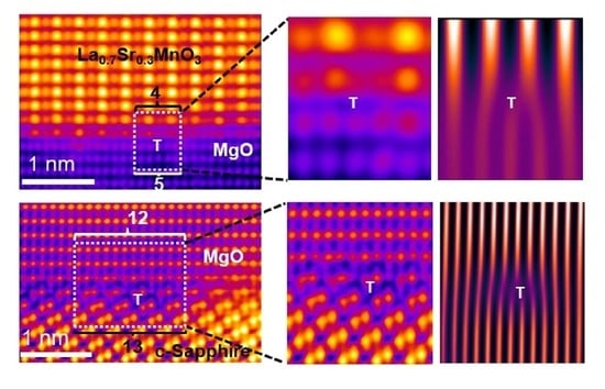
Understanding the interfaces in heterostructures at an atomic scale is crucial in enabling the possibility to manipulate underlying functional properties in correlated materials. This work presents a detailed study on the atomic structures of heterogeneous interfaces in La0.7Sr0.3MnO3 (LSMO) film grown epitaxially on c-Al2O3 (0001) with a buffer layer of MgO. Using aberration-corrected scanning transmission electron microscopy, we detected nucleation of periodic misfit dislocations at the interfaces of the large misfit systems of LSMO/MgO and MgO/c-Al2O3 following the domain matching epitaxy paradigm. It was experimentally observed that the dislocations terminate with 4/5 lattice planes at the LSMO/MgO interface and with 12/13 lattice planes at the MgO/c-Al2O3 interface. This is consistent with theoretical predictions. Using the atomic-resolution image data analysis approach to generate atomic bond length maps, we investigated the atomic displacement in the LSMO/MgO and MgO/c-Al2O3 systems. Minimal presence of residual strain was shown at the respective interface due to strain relaxation following misfit dislocation formation. Further, based on electron energy-loss spectroscopy analysis, we confirmed an interfacial interdiffusion within two monolayers at both LSMO/MgO and MgO/c-Al2O3 interfaces. In essence, misfit dislocation configurations of the LSMO/MgO/c-Al2O3 system have been thoroughly investigated to understand atomic-scale insights on atomic structure and interfacial chemistry in these large misfit systems.

Graphical Abstract

1. Introduction

The nature of epitaxial growth and the control of defects in the thin film heterostructure are considered of significant importance for tuning next-generation electrical, optical, and magnetic devices [1,2]. Unlike their bulk counterparts, oxide thin films have a heterogeneous structure suitable for these applications [3]. The coupling states such as charge, orbitals, and lattice strain are susceptible to heterostructure perturbation leading to the emergence of new properties [4]. Studies on oxide thin film confirm that the presence of dislocation in heterostructure can alter the film properties [5,6]. The lattice strain and defects at the interface control the quality of the thin film and its associated functionalities [6,7].
A particularly intriguing correlated metal–oxide material is Lanthanum strontium manganese oxide (La0.7Sr0.3MnO3 or LSMO) with a Curie temperature of 370 K [8,9]. Studies show that the presence of the strain in epitaxial film can greatly affect the properties of LSMO [10,11,12,13]. It is well established that the misfit present at the interface introduces strain in the system and is gradually relieved after a few hundred monolayers. This result ensures the thin film properties are directly influenced by the interfacial defects. Although LSMO is typically grown on SrTiO3 (STO) or MgO, the lattice misfit differs between LSMO/MgO and LSMO/STO. LSMO and STO share similar crystal structures with a fractional lattice misfit of ~0.79%. Notably, the reported lattice misfit between the LSMO and MgO is greater than ~7% [10]. In both cases, the lattice parameter of LSMO is smaller and expands the film along the in-plane direction, resulting in compression in the out-of-plane direction. It has been reported that STO affects titanium–oxygen octahedral rotations that influence LSMO growth [10]. In addition, STO induces electronic softening of the Mn-O bond, which scatters the spin and negatively affects the properties of LSMO thin film [14,15,16]. Contrarily, MgO does not exhibit these types of phenomena. Substrates such as MgO have large lattice misfits with film and are energetically favorable to containing misfit dislocations that accelerate film relaxation. The misfit dislocations are nucleated on the film surface at a critical thickness and then relocate towards the interface. Therefore, complete film relaxation can be achieved within a few monolayers, thus allowing the subsequent film to grow relaxed or strain free. This process can be explained by domain-matching epitaxy (DME), where integral multiples of lattice constants match across film–substrate interfaces [17,18]. Studies show that many systems with large lattice misfits can be grown successfully on MgO (001) following the DME process [19,20,21,22,23]. In essence, the coupling effect, film–substrate thermal expansion difference, and the step terrace may increase difficulty in understanding the interfacial structure [4,24].
The development of aberration-corrected electron microscopy made it possible to collect atomic resolution images [25]. An important characteristic of scanning transmission electron microscopy (STEM) is its capability of efficiently recording image contrast depending upon the lattice atomic number (Z). Interestingly, the atomic contrast does not vary with alterations in film thickness and imaging focus [26]. It is therefore simple to distinguish the atomic column and interfaces from the system. In this study, a detailed investigation of the interfacial structure was performed on the oxide heterostructure LSMO/MgO/c-Al2O3 utilizing aberration-corrected scanning transmission electron microscopy (STEM) together with electron energy-loss spectroscopy (EELS) measurements. Particularly, this study focuses on the misfit dislocations observed at the LSMO/MgO and MgO/c-Al2O3 interfaces. A detailed examination of the interface shows the additional misfit strain can be accommodated within the DME framework by changing the periodicity of the dislocations. Therefore, strain is relieved from the system within a couple of monolayers following the application of DME concepts. This allows the misfit strain to be confined and engineered near the interface. As a result, the remaining film can grow defect and strain free. In addition, the execution of atomic bond length mapping ensures the strain-free growth of the film with exception to a small number of monolayers near the interface. Finally, EELS was performed for investigation and clarification of the interfacial chemistry between the subsequent film and substrate. This study signifies the importance of misfit dislocations in thin film, thus representing a promising pathway in the improvement of film quality and its associated properties.

2. Experimental Methodology

The LSMO and MgO thin films were grown epitaxially on the (0001) c-Al2O3 substrate using pulsed laser deposition technique with a KrF (wavelength 248 nm) excimer laser. Base pressure inside the vacuum chamber prior to the deposition and sample-target distance were maintained at ~5 × 10⁻⁷ Torr and ~4.5 cm. Thin films of MgO and LSMO were deposited at laser energy densities ~3.1 J/cm2 and ~1.7 J/cm2, respectively, while maintaining a substrate temperature of ~680 °C (MgO) and ~800 °C (LSMO). The estimated thickness of deposited MgO and LSMO were ~25 nm and ~15 nm, respectively. During the deposition, the oxygen partial pressure was maintained at ~1 × 10⁻³ Torr. The films were grown over c-cut sapphire (0001) with epitaxial MgO growing in (111) out-of-plane direction followed by LSMO in (110) growth direction. A detailed description of thin film synthesis is provided elsewhere [10,27]. The atomic-resolution electron microscopic investigations were performed using a fifth-order aberration-corrected scanning transmission electron microscope (STEM) (Nion UltraSTEM 200) operated at 200 KeV. The high-angle annular dark field (HAADF) imaging was acquired with an inner collection semi-angle of 65 mrad at a probe current of 18 ± 2 pA. The convergence semi-angle of the electron probe was 30 mrad. The EELS data were acquired with a collection angle of 48 mrad. The samples for STEM analysis were prepared by conventional mechanical thinning, precision polishing, and ion-milling in a liquid N2 environment [28,29].

3. Results and Discussion

Figure 1a presents an overview HAADF image of the LSMO thin film grown on the c-Al2O3 substrate with a MgO buffer layer using pulsed laser deposition. The LSMO film of thickness ~15 ± 1 nm is grown uniformly on the MgO buffer layer (~25 nm). Figure 1b,c subsequently illustrates the interface between LSMO/MgO and MgO/c-Al2O3 illustrating the epitaxial film growth. The verification of epitaxial growth was performed by x-ray diffraction (θ–2θ and Φ-scan) studies on these films [10,27]. Figure 1b shows the atomic-resolution HAADF image consisting of LSMO film on MgO. Since the intensity of the HAADF image is proportionally related with the atomic number of the elements with a dependence of Zn, where n= 1.5 to 2 [30], the atoms with the brightest intensity presents the La/Sr atoms columns in the Figure 1b and the less bright atoms in between them are the Mn atoms. On the other hand, the Mg atoms show the least intensity. The atomic arrangement, here, is viewed in LSMO (110) and MgO (112) surface normal directions. Figure 1c exhibits the atomic resolution HAADF image of the interface between the MgO buffer layer on the c-Al2O3 substrate. The Mg atoms appear less luminous than the c-Al2O3 atoms. The interface between MgO and c-Al2O3 is well distinguished based on the atomic column intensity as well as the crystal structure orientations.
Subsequent investigations were performed focusing on understanding the highly resolved atomic arrangement and electronic structure insights of individual interfaces of LSMO/MgO and MgO/c-Al2O3. Figure 2a shows the HAADF image revealing the interface between the LSMO and MgO films. Figure 2b–d represent electron diffraction spots from the respective film and interface. The electron diffraction spots show epitaxial matching of (2 2 ¯ 0)LSMO‖(2 2 ¯ 0)MgO. Figure 2e further illustrates an atomic-resolution HAADF image of the LSMO/MgO interface indicating the formation of dislocations with periodicity. It is evident from the Figure 2e that the dislocation terminates at the LSMO/MgO interface with the 4/5 lattice plane periodically which confirms the heterostructure following the DME paradigm in this study. It is important to note here that the lattice mismatch between the matching planes of MgO and LSMO is estimated to be ~7.7%, given the d-spacing of (110)MgO and (110)LSMO is 0.297 nm and 0.2739 nm. The epitaxial growth of such a large lattice misfit (>7%) between two materials, such as MgO and LSMO, is explained by DME, which illustrates that the large mismatch favors the dislocation formation at the periodic interval at a critical film thickness. These dislocations glide to the interface and allow the formation of relaxed epitaxial film growth [18,31,32,33,34]. In addition, a Burger circuit formation has been constructed at the LSMO/MgO interface to identify the dislocation core, as shown in Figure 2e along with inverse fast Fourier transform (IFFT) analysis. The Burger vector shows the displacement of half a unit in the MgO buffer layer. Figure 2f shows the corresponding IFFTs along with the magnified HAADF images of the dislocation cores. It is clear from the images that the four lattice planes of LSMO are aligned with the five lattice planes of MgO periodically at the interface. The observed extra half plane in MgO causes the lattice distortion keeping the LSMO atomic column at equidistance. Additionally, it is observed that interdiffusion between Mg atoms from MgO and Mn atom from LSMO takes place to facilitate the lattice relaxation as there is an extra atom counted in the first LSMO layer at the dislocation core of these misfit dislocations. Based on the atom intensity in the HAADF image, one could eliminate the possibility of La atom diffusion across the interface. This is an important finding as such atomic rearrangement could alter the density of states, providing the traps or recombination centers for electrons and holes, thus directly affecting the electronic properties of the system [35]. The change in the density of states is directly correlated with the energy-loss function seen in the electron energy-loss spectroscopy which will be subsequently discussed in this study. These observations have been previously seen in other heterostructure systems such as ZnO/c-Al2O3 where the Zn atoms were rearranged at the core of misfit dislocations and diffused into c-Al2O3 substrate lattice [36].
To estimate the atomic displacement at/near the interface of LSMO and MgO, a script-based computational quantification is subsequently performed on the HAADF image data that identifies the atomic position coordinates based on the intensity of the atoms [37,38,39,40]. This quantification analysis provides a two-dimensional array of atomic positions. Figure 3a–c reveals the atomic displacement maps of DLa-La and DMg-Mg along the in-plane and out-of-plane directions, respectively. Atomic spacing along the horizontal axis is termed as “in-plane” direction, whereas atomic spacing along the vertical axis is termed as “out-of-plane” direction. The change in the atomic bond length of La-La (DLa-La) and Mg-Mg (DMg-Mg) has been calculated with respect to the atomic positions of Mg atoms in MgO to understand the pseudomorphic growth of LSMO film at the interface. The atomic displacement map in the in-plane direction shows the presence of residual strain at the interface after the formation of misfit dislocations. The maximum atomic displacement is obtained along the in-plane direction is ~0.006 nm, whereas the maximum atomic displacement in the out-of-plane is ~0.002 nm, which appears more relaxed.
Figure 3d–f further displays the atomic displacement mapping that is directly related to the increase/decrease in the bond length of La-La atoms in the LSMO thin film. The HAADF image in Figure 3d shows a region in LSMO thin film containing an antiphase boundary which is a planar defect with a (220) plane missing in the ordered arrangement. The nucleation of antiphase boundaries has been seen at the step on the substrate/film. For a single step, generated antiphase boundary tends to be perpendicular to the interface, as is seen in the LSMO film [41]. Figure 3e shows the decrease in the bond length between La-La atoms in the in-plane direction at the antiphase boundary. On the other hand, there is no observable change in the out-of-plane direction.
Figure 4 further shows the plane-by-plane EELS analysis to determine the interdiffusion of atoms across the LSMO/MgO interface and to understand the interfacial chemistry. Figure 4a again shows a HAADF image of LSMO film grown epitaxially on MgO. In Figure 4b, one can observe a region of interest over which atomic-resolution EELS analysis is performed. Figure 4c,d exhibit the EELS spectra for Mn and O-K edge, respectively, from each defined plane in Figure 4b. It is evident from Figure 4c that the Mn in the LSMO region (layers 1–3) has a characteristic L32 absorption edge that diminishes in the MgO substrate (lasers 6–7) [27]. The intensity of the Mn-L32 peak decreases by 10-fold in MgO; however, it still weakly appears due to possible minor diffusion or delocalization of EELS signal. Contrarily, the O-K edge behavior transforms from the LSMO to MgO according to its known characteristic nature as shown in Figure 4d. In O-K spectra, a pre-peak at 526 eV is observed in the LSMO region (layers 1–4), whereas no pre-peak is observed in the MgO region (layers 6–7). The evident pre-peak in the LSMO region indicates the stronger Mn-O hybridization. An additional observation worth noting is that the LSMO region (layers 1–3) has no post-peaks at 555 eV, as indicated in the Figure 4d whereas the MgO region has noticeable post-peak (layers 6–7). Both LSMO and MgO contribute to the O-K edge in close proximity at the interface (layers 4–5) due to interdiffusion, resulting in layers 4–5 displaying both pre-peaks and post-peaks.
We further analyzed the interface between MgO and c-Al2O3 substrate to appreciate the atomic-scale characteristics of this interface. The atomic-resolution HAADF image reveals the sharp interface between MgO and c-Al2O3, as shown in Figure 5a. Figure 5b–d demonstrate fast Fourier transform (FFT) from the respective film and the interface illustrating the diffraction spots from the individual crystal structures. The electron diffraction spots show epitaxial matching of (2 2 ¯ 0)MgO ( 3 3 ¯ 0 ) Al 2 O 3 (in Figure 5d). The d-spacing of (2 2 ¯ 0) in MgO and ( 3 3 ¯ 0 ) in c-Al2O3 are 0.1491 nm and 0.1374 nm give rise to a lattice misfit of ~8.5% in the MgO film. This leads to the nucleation of misfit dislocations with a 12/13 plane matching between MgO and c-Al2O3 following the DME paradigm. As shown in Figure 5e, these results are consistent with the experimental finding where periodic misfit dislocations are indicated with an average 12/13 lattice plane matching, confirming the validity of DME in this system. Figure 5f presents the respective inverse FFT images of various dislocation cores. The presence of an extra half plane in the c-Al2O3 causes lattice distortion, whereas the MgO buffer layer atomic columns maintain an equidistant column with relaxed lattice planes. The HAADF image reveals the step with atomic scale height at both interfaces rather than maintaining the same starting plane. This represents possible interdiffusion along the incident beam directions. The rise in the atomic step can shift the interfacial contact which changes the dislocation core configuration accordingly. The change in the configuration of the dislocation core contributes to the possible interfacial interaction between the film, buffer layer, and substrate. In addition, interdiffusion induces the compositional disparity at the interface, thus hampering the dislocation core spacing in addition to the periodicity. Interestingly, the interdiffusion most seemingly occurred at a position slightly away from the dislocation cores. Despite having the dislocation core at the LSMO/MgO and MgO/c-Al2O3 interface, the absence of defects such as pores confirms the excellent quality of deposited LSMO film and the MgO buffer layer.
Script-based computational quantification is performed on the HAADF image as shown in Figure 6a. This computational quantification approach bears similarity with the analysis shown in Figure 3. Here, the atomic bond length of DxMg-Mg/DAl-Al, and DzMg-Mg/D’Al-Al is investigated. These atomic bond length maps are shown in Figure 6b and Figure 6c, respectively. The relative color uniformity of the pixels, in Figure 6b,c, indicates relaxation of the deposited MgO film, maintaining the epitaxial matching of (2 2 ¯ 0)MgO ( 3 3 ¯ 0 ) Al 2 O 3 .
Figure 7a shows a HAADF image of the MgO buffer layer film grown epitaxially on c-Al2O3. Figure 7b shows a magnified HAADF image revealing possible interdiffusion at the interface of MgO and c-Al2O3. To further investigate the interface, a detailed plane-by-plane EELS analysis observing the nature of O-K edge is performed, as shown in Figure 7c. The main peak of O-K edge broadens as it transfers from MgO to c-Al2O3. Based on the nature of the O-K absorption edge, it is evident that layers 1–2 are characteristic of MgO and layers 5–7 of c-Al2O3. The arrows marked at 543 eV indicate the characteristic O-K post-peak in the MgO region, whereas there is no evidence of this post-peak in the c-Al2O3 region. Layers 3–4 indicate the interdiffusion between the MgO and c-Al2O3, as the spectrum shows characteristics of both MgO and c-Al2O3 in O-K edge. Overall, the investigations suggest that, in the large misfit system of MgO/c-Al2O3, strain is relaxed by the formation of misfit dislocations; however, we observe an interdiffusion within two monolayers at the interface.

4. Conclusions

In this research, we have utilized atomic-resolution STEM imaging together with EELS analysis to recognize the characteristics of the heterogeneous interface of the LSMO/MgO and MgO/c-Al2O3 system. Each system shows excellent consistency with the DME paradigm accompanied by misfit dislocations configurations. In both cases, periodic misfit dislocations were observed with 4/5 and 12/13 plane matching, respectively, for LSMO/MgO and MgO/c-Al2O3 interfaces. Further investigation of the heterogeneous interface reveals geometrical (atomic height step) and compositional (interdiffusion) changes, thus affecting the interface chemistry and dislocation core structure. The characterization and experimental observations bolster a clearer understanding of the heterogeneous interface and defects in structures on an atomic scale. These findings support an opportunity to tune the next generation of devices.

Author Contributions

Conceptualization, R.S. and J.N.; methodology, S.M., A.K.G., B.H.B., V.S. and R.S.; software, S.M., A.K.G., B.H.B., V.S. and R.S.; validation, R.S. and J.N.; writing—original draft preparation, S.M. and A.K.G.; writing—review and editing, S.M., A.K.G., R.S. and J.N.; supervision, R.S. and J.N. All authors have read and agreed to the published version of the manuscript.

Funding

R.S. acknowledges the support of faculty start-up funding at Oklahoma State University.

Institutional Review Board Statement

Not applicable.

Informed Consent Statement

Not applicable.

Data Availability Statement

Not applicable.

Acknowledgments

The electron microscopic data acquisition in this research was conducted at the Center for Nanophase Materials Sciences, which is a DOE Office of Science User Facility. Authors also want to thank Daniel Rasic for growing the heterostructures and John Prater for useful discussions.

Conflicts of Interest

The authors declare no conflict of interest.

References

  1. Queisser, H.J.; Haller, E.E. Defects in Semiconductors: Some Fatal, Some Vital. Am. Assoc. Adv. Sci. 1998, 281, 945–950. [Google Scholar] [CrossRef] [PubMed]
  2. Mahajan, S. Defects in semiconductors and their effects on devices. Acta Mater. 2000, 48, 137–149. [Google Scholar] [CrossRef]
  3. Dagotto, E. Complexity in Strongly Correlated Electronic Systems. Science 2005, 309, 257–262. [Google Scholar] [CrossRef] [PubMed] [Green Version]
  4. Reiner, J.W.; Walker, F.J.; Ahn, C.H. Atomically Engineered Oxide Interfaces. Science 2009, 323, 1018–1019. [Google Scholar] [CrossRef]
  5. Jia, C.L.; Thust, A.; Urban, K. Atomic-scale analysis of the oxygen configuration at a SrTiO3 dislocation core. Phys. Rev. Lett. 2005, 95, 1–4. [Google Scholar] [CrossRef] [PubMed] [Green Version]
  6. Szot, K.; Speier, W.; Bihlmayer, G.; Waser, R. Switching the electrical resistance of individual dislocations in single-crystalline SrTiO3. Nat. Mater. 2006, 5, 312–320. [Google Scholar] [CrossRef] [PubMed]
  7. Schlom, D.G.; Chen, L.-Q.; Eom, C.-B.; Rabe, K.M.; Streiffer, S.K.; Triscone, J.-M. Strain Tuning of Ferroelectric Thin Films. Annu. Rev. Mater. Res. 2007, 37, 589–626. [Google Scholar] [CrossRef] [Green Version]
  8. Bowen, M.; Bibes, M.; Barthélémy, A.; Contour, J.P.; Anane, A.; Lemaıtre, Y.; Fert, A. Nearly total spin polarization in La2/3Sr1/3MnO3 from tunneling experiments. Appl. Phys. Lett. 2003, 82, 233–235. [Google Scholar] [CrossRef] [Green Version]
  9. Hwang, H.Y.; Cheong, S.-W.; Radaelli, P.G.; Marezio, M.; Batlogg, B. Lattice Effects on the Magnetoresistance in Doped LaMnO3. Phys. Rev. Lett. 1995, 75, 914–917. [Google Scholar] [CrossRef] [PubMed] [Green Version]
  10. Rasic, D.; Sachan, R.; Temizer, N.K.; Prater, J.; Narayan, J. Oxygen Effect on the Properties of Epitaxial (110) La0.7Sr0.3MnO3 by Defect Engineering. ACS Appl. Mater. Interfaces 2018, 10, 21001–21008. [Google Scholar] [CrossRef] [PubMed]
  11. Kim, B.; Kwon, D.; Song, J.H.; Hikita, Y.; Kim, B.G.; Hwang, H.Y. Finite size effect and phase diagram of ultra-thin La0.7Sr0.3MnO3. Solid State Commun. 2010, 150, 598–601. [Google Scholar] [CrossRef]
  12. Tsui, F.; Smoak, M.C.; Nath, T.K.; Eom, C.B. Strain-dependent magnetic phase diagram of epitaxial La0.67Sr0.33MnO3 thin films. Appl. Phys. Lett. 2000, 76, 2421–2423. [Google Scholar] [CrossRef]
  13. Vailionis, A.; Boschker, H.; Siemons, W.; Houwman, E.P.; Blank, D.H.A.; Rijnders, G.; Koster, G. Misfit strain accommodation in epitaxial ABO3 perovskites: Lattice rotations and lattice modulations. Phys. Rev. B Condens. Matter Mater. Phys. 2011, 83, 1–10. [Google Scholar] [CrossRef] [Green Version]
  14. Sun, J.Z.; Abraham, D.W.; Rao, R.A.; Eom, C. Thickness-dependent magnetotransport in ultrathin manganite films. Appl. Phys. Lett. 1999, 74, 3017–3019. [Google Scholar] [CrossRef] [Green Version]
  15. Boschker, H.; Kautz, J.; Houwman, E.P.; Siemons, W.; Blank, D.H.A.; Huijben, M.; Koster, G.; Vailionis, A.; Rijnders, G. High-Temperature Magnetic Insulating Phase in Ultrathin La0.67Sr0.33MnO3 Films. Phys. Rev. Lett. 2012, 109, 157207. [Google Scholar] [CrossRef] [Green Version]
  16. Sandiumenge, F.; Santiso, J.; Balcells, L.; Konstantinovic, Z.; Roqueta, J.; Pomar, A.; Espinos, J.P.; Martínez, B. Competing Misfit Relaxation Mechanisms in Epitaxial Correlated Oxides. Phys. Rev. Lett. 2013, 110, 107206. [Google Scholar] [CrossRef] [PubMed] [Green Version]
  17. Narayan, J.; Larson, B.C. Domain epitaxy: A unified paradigm for thin film growth. J. Appl. Phys. 2003, 93, 278–285. [Google Scholar] [CrossRef]
  18. Narayan, J.; Tiwari, P.; Chen, X.; Singh, J.; Chowdhury, R.; Zheleva, T. Epitaxial growth of TiN films on (100) silicon substrates by laser physical vapor deposition. Appl. Phys. Lett. 1992, 61, 1290–1292. [Google Scholar] [CrossRef]
  19. Yang, S.; Kuang, W.; Liou, Y.; Tse, W.; Lee, S.; Yao, Y. Growth and characterization of La0.7Sr0.3MnO3 films on various substrates. J. Magn. Magn. Mater. 2004, 268, 326–331. [Google Scholar] [CrossRef]
  20. Špankova, M.; Chromik, Š.; Vávra, I.; Sedláčková, K.; Lobotka, P.; Lucas, S.; Stanček, S. Epitaxial LSMO films grown on MgO single crystalline substrates. Appl. Surf. Sci. 2007, 253, 7599–7603. [Google Scholar] [CrossRef]
  21. Wang, C.; Zhu, Y.; Li, L.; Shen, Q.; Zhang, L. Epitaxial growth and transport property of La0.9Sr0.1MnO3 thin films deposited on MgO, LaAlO3 and SrTiO3 substrates. J. Alloy. Compd. 2017, 693, 832–836. [Google Scholar] [CrossRef]
  22. Štrbík, V.; Reiffers, M.; Dobročka, E.; Soltys, J.; Špankova, M.; Chromik, Š. Epitaxial LSMO thin films with correlation of electrical and magnetic properties above 400K. Appl. Surf. Sci. 2014, 312, 212–215. [Google Scholar] [CrossRef]
  23. Ranno, L.; Llobet, A.; Hunt, M.; Pierre, J. Influence of substrate temperature on magnetotransport properties of thin films of La0.7Sr0.3MnO3. Appl. Surf. Sci. 1999, 138-139, 228–232. [Google Scholar] [CrossRef]
  24. Chu, M.W.; Szafraniak, I.; Scholz, R.; Harnagea, C.; Hesse, D.; Alexe, M.; Gösele, U. Impact of misfit dislocation on the po-larization instability of epitaxial nanostructured ferroelectric perovskites. Nat. Mater. 2004, 3, 87–90. [Google Scholar] [CrossRef] [PubMed]
  25. Erni, R.; Rossell, M.D.; Kisielowski, C.; Dahmen, U. Atomic-Resolution Imaging with a Sub-50-pm Electron Probe. Phys. Rev. Lett. 2009, 102, 096101. [Google Scholar] [CrossRef] [PubMed] [Green Version]
  26. Klenov, D.O.; Stemmer, S. Contributions to the contrast in experimental high-angle annular dark-field images. Ultramicroscopy 2006, 106, 889–901. [Google Scholar] [CrossRef] [PubMed]
  27. Rasic, D.; Sachan, R.; Prater, J.; Narayan, J. Structure-property correlations in thermally processed epitaxial LSMO films. Acta Mater. 2019, 163, 189–198. [Google Scholar] [CrossRef]
  28. Moatti, A.; Sachan, R.; Gupta, S.; Narayan, J. Vacancy-Driven Robust Metallicity of Structurally Pinned Monoclinic Epitaxial VO2 Thin Films. ACS Appl. Mater. Interfaces 2019, 11, 3547–3554. [Google Scholar] [CrossRef] [PubMed]
  29. Moatti, A.; Sachan, R.; Narayan, J. Mechanism of strain relaxation: Key to control of structural and electronic transitions in VO2 thin-films. Mater. Res. Lett. 2019, 8, 16–22. [Google Scholar] [CrossRef] [Green Version]
  30. Krivanek, O.; Chisholm, M.F.; Nicolosi, V.; Pennycook, T.J.; Corbin, G.J.; Dellby, N.; Murfitt, M.F.; Own, C.S.; Szilagyi, Z.S.; Oxley, M.P.; et al. Atom-by-atom structural and chemical analysis by annular dark-field electron microscopy. Nature 2010, 464, 571–574. [Google Scholar] [CrossRef] [PubMed] [Green Version]
  31. Moatti, A.; Sachan, R.; Prater, J.; Narayan, J. Control of Structural and Electrical Transitions of VO2 Thin Films. ACS Appl. Mater. Interfaces 2017, 9, 24298–24307. [Google Scholar] [CrossRef] [PubMed]
  32. Rasic, D.; Sachan, R.; Chisholm, M.F.; Prater, J.; Narayan, J. Room Temperature Growth of Epitaxial Titanium Nitride Films by Pulsed Laser Deposition. Cryst. Growth Des. 2017, 17, 6634–6640. [Google Scholar] [CrossRef]
  33. Gupta, S.; Sachan, R.; Narayan, J. Evidence of weak antilocalization in epitaxial TiN thin films. J. Magn. Magn. Mater. 2020, 498, 166094. [Google Scholar] [CrossRef]
  34. Liu, X.-Y.; Arslan, I.; Arey, B.W.; Hackley, J.; Lordi, V.; Richardson, C.J.K. Perfect Strain Relaxation in Metamorphic Epitaxial Aluminum on Silicon through Primary and Secondary Interface Misfit Dislocation Arrays. ACS Nano 2018, 12, 6843–6850. [Google Scholar] [CrossRef]
  35. Zhang, Z.; Yates, J.T., Jr. Band bending in semiconductors: Chemical and physical consequences at surfaces and interfaces. Chem. Rev. 2012, 10, 5520–5551. [Google Scholar] [CrossRef] [PubMed]
  36. Narayan, J. Recent progress in thin film epitaxy across the misfit scale. Acta Mater. 2013, 61, 2703–2724. [Google Scholar] [CrossRef]
  37. Zhang, Y.; Xue, H.; Zarkadoula, E.; Sachan, R.; Ostrouchov, C.; Liu, P.; Wang, X.-L.; Zhang, S.; Wang, T.S.; Weber, W.J. Coupled electronic and atomic effects on defect evolution in silicon carbide under ion irradiation. Curr. Opin. Solid State Mater. Sci. 2017, 21, 285–298. [Google Scholar] [CrossRef]
  38. Gupta, A.K.; Arora, G.; Aidhy, D.S.; Sachan, R. ∑3 Twin Boundaries in Gd2Ti2O7 Pyrochlore: Pathways for Oxygen Migration. ACS Appl. Mater. Interfaces 2020, 12, 45558–45563. [Google Scholar] [CrossRef] [PubMed]
  39. Aidhy, D.S.; Sachan, R.; Zarkadoula, E.; Pakarinen, O.; Chisholm, M.F.; Zhang, Y.; Weber, W.J. Fast ion conductivity in strained defect-fluorite structure created by ion tracks in Gd2Ti2O7. Sci. Rep. 2015, 5, 16297. [Google Scholar] [CrossRef] [PubMed] [Green Version]
  40. Gupta, A.K.; Gupta, S.; Sachan, R. Laser Irradiation Induced Atomic Structure Modifications in Strontium Titanate. JOM 2021, 1–8. [Google Scholar] [CrossRef]
  41. Wang, Z.; Guo, H.; Shao, S.; Saghayezhian, M.; Li, J.; Fittipaldi, R.; Vecchione, A.; Siwakoti, P.; Zhu, Y.; Zhang, J.; et al. Designing antiphase boundaries by atomic control of heterointerfaces. Proc. Natl. Acad. Sci. USA 2018, 115, 9485–9490. [Google Scholar] [CrossRef] [PubMed] [Green Version]
Figure 1. (a) A representative HAADF image of LSMO thin film grown on the c-Al2O3 substrate where MgO is the buffer layer. Atomic resolution HAADF image showing the interface between the (b) LSMO thin film and the MgO buffer layer, (c) the MgO buffer layer, and c-Al2O3 substrate.
Crystals 11 01493 g001
Figure 2. (a) HAADF image of LSMO over MgO showing the defined La-La and Mg-Mg bond length, (bd) shows electron diffraction spots for the respective film, (e) HAADF image showing a 4/5 planes lattice matching and the dislocation periodicity, and (f) IFFTs along (110) with the magnified images of dislocation cores. Vertical arrows of different colors point to the extra-half planes at each dislocation core. The corresponding matching domains were bracketed with their sizes noted by the number of the LSMO and MgO lattice planes, respectively. A Burgers circuit is applied on a dislocation core to identify the Burgers vector along the edge dislocation.
Crystals 11 01493 g002
Figure 3. (a) HAADF image and (b,c) atomic bond length maps of DLa-La and DMg-Mg along the in-plane, and out-of-plane directions, respectively, (d) HAADF image of grown LSMO film with an indicated antiphase boundary, (e,f) atomic bond length maps of DLa-La along in-plane and out-of-plane directions, respectively.
Crystals 11 01493 g003
Figure 4. (a,b) Atomic-resolution HAADF image of LSMO grown on MgO, (c,d) atomic plane-by-plane EELS spectra for Mn and O-K edge within the LSMO and MgO corresponding to planes shown in (b).
Crystals 11 01493 g004
Figure 5. (a) HAADF image of MgO film grown on c-Al2O3. Mg and Al atoms are indicated in orange and red colors. (bd) Electron diffraction spots shown for the respective film, (e) HAADF image revealing periodic dislocation formation at the MgO and c-Al2O3 interface with 12/13 planes lattice matching, (f) IFFTs along (110) with magnified images of dislocation cores.
Crystals 11 01493 g005
Figure 6. (a) HAADF image containing MgO/c-Al2O3 interface, (b,c) atomic bond length maps of DxMg-Mg/DAl-Al, and DzMg-Mg/D’Al-Al, respectively.
Crystals 11 01493 g006
Figure 7. (a,b) Atomic-resolution HAADF image of MgO grown on c-Al2O3, (c) plane-by-plane EELS spectra of O-K from the MgO, and c-Al2O3 corresponding to planes shown in (b).
Crystals 11 01493 g007
Publisher’s Note: MDPI stays neutral with regard to jurisdictional claims in published maps and institutional affiliations.