Article

Error Analysis and Correction for Quantitative Phase Analysis Based on Rietveld-Internal Standard Method: Whether the Minor Phases Can Be Ignored?

1
Shandong Provincial Key Laboratory of Preparation and Measurement of Building Materials, University of Jinan, Jinan 250022, China
2
School of Materials Science and Engineering, Tongji University, Shanghai 201804, China
3
Departamento de Química Inorgánica, Cristalografía y Mineralogía, Universidad de Málaga, 29071 Málaga, Spain
*
Author to whom correspondence should be addressed.
Crystals 2018, 8(3), 110; https://doi.org/10.3390/cryst8030110
Submission received: 26 December 2017 / Revised: 14 February 2018 / Accepted: 19 February 2018 / Published: 27 February 2018
(This article belongs to the Special Issue Rietveld Refinement in the Characterization of Crystalline Materials)

Abstract

:
The Rietveld-internal standard method for Bragg-Brentano reflection geometry (θ/2θ) X-ray diffraction (XRD) patterns is implemented to determine the amorphous phase content. The effect of some minor phases on quantitative accuracy is assessed. The numerical simulation analysis of errors and the related corrections are discussed. The results reveal that high purity of crystalline phases in the standard must be strictly ensured. The minor amorphous or non-quantified crystalline phases exert significant effect on the quantitative accuracy, even with less than 2 wt% if ignored. The error levels are evaluated by numerical simulation analysis and the corresponding error-accepted zone is suggested. To eliminate such error, a corrected equation is proposed. When the adding standard happens to be present in sample, it should be also carefully dealt with even in low amounts. Based on that ignorance, the absolute and relative error equations (ΔAE, ΔRE) are derived, as proposed. The conditions for high quantitative accuracy of original equation is strictly satisfied with a lower amount of standard phase present in sample, less than 2 wt%, and a higher dosage of internal standard, larger than 20 wt%. The corrected equation to eliminate such quantitative error is suggested.

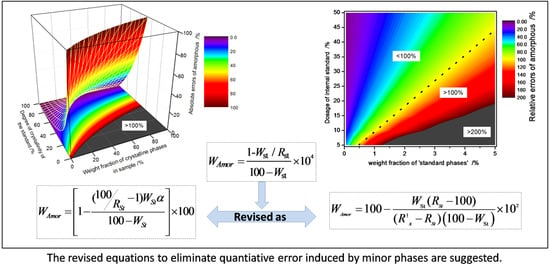
Graphical Abstract

1. Introduction

Quantitative phase analysis based on X-ray diffraction (XRD) can be traced back to as early as 1919 [1]. Hull firstly proposed that this technique had the potential to perform accurately quantitative analysis. Subsequently, Alexander developed practical XRD methods and derived the related theoretical basis [2]. With the improvements of XRD analysis, different quantitative methods have been presented in succession, such as reference intensity ratio (RIR) method [3], external standard method [4], matrix-flushing method [5], non-standard method [6], doping method [7], and Rietveld method [8]. The Rietveld method, as proposed by Hugo M. Rietveld, is widely accepted due to its whole-pattern fitting approach instead of single-peak analysis. The main advantage is that it can effectively minimize or eliminate the inaccuracies arising from preferred orientation, particle statistics, microabsorption, peaks overlapping, and detection of amorphous phase and trace phases [9,10,11]. Over the last two decades, it has become widely accepted by scientific community, gradually being a standard practice, as it is possible to solve those problems associated with crystalline materials. However, there are two preconditions: (1) the quantified phase is the crystalline phase, and (2) the crystal structure is known [12]. Therefore, if amorphous material is present, quantitative results could not be obtained directly. In such case, it is normal to use a reference material for the recalculation of the phase contents [13]. This reference material is either mixed in the sample as internal standard [10] or measured separately under identical conditions as external standard [14]. The internal standard method derives the amorphous content from the comparison between the actual dosage and Rietveld result of the internal standard, Equation (1) [13]:
W A m o r = 1 W S t / R S t 100 W S t × 10 4
where WSt stands for the actual dosage of the internal standard, and RSt stands for the Rietveld results of the internal standard.
Meanwhile, the external standard method focuses on determining the diffractometer constant with an appropriate standard, which is used to determine the weight fraction of each crystalline phase. From the difference between 100 wt% and the sum of the crystalline phase contents, an overall weight percentage of amorphous can be subsequently derived.
G = K e = S S t · ρ S t V S t 2 μ S t / W S t
W A m o r = 100 S α ρ α V α 2 μ s / G
Where SSt and Sα stands for the scale factors of the external standard and each phase in the mixture, respectively, ρ is the density, V is the unit cell volume, and µSt and µs are the mass absorption coefficient of the external standard and the sample, respectively.
Besides these two methods, the amorphous content could also be quantified by the ‘PONKCS’ (Partial Or No Known Crystal Structure) method that relies upon treating a set of peaks of amorphous phase as a single entity [15]. Amorphous phase is characterized by measured rather than a calculated structure factor.
The Rietveld-internal standard method, as the most widely used technique, is a relatively easy and direct strategy, no other measurements or calibrations are needed. If the analyses are carefully performed and the amount of amorphous content to be determined over 15 wt%, the accuracy is satisfactory, close to 1% [10,13,16,17,18]. On the contrary, the external and ‘PONKCS’ methods are more complicated and some other preconditions need to be met in the meantime. For instance, the external standard method strictly requires the identical conditions of XRD data acquisition and different mass attenuation coefficient correction between sample and standard. Though ‘PONKCS’ strategy may solve the problem of undistinguished ability between different amorphous phases, it depends on accurate identification and the calibration of amorphous. A problem may occur because of the complexity of this analyzing process, especially when the amorphous phases are less evident or difficult for characterization [19].
Beside the advantage of the internal method mentioned above, the determination of amorphous contents is also a very challenging operation indeed. First of all, the effects of internal standard should be well considered. A significant error may occur when the selected standard is not homogeneously mixed into the sample or when an obvious absorption contrast exists between sample and standard [20]. Furthermore, use of an appropriate amount of internal standard is also a key point to guarantee the accuracy. Discussions about that influence on accuracy of amorphous phase quantitation have been reported [21,22]. The results illustrated that the quantitative accuracy of amorphous phase follows a nonlinear function by Rietveld-internal standard method, which in turn leads to a serious error in determining the minor amount of amorphous content. Most of these issues can be mitigated based upon the above results by adequate sample preparation and correct data acquisition [23,24,25,26]. However, there are still some factors that need further attention. Here, we study the error analysis that was introduced by some minor phases that are related to the internal standard. They include the minor impurity phases of the internal standard and the sample containing the same crystalline phase as the internal standard. There is often an overlook about contribution of these minor phases. The basic goal is to understand such effect on the quantitative accuracy. In order to do so, a three-dimensional numerical simulation database, where the information includes absolute and relative errors is conducted, and relative corrected equations have been proposed. This study is a step forward to better understand quantitative phase analysis based on Rietveld-internal standard method.

2. Materials and Methods

2.1. Raw Materials

The Powders of SiO2 [ABCR GmbH. Co. KG (Karlsruhe, Germany)] and ZnO [Sigma-Aldrich, Co. LLC (St. Louis, MO, USA)], purity of 99.9%, are chosen as the internal standard in this work. They were sieved firstly through 74 μm prior to be used. The glass powder was adopted as amorphous component, ground with particle sizes less than 20 μm (80% in number statistics), and surface area 500 m2/kg determined by specific surface area measuring instrument. The glass has a chemical composition (wt% of oxides) determined by XRF, of SiO2 (72.5), Al2O3 (1.8), CaO (5.8), Na2O (13.3), K2O (1.6), MgO (3.8), and ZnO (0.8).

2.2. Sample Preparation

The powders of SiO2, ZnO and glass were weighed, with a designed mass ratio of 45%:45%:10%, as 4.507 g, 4.473 g, and 1.033 g, respectively. The above powders were subsequently mixed and homogenized by hand for 30 min in an agate mortar. Finally, they were uniformly filled into the holder and slightly leveled for XRD measurement. That mixtures were prepared and then underwent XRD tests by triplicate. XRD patterns didn’t show significant differences.

2.3. Data Collection and Processing

Chemical composition of glass was determined by the X-Ray Fluorescence (SRS3400, Bruker AXS Corporation, Karlsruhe, Germany). Particle statistic of powders was measured by laser particle size analyzer (LS 230 from Beckman Coulter, Brea, CA, USA). The X-ray diffraction patterns of mixtures were measured in Bragg-Brentano reflection geometry (θ/2θ) on an X’Pert MPD PRO diffractometer (PANalytical International Corporation, Almelo, Netherland) and GSAS-EXPGUI software (Los Alamos National Laboratory, Los Alamos, NM, USA) [27]. The detailed instrument settings for XRD are summarized in Table 1.

3. Results and Discussion

3.1. Quantitative Error Induced by Minor Impurity Phase of Internal Standard

The XRD pattern of that mixture was analyzed by Rietveld whole-pattern fitting based upon GSAS-EXPGUI software. The starting crystalline structure models of SiO2 [28] and ZnO [29] were imported from literature. The instrument function file was chosen based on CuKα1 as the incident X-ray and Germanium as the monochromator (monochromatic model with wavelength of 1.54056 and polarization fraction value of 0.8). For the refinement of peak shape parameters, pseudo-Voigt function [30] with asymmetry correction [31] was chosen and the peak width and asymmetry factor, such as LY, GW, H/L, and S/L were initially set to 12 (0.01°), 5 (0.01°), 0.02, and 0.02, respectively. The refined parameters included unit-cell parameters, zero-shift correction, background parameters, phase fractions, and peak shape parameters (LY and GW). A linear interpolation function was chosen to fit the background with polynomial term gradually increasing to 36. Peak shapes were fitted by refining the Gaussian contribution and Lorentzian contribution separately when appropriated. During the process of Rietveld refinement, the refined parameters had regular convergence and least-square R factors, assessing the fitness of pattern, decreased gradually. Figure 1 shows the Rietveld refinement pattern of the artificial mixture. The stable refinements and satisfactory fits, as indicated by the smoothness of the Yobs-Ycalc curve illustrated the Rietveld refinement was reliable. Moreover, the analysis was performed by triplicate in order to assess the precision. The results are close but not identical with relative errors lower than 1%. The quantitative results are listed in Table 2, which also includes the quantitative phase analysis corrected by taking into account the microabsorption effect [32,33]. This is important in this mixture as linear absorption coefficient for SiO2 is 92 cm−1, while that for ZnO is 290 cm−1. Both standards have very similar particle size (~4 μm); consequently, ZnO will always be underestimated.
The middle column contains direct Rietveld results, assuming that all of the phases in the sample are crystalline phases; the right-most column contains Rietveld results corrected for microabsorption.
To obtain the quantitative results of the amorphous phase (Glass), SiO2 and ZnO were considered as internal standards, respectively. If microabsorption effect is not considered, the errors in amorphous determination are inevitably higher. However, even when considering that factor, the quantitative results are still not well reproducible with a significant relative deviation in the three different analysis, independently of the internal standard used (Table 3). However, when compared with different recorded XRD pattern analysis based on the same internal standard, the quantitative results show good consistency with absolute deviation of phases less than 1%.
‘1’ and ‘2’ represents quantitative results included amorphous phase (glass), taking SiO2 and ZnO as internal standard, respectively.
To further verify this conjecture, Rietveld quantitative phase analysis of that mixture was performed by external standard method (G-factor method). For calculating the G-factor, the polished polycrystalline quartz rock was firstly considered as the standard. It has the advantage of avoiding the error induced by powder standard during the sample preparation, such as fluctuation of surface roughness, packing density, and so on. XRD data collection of the quartz rock standard was as close in time and identical in diffractometer configuration as possible to the artificial mixture sample, which could make sense of the formula of ‘G_SiO2 = G_sample’. The mass absorption coefficient of artificial mixture sample under the condition of CuKα1 radiation was determined as 42.34 cm2/g by Highscore Plus software (PANalytical, Almelo, Netherland). After correlative parameters obtained from Rietveld refinement and crystallinity of quartz rock obtained from former analysis, G value was calculated as 5.51 × 10⁻²⁰ (Table 4). Based on this G value, quantitative results of crystalline and amorphous phases were given in Table 5. The average quantitative result of ‘Glass’ phase among three calculations is 13.08%, which is about 3% larger than the original weighed fraction. The extra part is contributed by amorphous or non-quantified crystalline phases (ACn) in SiO2 and ZnO.
To eliminate such quantitative errors induced by ACn in the internal standard, the original Equation (1) needs to be improved. Using Rietveld refinement, the improved equation for weight fraction of amorphous phase in original sample (WAmor) can be derived as Equation (4), and the intermediate derivation process was shown in the supplementary materials.
W A m o r = [ 1 ( 100 R S t 1 ) × W S t × α 100 W S t ] × 100
where ‘α’ is defined as the crystallinity in that internal standard. The equations of absolute error are successively derived for theoretical calculation of the error level between the original (Equation (1)) and improved equation (Equation (4)).
W A m o r ( i m p r ) W A m o r ( origi ) = ( 1 R S t 1 100 ) ( 1 W S t 1 100 ) × ( 1 α )
To simplify Equation (5), RSt is firstly derived and expressed by crystallinity of internal standard (α) and the original sample (β) as Equation (6). The absolute error (ΔAE) can be finally converted as Equation (7). Moreover, the relative error (ΔRE) can be proposed as Equation (8).
R st = W S t α W S t α + ( 100 W S t ) β × 100
Δ A E = W A m o r ( i m p r ) W A m o r ( origi ) = 1 α α × β
Δ R E = W A m o r ( i m p r ) W A m o r ( origi ) W A m o r ( i m p r ) = 1 α α × β 1 β
To evaluate the error level, the numerical simulation analysis of absolute and relative errors was performed. The results, corresponding to crystallinity of the internal standard (α) and the weight fractions of crystalline phases in original sample (β), were displayed in Figure 2. The three-dimensional (3D) pattern of error distributions was restricted to be under 100% for absolute error and 500% for relative error. The corresponding two-dimensional (2D) error areas were depicted at the bottom with color bars to distinguish the different error levels. It reveals the error becomes significant with the increase of weight fraction of crystalline phases in original sample and decrease of crystallinity of internal standard (bottom right corner), which means the original amorphous phase calculation (Equation (1)) is not applicable any more. On the contrary, the data located at the top left of 2D error areas, as shown in purple and blue colors, can be accepted and applied to original equation. When the preparatory conditions were set as ‘ Δ A E 10 % Δ A E 50 % ’, the error-accepted zone is displayed in Figure 3. It is from the intersection operation between ‘ α 10 β / ( 1 + 10 β ) ’ and ‘ α 2 β / ( 1 + β ) ’. An intersection point (8/9, 4/5) can be calculated from the above equations. Therefore, α and β should satisfy the relationship of ‘ α 10 β / ( 1 + 10 β ) ’ when ‘ β ( 0 , 80 % ) ’ and ‘ α 2 β / ( 1 + β ) ’ when ‘ β ( 80 % , 100 % ) ’.

3.2. Quantitative Error Induced by Minor Standard Phase Present in Sample (SPS)

For the Rietveld/internal standard quantitative analysis, there is another problem that is often overlooked. It is also related to the standard besides the effect of its minor impurity phases above. The normal standards such as SiO2, Al2O3, ZnO, and TiO2 are preferably chosen due to their simple and known crystal structure, characteristic-sharp diffraction peaks and a variable availability of linear absorption coefficient. However, it is also important to highlight that such standards to be added for amorphous content determination may be present previously in the sample. In this work, we refer such a standard phase that is present in the sample as SPS. If SPS is minor phase and there is no better standard, they are often overlooked and assuming that contribution to quantitative accuracy is negligible. Here, we study the error analysis introduced by that extreme case and its effect on quantitative accuracy is discussed. The equations of absolute and relative errors are finally derived as Equations (9) and (10), and the intermediate derivation process was shown in the supplementary materials.
Δ A E = 100 x y + 100 100 x z + 1
Δ R E = [ 100 x y + 100 ( 100 x z + 1 ) y ] × 100
where x is set as ‘WSt/100 − WSt’ while y and z stand for the weight fraction of amorphous phase(WAmor) and the SPS phase (WX) in original sample, respectively.
The numerical simulation analysis of absolute and relative errors was successively performed. The results, corresponding to WSt and WX, were displayed in Figure 4. Figure 4a,c,e shows the absolute error distribution in which the content of amorphous phase is assumed to be 10%, 20%, and 50%, respectively. The corresponding 2D error patterns, reported by Figure 4b,d,f, displayed the relative errors distributions. It is concluded that the error level of amorphous phase quantitation is positively correlated with WX and negatively correlated with WSt. The amorphous phase error becomes more sensitive with smaller of WSt or larger of WX. When compared with the results from 10 wt% to 50 wt% of the amorphous content, the conclusion can be drawn that quantitative error gradually dropped down at a higher dosage of internal standard. The relative errors displayed in Figure 4f are all less than 100% when the weight fraction of amorphous phase reaches 50 wt%. Likewise, the error is related with the weight fraction of standard phase present in sample and dosage of internal standard. The conditions for high quantitative accuracy of original equation are strictly satisfied with a lower amount of SPS phase, less than 2 wt%, and a higher dosage of internal standard, larger than 20 wt%. For example, if the weight percentage of SPS phase is set as 2 wt%, the absolute and relative errors are as high as 8% and 39%, respectively, when the amorphous content and dosage of internal standard are less than 20%.
For the above quantitative errors reduction, the corrected equation for amorphous phase can be finally proposed as Equation (11), with the intermediate derivation process shown in the supplementary materials.
W A m o r = 100 W S t ( R S t 100 ) ( R X R S t ) ( 100 W S t ) × 10 2
where RSt and RX stand for the Rietveld quantitative result of internal standard and the standard phase present in sample.

4. Conclusions

For Bragg-Brentano diffraction, the ignorance of the amorphous or non-quantified crystalline phases (ACn) in standard exerts significant effect on the quantitative accuracy by the Rietveld-internal standard method. The error level is only related to the crystallinity of internal standard (α) and weight percentage of amorphous phase in original sample (β), and the equations of absolute/relative errors (ΔAERE) are derived as proposed. The numerical simulation analysis reveals the original equation is not applicable in most of the cases and the corresponding error-accepted zone is suggested from the relationship between α and β. When the preparatory conditions are set as ‘ΔAE ≤ 10% ∩ ΔRE ≤ 50%’, then the error-accepted zone can be expressed as ‘α ≥ 10β/(1 + 10β)’, ‘β (0.80%)’ and ‘α ≥ 2β/(1 + β)’, ‘β (80%, 100%)’. For the above quantitative error elimination, the equation for ACn has been improved as proposed.
Based upon Bragg-Brentano diffraction, the ignorance of minor SPS phase in original sample also has significant effect on the quantitative accuracy by the Rietveld-internal standard method. The effect is evaluated from absolute/relative errors (ΔAERE) analysis with error-equations derived as proposed. The numerical simulation analysis reveals that the quantitative error has a positive relationship with the content of the SPS phase (WX) and negative relationship with internal standard (WSt). The error becomes more sensitive with smaller of WSt or larger of Wx. The error-zone demonstrates that original ACn equation is inapplicable (ΔAE > 7.8%, ΔRE> 38.9%) under the condition of higher content of SPS phase (WX > 2wt%), while the internal standard is under 20 wt%. The improved equation for such quantitative error elimination is suggested.

Supplementary Materials

The following are available online at https://www.mdpi.com/2073-4352/8/3/110/s1. Part S1: the intermediate derivation process of Equation (4), Part S2: the intermediate derivation process of Equations (9) and (10), Part S3: the intermediate derivation process of Equation (11).

Acknowledgments

This Research is supported by the National Natural Science Foundation of China (No. 51602126), the National Key Research and Development Plan of China (No. 2016YFB0303505), the 111 Project of International Corporation on Advanced Cement-based Materials (No. D17001) and China and University of Jinan Postdoctoral Science Foundation funded project (No. 2017M622118, No. XBH1716). AG De la Torre thanks Spanish MINECO BIA2014-57658-C2-2-R(co-funded by FEDER) grant.

Author Contributions

Piqi Zhao, Lingchao Lu and Xin Cheng conceived and designed the experiments; Piqi Zhao performed the experiments; Piqi Zhao and Angeles G. De la Torre analyzed the data; Piqi Zhao, Xianping Liu and Lingchao Lu participated in manuscript writing and revision; All authors collaborated on the interpretation of the results and on the preparation of the manuscript.

Conflicts of Interest

The authors declare no conflict of interest.

References

  1. Hull, A.W. A new method of chemical analysis. J. Am. Chem. Soc. 1919, 41, 1168–1175. [Google Scholar] [CrossRef]
  2. Alexander, L.; Klug, H.P. Basic aspects of X-ray absorption in quantitative diffraction analysis of powder mixtures. Powder Diffr. 1948. [Google Scholar] [CrossRef]
  3. Chung, F.H. Quantitative interpretation of X-ray diffraction patterns of mixtures. II. Adiabatic principle of X-ray diffraction analysis of mixtures. J. Appl. Crystallogr. 1974, 7, 526–531. [Google Scholar] [CrossRef]
  4. LeRoux, J.; Lennox, D.H.; Kay, K. Direct quantitative X-ray analysis by diffraction-absorption technique. Anal. Chem. 1953, 25, 740–743. [Google Scholar] [CrossRef]
  5. Chung, F.H. Quantitative interpretation of X-ray diffraction patterns of mixtures. I. Matrix-flushing method for quantitative multicomponent analysis. J. Appl. Crystallogr. 2010, 7, 519–525. [Google Scholar] [CrossRef]
  6. Zevin, L.S. A method of quantitative phase analysis without standards. J. Appl. Crystallogr. 1977, 10, 147–150. [Google Scholar] [CrossRef]
  7. Popović, S.; Gržeta-Plenković, B. The doping method in quantitative X-ray diffraction phase analysis. J. Appl. Crystallogr. 1983, 16, 505–507. [Google Scholar] [CrossRef]
  8. Rietveld, H.M.A. profile refinement method for nuclear and magnetic structures. J. Appl. Crystallogr. 2010, 2, 65–71. [Google Scholar] [CrossRef]
  9. Bish, D.L.; Howard, S.A. Quantitative phase analysis using the Rietveld method. J. Appl. Crystallogr. 1988, 21, 86–91. [Google Scholar] [CrossRef]
  10. Winburn, R.S.; Grier, D.G.; Mccarthy, G.J.; Peterson, R.B. Rietveld quantitative X-ray diffraction analysis of NIST fly ash standard reference materials. Powder Diffr. 2000, 15, 163–172. [Google Scholar] [CrossRef]
  11. Gualtieri, A.; Artioli, G. Quantitative determination of chrysotile asbestos in bulk materials by combined Rietveld and RIR methods. Powder Diffr. 1995, 10, 269–277. [Google Scholar] [CrossRef]
  12. Snellings, R.; Salze, A.; Scrivener, K.L. Use of X-ray diffraction to quantify amorphous supplementary cementitious materials in anhydrous and hydrated blended cements. Cem. Concr. Res. 2014, 64, 89–98. [Google Scholar] [CrossRef]
  13. De la Torre, A.G.; Bruque, S.; Aranda, M.A.G. Rietveld quantitative amorphous content analysis. J. Appl. Crystallogr. 2001, 34, 196–202. [Google Scholar] [CrossRef]
  14. Jansen, D.; Stabler, C.; Goetz-Neunhoeffer, F.; Dittrich, S.; Neubauer, J. Does Ordinary Portland Cement contain amorphous phase? A quantitative study using an external standard method. Powder Diffr. 2011, 26, 31–38. [Google Scholar] [CrossRef]
  15. Scarlett, N.V.Y.; Madsen, I.C. Quantification of phases with partial or no known crystal structures. Powder Diffr. 2006, 21, 278–284. [Google Scholar] [CrossRef]
  16. Álvarez-Pinazo, G.; Cuesta, A.M.; García-Maté, M.; Santacruz, I.; Losilla, E.R.; De la Torre, A.G.; León-Reina, L.; Aranda, M.A.G. Rietveld quantitativephaseanalysis of Yeelimite-containingcements. Cem. Concr. Res. 2012, 42, 960–971. [Google Scholar] [CrossRef]
  17. Li, H.; Xu, W.; Yang, X.; Wu, J. Preparation of Portland cement with sugar filter mud as lime-based raw material. J. Clean. Prod. 2014, 66, 107–112. [Google Scholar] [CrossRef]
  18. Zhao, P.; Wang, P.; Liu, X. Influence of particle size distribution on accurate X-ray quantitative analysis of tetracalcium aluminoferrite. Adv. Cem. Res. 2015, 27, 364–370. [Google Scholar] [CrossRef]
  19. Madsen, I.C.; Scarlett, N.V.Y.; Kern, A. Description and survey of methodologies for the determination of amorphous content via X-ray powder diffraction. Z. Kristallogr. Cryst. Mater. 2011, 12, 944–955. [Google Scholar] [CrossRef]
  20. Hermann, H.; Ermrich, M. Microabsorption Correction of X-Ray Intensities Diffracted by Multiphase Powder Specimens. Powder Diffr. 1989, 4, 189–195. [Google Scholar] [CrossRef]
  21. Westphal, T.; Füllmann, T.; Pöllmann, H. Rietveld quantification of amorphous portions with an internal standard—Mathematical consequences of the experimental approach. Powder Diffr. 2009, 24, 239–243. [Google Scholar] [CrossRef]
  22. Zhao, P.; Liu, X.; De la Torre, A.G.; Lu, L.; Sobolev, K. Assessment of quantitative accuracy of Rietveld/XRD analysis of the crystalline and amorphous phases in fly ash. Anal. Method 2017, 9, 2415–2424. [Google Scholar] [CrossRef]
  23. De la Torre, A.G.; Aranda, M.A.G. Accuracy in Rietveld quantitative phase analysis of Portland cements. J. Appl. Crystallogr. 2003, 36, 1169–1176. [Google Scholar] [CrossRef]
  24. Zhao, P.Q.; Liu, X.P.; Wu, J.G.; Wang, P. Rietveld quantification of γ-C2S conversion rate supported by synchrotron X-ray diffraction images. J. Zhejiang Univ. Sci. A 2013, 14, 815–821. [Google Scholar] [CrossRef]
  25. Guirado, F.; Galí, S. Quantitative Rietveld analysis of CAC clinker phases using synchrotron radiation. Cem. Concr. Res. 2006, 36, 2021–2032. [Google Scholar] [CrossRef]
  26. León-Reina, L.; De la Torre, A.G.; Porras-Vázquez, J.M.; Cruz, M.; Ordonez, L.M.; Alcobé, X.; Gispert-Guirado, F.; Larrañaga-Varga, A.; Paul, M.; Fuellmann, T.; et al. Round robin on Rietveld quantitative phase analysis of Portland cements. J. Appl. Crystallogr. 2009, 42, 906–916. [Google Scholar] [CrossRef]
  27. Larson, A.C.; Von Dreele, R.B. General Structure Analysis System (GSAS); Los Alamos National Laboratory Report LAUR86-748; Los Alamos National Laboratory: Los Alamos, NM, USA, 2004.
  28. Will, G.; Bellotto, M.; Parrish, W.; Hart, M. Crystal structures of quartz and magnesium germanate by profile analysis of synchrotron-radiation high-resolution powder data. J. Appl. Crystallogr. 1988, 21, 182–191. [Google Scholar] [CrossRef]
  29. Kihara, K.; Donnay, G. Anharmonic thermal vibrations in ZnO. Can. Mineral. 1985, 23, 647–654. [Google Scholar]
  30. Thompson, P.; Cox, D.E.; Hastings, J.B. Rietveld refinement of Debye–Scherrer synchrotron X-ray data from Al2O3. J. Appl. Crystallogr. 1987, 20, 79–83. [Google Scholar] [CrossRef]
  31. Finger, L.W.; Cox, D.E.; Jephcoat, A.P. A correction for powder diffraction peak asymmetry due to axial divergence. J. Appl. Crystallogr. 1994, 27, 892–900. [Google Scholar] [CrossRef]
  32. Brindley, G.W. The effect of grain or particle Size on X-ray reflections from mixed powders and alloys, considered in relation to the quantitative determination of crystalline substances by X-ray methods. Philos. Mag. 1977, 36, 347–369. [Google Scholar] [CrossRef]
  33. Taylor, J.C.; Matulis, C.E. Absorption contrast effects in the quantitative XRD analysis of powders by full multiphase profile refinement. J. Appl. Crystallogr. 1991, 24, 14–17. [Google Scholar] [CrossRef]
Figure 1. Rietveld XRD pattern of an artificial mixture (The circles correspond to observed data, the thin line is the calculated patterns by the Rietveld method. The Yobs-Ycal stands for the difference pattern plotted as blue line at the bottom). Main peaks due to each phase have been labeled Δ:SiO2; Θ:ZnO.
Crystals 08 00110 g001
Figure 2. Error analysis between the original and improved equation (a: the absolute error (ΔAE); b: the relative error (ΔRE)).
Crystals 08 00110 g002
Figure 3. Accepted error-zone under the condition of ‘ Δ A E 10 % Δ A E 50 % ’.
Crystals 08 00110 g003
Figure 4. Absolute and relative errors analysis of amorphous phase. (a,c,e): the absolute error distribution in which the content of amorphous phase is assumed to be 10%, 20%, and 50%, respectively; (b,d,f) The corresponding 2D relative errors patterns of (a,c,e), respectively.
Crystals 08 00110 g004
Table 1. The Instrument Settings for X-ray Diffraction (XRD).
Scanning Type
Detector
Continuous Scanning
X’Celerator Detector
GeometryReflection/flat sample
X-ray radiation/tube working conditionsCuKα1, 45 kV/40 mA
Primary MonochromatorGe (111)
Anti-scatter slit/°1/2
Soller slit (rad)0.04
Divergence slit/°1/2
Angular range, 2θ/°5–70
Step width/°0.0167
Measure time/h2
Sample spinning speed (r.p.m)15
Table 2. Rietveld Quantitative Phase Analysis of the Mixture.
PhasesWeighed/wt% SiO2_ZnO_Glass
Riet/wt%, UncorrectedRiet/wt%, Microabsorption Corrected
SiO245.01 51.1 48.4
ZnO44.67 48.9 51.6
Glass 10.32 ————
Table 3. Rietveld quantitative results, including amorphous component derived by internal method for the artificial mixture, using the corrected values of Table 2.
PhasesWeighed/wt% SiO2_ZnO_Glass
1Sta(SiO2)/wt%2Sta(ZnO)/wt%
SiO245.01 Fixed41.90
ZnO44.67 47.99Fixed
Glass 10.32 7.0013.43
Table 4. G Value Calculation Based on Rietveld Refinement of Quartz Rock.
CquartzDensity
[g/cm3]
Refined Unit Cell Volume [cm3]Total Mass Absorption Coefficient [cm2/g]G Value
87.92.6461.13 × 10−2234.845.51 × 10−20
Table 5. Comparison of the Weight Fractions and Rietveld Quantitative Result (G-factor Method) of Artificial Mixture.
PhasesWeighed/% SiO2_ZnO_Glass
SiO245.01 44.16
ZnO44.67 42.75
Glass 10.32 13.08