Article

Design and Crystal Structures of Two New Compounds Fused with 3,4,5-Trimethoxybenzyl Group and 6,10-Dioxaspiro Group

Department of Chemistry, Chemical Engineering and Environmental Engineering, Weifang University, Weifang 261061, China
*
Authors to whom correspondence should be addressed.
Crystals 2018, 8(4), 146; https://doi.org/10.3390/cryst8040146
Submission received: 7 March 2018 / Revised: 14 March 2018 / Accepted: 15 March 2018 / Published: 23 March 2018
(This article belongs to the Section Crystal Engineering)

Abstract

:
Two new compounds, 8-(3,4,5-trimethoxybenzylidene)-6,10-dioxaspiro[4,5]decane-7,9-dione (J1) and 8-(3,4,5-trimethoxybenzyl)-6,10-dioxaspiro[4,5]decane-7,9-dione (J2) have been designed and generated by the reaction of 3,4,5-trimethoxybenzaldehyde with 6,10-dioxaspiro[4,5]decane-7,9-dione in different solvents. The 3,4,5-trimethoxybenzyl group of the two compounds connects with the dioxaspiro group via double bond (J1) and single bond (J2), respectively. The crystal structure determinations show that Compound J1 belongs to the triclinic system, space group P-1 with a = 5.4056(11) Å, b = 11.465(2) Å, c = 13.502(3) Å, α = 92.06(3)° Å, β = 91.35(3)°, γ = 93.50(3)°, C18H20O7, Mr = 348.34, V = 834.4(3) Å3, Z = 2, Dc = 1.386 g/cm3, F(000) = 368. Compound J2 is also of the triclinic system, space group P-1 with a = 8.8778(18) Å, b = 9.5285(19) Å, c = 11.082(4) Å, α = 89.69(3)°, β = 67.38(3)°, γ = 81.68(3)°, C18H22O7, Mr = 250.36, V = 854.9(3) Å3, Z = 2, Dc = 1.361 g/cm3, F(000) = 372. Both compounds form three-dimensional network structures via C–H···O intra- and intermolecular hydrogen bonds.

Graphical Abstract

1. Introduction

The design and synthesis of compounds containing the spiro ring framework have received much attention owing to their wide applications in medicine. For example, spiro-iminosugars with 1,2,3-triazole have shown glycosidase inhibitory activity as well as antifungal activity [1]. Spiro-oxindoles were identified as anti-breast cancer and potent antiproliferative agents [2,3,4]. Spirombandakamines possess strong antiprotozoal activity [5]. Spiro-thiazine derivatives act as antimycobacterial activity [6]. Recently, spiro compounds have also drawn much attention due to diverse applications in organic functional materials such as circularly polarized luminescence (CPL) materials [7], a new rhodamine-based chemosensor [8], organic light emitting diodes (OLEDs) [9], a high-performance electrophosphorescent device [10], a fluorescent switch [11], optical data storage [12], and so on.
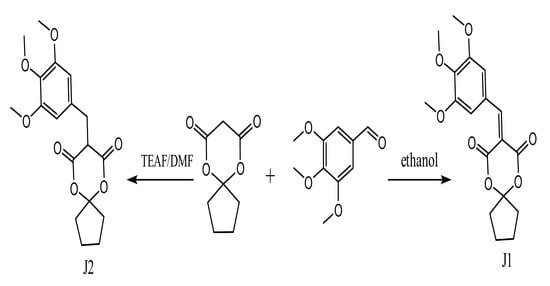
For these reasons, for 10 years, our group has exploited several approaches to synthesize all kinds of spiro compounds [13,14,15,16]. In continuation of our work, two new compounds: 8-(3,4,5-trimethoxybenzylidene)-6,10-dioxaspiro[4,5]decane-7,9-dione (J1) and 8-(3,4,5-trimethoxybenzyl)-6,10-dioxaspiro[4,5]decane-7,9-dione (J2) have been prepared via different synthetic routes (Scheme 1). In comparison to our previously synthetic route of Compound J1, Compound J2 was linked via single bond instead of double bond. In addition, their structures are also characterized by elemental analysis, IR, 1H and 13C NMR analysis, and single-crystal X-ray diffraction.

2. Materials and Methods

2.1. Physical Measuremints

The IR spectra were recorded as KBr pellets with a Nicolet FT-IR 510P Spectrometer (Nicolet Instrument Inc., Madison, WI, USA). The analyses of C, H, and N were made on an Elementar Vario EL III elemental analyzer (Elementar, Hanau, Germany). 1H NMR and 13C NMR spectra were recorded on a Bruker Avance-500 spectrometer (Bruker, Elisabethhof, The Netherlands) with CD3COCD3 as the solvent. X-ray diffraction for Compounds J1 and J2 were performed on a Spider diffractometer (Rigaku, Tokyo, Japan).

2.2. Preparation of J1 and J2

J1 is easily obtained by a literature known method [14]. A 100 mL round-bottomed flask was charged with 3,4 5-trimethoxybenzaldehyde (0.98 g, 5 mL), 6,10-dioxaspiro[4,5]decane-7,9-dione(0.85 g, 5 mL), and ethanol (15 mL). The mixture was stirred and refluxed for 2.5 h, and the solution was then cooled to generate the precipitate. Yield 31.3%, m.p.: 96.9~98.1 °C. Anal. Calcd. for C18 H20 O7: C, 62.06; H, 5.79; Found: C, 62.18; H, 5.68. 1H NMR (500 MHz, CD3OCD3): 7.65 (s, 2H, Ar–H), 8.19 (s, 1H, =CH–), 3.83 (s, 9H,–OCH3), 2.0~2.15 (m, 4H, cyclopentane–H), 1.77~1.80 (s, 4H, cyclopentane–H). 13C NMR (500 MHz, CD3OCD3): δ = 22.9 (CH2, C16/C17, cyclopentane), 29.01 (CH2, C15/C18, cyclopentane), 55.7 (CH3, C1/C3, trimethoxybenzyl ring), 78.32 (CH3, C2, trimethoxybenzyl ring), 106.57 (CH, C7/C9, trimethoxybenzyl ring), 112.24 (C, C14, 1,3-dioxane ring) 114.39 (C, C12, 1,3-dioxane ring), 126.9 (C, C8, trimethoxybenzyl ring), 143.37 (C, C5 trimethoxybenzyl ring), 152.73 (C, C4/C6 trimethoxybenzyl ring), 156.63 (CH, C10, =CH–), 163.72 (C, C11/C13, 1,3-dioxane ring). Evaporation of a solution in petroleum ether/ethyl acetate (2:1 = v/v) at room temperature yielded yellow crystals of 8-(3,4,5-trimethoxybenzylidene)-6,10-dioxaspiro[4,5]decane-7,9-dione (J1).
A solution of TEAF was prepared dropwise adding triethylamine (40 mL) to methanoic acid (10 mL). A 100 mL round-bottomed flask was charged with 3,4,5-trimethoxybenzaldehyde (0.98 g, 5 mL), 6,10-dioxaspiro[4,5]decane-7,9-dione (0.85 g, 5 mL), TEAF (5 mL), and DMF (5 mL). The mixture was stirred at room temperature for half an hour and was then heated at 50 °C for 12 h. After the mixture was cooled to room temperature and water was added (15 mL), the solution was placed at 5~10 °C for another 16 h. The precipitate was washed twice with ethanol (12 mL) and three times with water and dried. Yield 18.5%. m.p.: 134.9–135.9 °C. Anal. Calcd. For C18H22O7: C, 61.71; H, 6.33. Found: C, 61.63; H, 6.35. 1H NMR (500 MHz, CD3OCD3): 7.14 (s, 2H, Ar–H), 3.22 (s, 2H, –CH2–), 3.67 (s, 1H, 1,3-dioxane ring–H), 4.27 (s, 9H,–OCH3), 2.04~2.31 (m, 4H, cyclopentane–H), 1.74~1.82 (s, 4H, cyclopentane–H). 13C NMR (500 MHz, CD3OCD3): δ = 22.3 (CH2, C16/C17, cyclopentane), 23.83 (CH2, C10, –CH2–), 28.9 (CH2, C15/C18, cyclopentane), 49.1 (CH, C12, 1,3-dioxane ring), 55.6 (CH3, C1/C3, trimethoxybenzyl ring), 78.3 (CH3, C2, trimethoxybenzyl ring), 113.72 (C, C14, 1,3-dioxane ring), 115.1 (CH, C7/C9, trimethoxybenzyl ring), 129.7 (C, C8, trimethoxybenzyl ring), 131.94 (C, C5, trimethoxybenzyl ring), 156.7 (C, C4/C6 trimethoxybenzyl ring), 170.12 (C, C11/C13, 1,3-dioxane ring). The evaporation of a solution in petroleum ether/ethyl acetate (4:1 = v/v) yielded yellow crystals of 8-(3,4,5-trimethoxybenzyl)-6,10-dioxaspiro[4,5]decane-7,9-dione (J2).

2.3. Crystallography

The X-ray data of the two compounds were collected on Rigaku Spider Rapid IP area detector at 293(2) K. The structure of the two compounds was determined by SHELXL-97 and SHELXT-97 [17]. The final cycle of refinement gave R = 0.0651 and wR = 0.1847 (R = 0.0578 and wR = 0.1682 for J2) with w = 1/(σ2(Fo2) + (0.1185P)2 + 0.1481P) (w = 1/(σ2(Fo2) + (0.1213P)2 + 0.0698P) for J2), where P = (Fo2 + 2Fc2)/3. The key parameters of J1 and J2 are summarized in Table 1. Crystallographic data for the structures reported in this paper have been deposited with the Cambridge Crystallographic Data Centre as supplementary publication No. CCDC-1567472 and CCDC-1567470. Copies of available material can be obtained, free of charge, on application to the Director, CCDC, 12 Union Road, Cambridge CB2 1EZ, UK (Fax: 44-(0)-1223-336033 or e-mail: [email protected]).

3. Results and Discussion

3.1. Crystal Structures

Selected bond lengths and angles of the two compounds are listed in Table 2. The molecular structures of J1 and J2 are shown in Figure 1. Packing arrangement in the unit cell of J1 and J2 are displayed in Figure 2, respectively.
As can be seen from Figure 1, two compounds both comprise the 3,4,5-trimethoxybenzyl group and the dioxaspiro group. From the bond length data, the C10–C12 bond length of 1.535(2) Å (J2) is longer that of 1.359(4) (J1), which indicates that the 3,4,5-trimethoxybenzyl group connects with the dioxaspiro group by the central C(10) atoms via double bond (J1) and single bond (J2), respectively. At the same time, the C8–C10 bond lengths of 1.516(2) (J2) and 1.445(4) (J1) Å are also typical of a C–C single bond. The C(12)–C(10)–C(8) bond angle of 114.13(13)° (J2) is smaller than that of 137.3(3)° (J1), which is in agreement with similar structures (137.76(2)° and 129.67(2)°) [14]. The double bond lengths of the 1,3-dioxane ring in J1 (O(4)–C(11) 1.206(4) Å, O(5)–C(13), and 1.197(4) Å) are similar to those of J2 (O(4)–C(11) 1.193(2) Å, O(5)–C(13), and 1.199(2) Å). In J1, the five atoms (C8 and C10–C13) are fairly planar, (plane equation: −3.0923x − 7.7068y − 5.0336z = 5.6522), with a maximum deviation from the mean plane of 0.016 Å. However, the five atoms (C8, C10–C13) of J2 are not planar. The dihedral angle of the plan (C4–C10) and the plan (O4, O6, C11, and C12) is 16.39° (J1) and 61.51(9)° (J2), respectively.
Table 3 shows that the two compounds both comprise some C–H···O intra- and intermolecular hydrogen bonds. The main difference of them is C(9)–H(9A)····O(4) hydrogen bonds. The intramolecular hydrogen bands of J1 occurs at C(9)–H(9A)····O(4), and the donor and acceptor distances are 2.9287(2) Å. However, the intermolecular hydrogen bands of J2 occurs at C(9)–H(9A)····O(4), and the donor and acceptor distances are 3.170(2) Å, with symmetry codes 1 − x, −1 − y, 1 − z, which is in accord with their structures. All above hydrogen bonds generate the three-dimensional network structures of both compounds (Figure 2).

3.2. IR Spectra

IR spectra (KBr pellet) of J1 and J2 are similar (Figure 3). Weak absorption peaks both can be found at 2956 cm−1, which is due to the stretching vibration C–H of the phenyl ring. Peaks at 1753, 1716, 1255, and 1124 cm−1 (J1) and 1781, 1753, 1246, and 1124 cm−1 (J2) are attributed to the stretching vibrations of C=O and C–O bands of the 1,3-dioxane ring. Five absorption peaks in J1 and four absorption peaks in J2 can be observed at 1600~1400 cm−1, indicating a C=C stretching vibration of the 3,4,5-trimethoxybenzyl group and the dioxaspiro group by central C(10) atoms via the double bond in J1. These are in accordance with the results of the crystal structure analysis.

4. Conclusions

Two new compounds containing 3,4,5-trimethoxybenzyl and 6, 10-dioxaspiro groups, C18H20O7 (J1) and C18H22O7 (J2), have been designed and synthesized via two different routes. Their structures have been determined via single-crystal X-ray diffraction. The crystal structure determinations show that they are both of the triclinic, P-1 space group. The main difference is that the 3,4,5-trimethoxybenzyl group connects with the dioxaspiro group by the central C(10) atoms via double bond in J1 and via single bond in J2, respectively. Both compounds form three-dimensional network structures via C–H···O intra- and intermolecular hydrogen bonds.

Acknowledgments

This project was supported by the Foundation of State Key Laboratory of High-Efficiency Utilization of Coal and Green (No. 2017-K29) and the Project of Shandong Province Higher Educational Science and Technology Program (No. J11LC62).

Author Contributions

Jinhe Jiang synthesized Compounds J1 and J2. Xia Wang conceived and designed the experiments. Wulan Zeng wrote the paper.

Conflicts of Interest

The authors confirm that there are no conflicts of interest.

References

  1. Chavan, S.R.; Gavale, K.S.; Khan, A.; Joshi, R.; Kumbhar, N.; Chakravarty, D.; Dhavale, D.D. Iminosugars spiro-linked with morpholine-fused 1,2,3-triazole: Synthesis, conformational analysis, glycosidase inhibitory activity, antifungal assay, and docking studies. ACS Omega 2017, 2, 7203–7218. [Google Scholar] [CrossRef]
  2. Gupta, A.K.; Bharadwaj, M.; Kumar, A.; Mehrotra, R. Spiro-oxindoles as a promising class of small molecule inhibitors of p53–MDM2 interaction useful in targeted cancer therapy. Top. Curr. Chem. 2017, 375, 3. [Google Scholar] [CrossRef] [PubMed]
  3. Hong, H.; Huang, L.; Teng, D.W. Aspirocyclic oxindole analogue: Synthesis and antitumor activities. Chin. Chem. Lett. 2011, 22, 1009–1012. [Google Scholar] [CrossRef]
  4. Yu, B.; Shi, X.J.; Qi, P.P.; Yu, D.Q.; Liu, H.M. Design, synthesis and biological evaluation of novel steroidal spiro-oxindoles as potent antiproliferative agents. J. Steroid Biochem. Mol. Biol. 2014, 14, 121–134. [Google Scholar] [CrossRef] [PubMed]
  5. Lombe, B.K.; Bruhn, T.; Feineis, D.; Mudogo, V.; Brun, R.; Bringmann, G. Antiprotozoal spirombandakamines A1 and A2, fused naphthylisoquinoline dimers from a congolese ancistrocladus. Plant. Org. Lett. 2017, 19, 6740–6743. [Google Scholar] [CrossRef] [PubMed]
  6. Dandia, A.; Singh, R.; Saini, D. Ionic liquid-mediated three-component synthesis of fluorinated spiro-thiazine devivatives and their antimycobacterial and DNA cleavage activities. J. Chem. Sci. Vol. 2013, 125, 1045–1053. [Google Scholar] [CrossRef]
  7. Takase, K.; Noguchi, K.; Nakano, K. Circularly polarized luminescence from chiral spiro molecules: Synthesis and optical properties of 10, 10’-Spirobi(indeno[1,2-b][1]benzothiophene) derivatives. Org. Lett. 2017, 19, 5082–5085. [Google Scholar] [CrossRef] [PubMed]
  8. Majumdar, A.; Lim, C.S.; Kim, H.M.; Ghosh, K. New six-membered pH-Insensitive rhodamine spirocycle in selective sensing of Cu2+ through C−C bond cleavage and its application in cell imaging. ACS Omega 2017, 2, 8167–8176. [Google Scholar] [CrossRef]
  9. Nakagawa, T.; Ku, S.Y.; Wong, K.T.; Adachi, C. Electroluminescence based on thermally activated delayed fluorescence generated by a spirobifluorene donor–acceptor structure. Chem. Commun. 2012, 48, 9580–9582. [Google Scholar] [CrossRef] [PubMed]
  10. Takizawa, S.; Kiriyama, K.; Ieki, K.; Sasai, H. A bifunctional spiro-type organocatalyst with high enantiocontrol: Application to the aza-Morita–Baylis–Hillman reactions. Chem. Commun. 2011, 47, 9227–9229. [Google Scholar] [CrossRef] [PubMed]
  11. Doddi, S.; Narayanaswamy, K.; Ramakrishna, B.; Singh, S.P.; Banga, P.R. Synthesis and spectroscopic investigation of diketopyrrolopyrrole-spiropyran dyad for fluorescent switch application. J. Fluoresc. 2016, 26, 1939–1949. [Google Scholar] [CrossRef] [PubMed]
  12. Yang, X.L.; Zhou, L.H.; Lv, L.; Zhao, X.; Hao, L.Y. Multi-stimuli-responsive poly(NIPA-co-HEMA-co-NVP) with spironaphthoxazine hydrogel for optical data storage application. Colloid Polym. Sci. 2016, 294, 1623–1632. [Google Scholar] [CrossRef]
  13. Zeng, W.L. 5,5-[(2,4-Dichlorophenyl)methylene]bis(2,2-dimethyl-1,3dioxane-4,6-dione). Acta Cryst. 2011, 67, o1894. [Google Scholar]
  14. Jiang, J.H.; Zeng, W.L. Synthesis and Crystal Structures of Two New Oxaspirocyclic Compounds. Crystals 2016, 6, 134. [Google Scholar] [CrossRef]
  15. Zeng, W.L.; Liu, H.L. Synthesis and Crystal Structure of 3-((m-Tolylamino)methylene)-1,5-dioxaspiro[5.5]undecane-2,4-dione. Asian J. Chem. 2014, 26, 4356–4358. [Google Scholar]
  16. Zeng, W.L.; Jiang, J.H. Synthesis and Crystal Structures of Two Novel O, N-Containing Spiro Compounds. Crystals 2016, 6, 69. [Google Scholar] [CrossRef]
  17. Sheldrick, G.M. A short history of SHELX. Acta Cryst. 2008, A64, 112–122. [Google Scholar] [CrossRef] [PubMed]
Scheme 1. Synthetic routes of J1 and J2.
Crystals 08 00146 sch001
Figure 1. The molecular structures of J1 and J2 with an atomic numbering scheme.
Crystals 08 00146 g001
Figure 2. Perspective views of three-dimensional packing structures of J1 and J2.
Crystals 08 00146 g002
Figure 3. Infrared spectra of J1 and J2.
Crystals 08 00146 g003
Table 1. The key parameters for J1 and J2.
CompoundsJ1J2
FormulaC18 H20 O7C18 H22 O7
CCDC No.15674721567470
Color/shapered/blockRed/needle
Mr348.34350.36
Crystal System, Space groupTriclinic, P-1Triclinic, P-1
a, b, c (Å)5.4056(11), 11.465(2), 13.502(3)8.8778(18), 9.5285(19), 11.082(2)
α, β, γ (°)92.06(3), 91.35(3), 93.50(3)89.69(3), 67.38(3), 81.68(3)
Crystal Size (mm)0.24 × 0.16 × 0.120.14 × 0.10 × 0.06
Wavelength (Å)0.710730.71073
θ Ranges (°)3.02–27.483.02–27.48
V(Å3)3135.5(2)854.9(3)
Z22
F(000)368372
D/g·cm−31.3861.361
μ/ mm−10.1070.105
−h, h/−k, k/−l, l−7:6; −14:14; −17:17−11:11; −10:12 ; −14:14
Total, unique and [I > 2σ(I)] reflections8107, 3785, 17868389, 3894, 2870
No. of reflections, restraints, parameters3785, 0, 2263894, 0, 226
R(int)0.03120.0261
R, wR, S0.0651, 0.1847, 1.1290.0578, 0.1682, 1.105
(∆ρ)max, (∆ρ)min (e/Å3)0.595, −0.4270.630, −0.334
Table 2. Selected bond lengths (Å) and bond angles (°) of J1 and J2.
J1J2
BondDist.Dist.
C(8)–C(10)1.445(4)1.516(2)
C(10)–C(12)1.359(4)1.535(2)
C(8)–C(7)1.407(4)1.380(3)
C(9)–C(8)1.394(4)1.391(2)
O(4)–C(11)1.206(4)1.193(2)
O(5)–C(13)1.197(4)1.199(2)
Angle(°)(°)
C(12)–C(10)–C(8)137.3(3)114.13(13)
C(9)–C(8)–C(7)119.1(3)119.28(15)
C(9)–C(8)–C(10)126.7(3)122.19(15)
C(7)–C(8)–C(10)114.2(2)118.51(16)
C(10)–C(12)–C(13)128.22(15)109.48(13)
C(10)–C(12)–C(11)114.83(14)113.71(15)
C(13)–C(12)–C(11)116.91(14)112.53(13)
Table 3. Intra- and intermolecular hydrogen bond in J1 and J2.
D–H···ASymmetryD–H(Å)H···A(Å)D···A(Å)∠D–H···A (°)
C(2)–H(2B)···O(3) (J1)intra0.96002.44813.0097(2)117.13
C(9)–H(9A)···O(4) (J1)intra0.93002.15322.9287(2)140.18
C(10)–H(10A)···O(5) (J1)intra0.93002.30802.7619(2)109.55
C(1)–H(1A)···O(4) (J1)−1 − x,2 − y,−z0.96002.59463.4547(2)149.25
C(7)–H(7A)···O(5) (J1)1 − x,1 − y,−z0.93002.39113.3017(3)166.21
C(18)–H(18B)···O(1) (J1)−x,2 − y,−z0.97002.55973.4643(2)155.20
C(10)–H(10B)···O(4) (J2)intra0.97002.43932.843(2)104.60
C(7)–H(7A)···O(5) (J2)intra0.93002.58403.118(2)116.97
C(2)–H(2B)···O(3) (J2)intra0.95992.30902.929(3)121.63
C(12)–H(12A)···O(1) (J2)1 − x,−y,1 − z0.98042.38543.300(2)154.88
C(9)–H(9A)····O(4) (J2)1 − x,−1 − y,1 − z0.92992.57233.170(2)122.45