
The Influence of Tanning Chemical Agents on DNA Degradation: A Robust Procedure for the Analysis of Tanned Animal Hide—A Pilot Study

 ,
, 
Abstract
:1. Introduction
2. Materials and Methods
2.1. Materials
2.2. Tanning
- Stage L1
- Stage L2
- Stage L3
- Stage L4
- Stage L5
- Stage L6
- Stage L7
- Stage L8
- Stage L9
2.3. Sampling
2.4. DNA Extraction
- Each hide sample obtained by biopsy was placed into a 2 mL ZR BashingBead Lysis Tube (Zymo Research, USA) filled with 600 µL of DNA/RNA Shield (Zymo Research, USA).
- The lysis tubes were then placed in a homogenizer (Disruptor Genie, Scientific Industries, Bohemia, NY, USA), and the mixture was homogenized for 6–10 min.
- After homogenization was completed, the tubes were briefly centrifuged (for approximately 10 s) at room temperature.
- Then, 300 μL of lysis buffer (Solid Tissue Buffer, Zymo Research), 20 μL of proteinase K, and 20 μL of dithiothreitol (DTT) were added.
- The mixture was vortexed for 10–15 s and incubated at 55 °C overnight.
- After the incubation was completed, centrifugation was performed for 1 min at 10,000 g and room temperature.
- The supernatant was transferred to a new 2 mL microcentrifuge tube.
2.5. DNA Quantification
2.5.1. qPCR
2.5.2. Fluorometry
2.6. Calculation of the Degradation Index
2.7. DNA Typing
2.8. XRF
3. Results
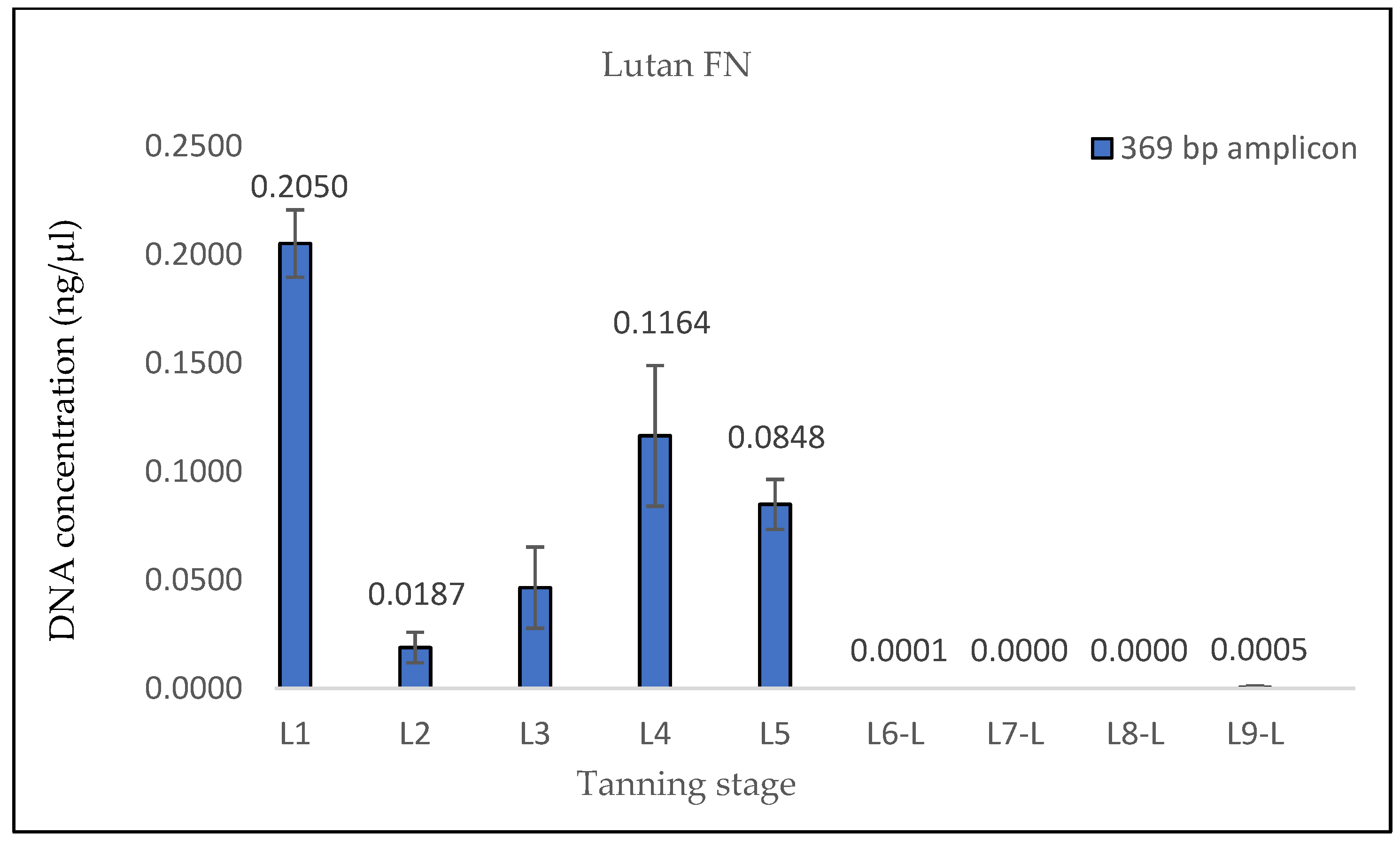
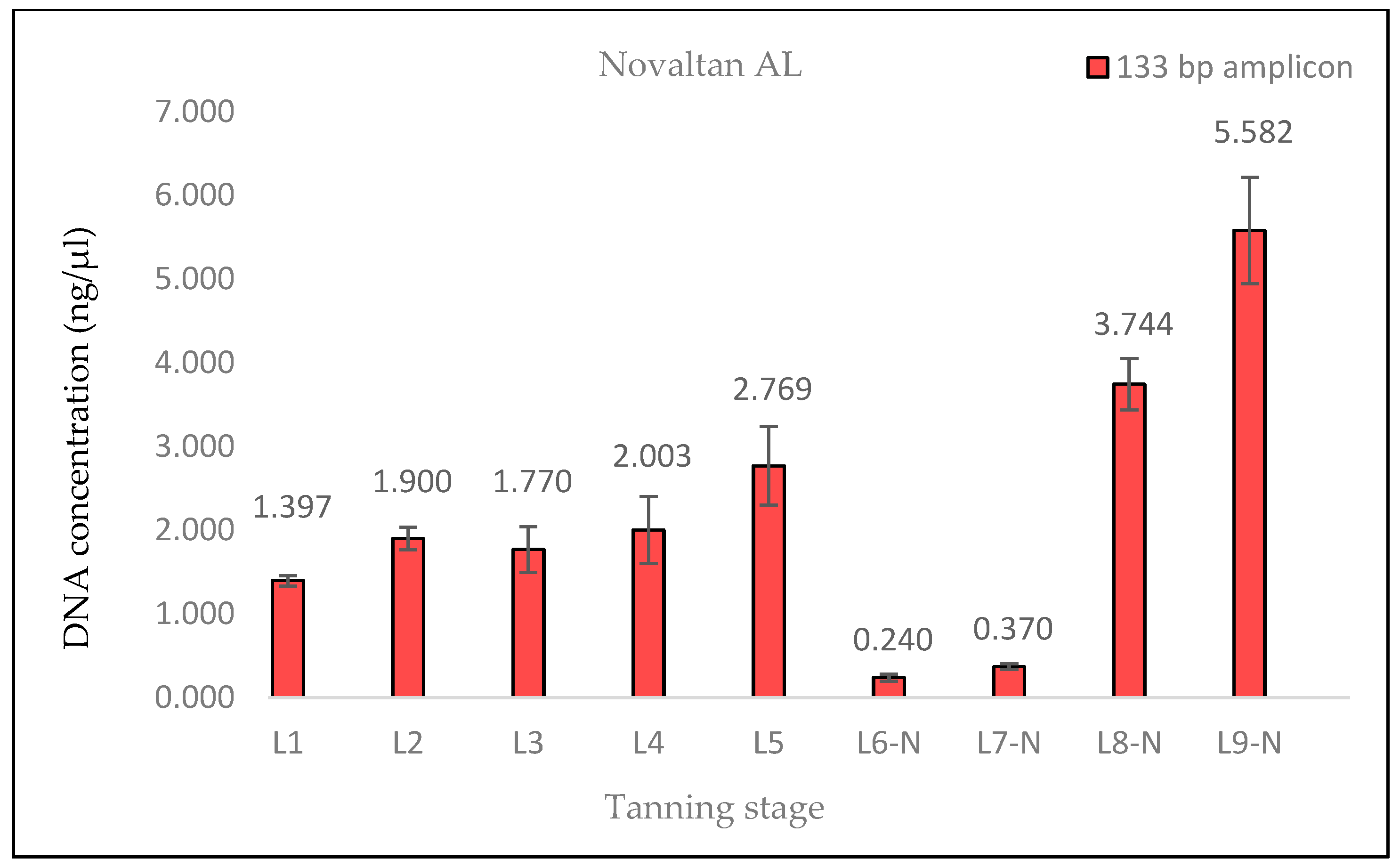
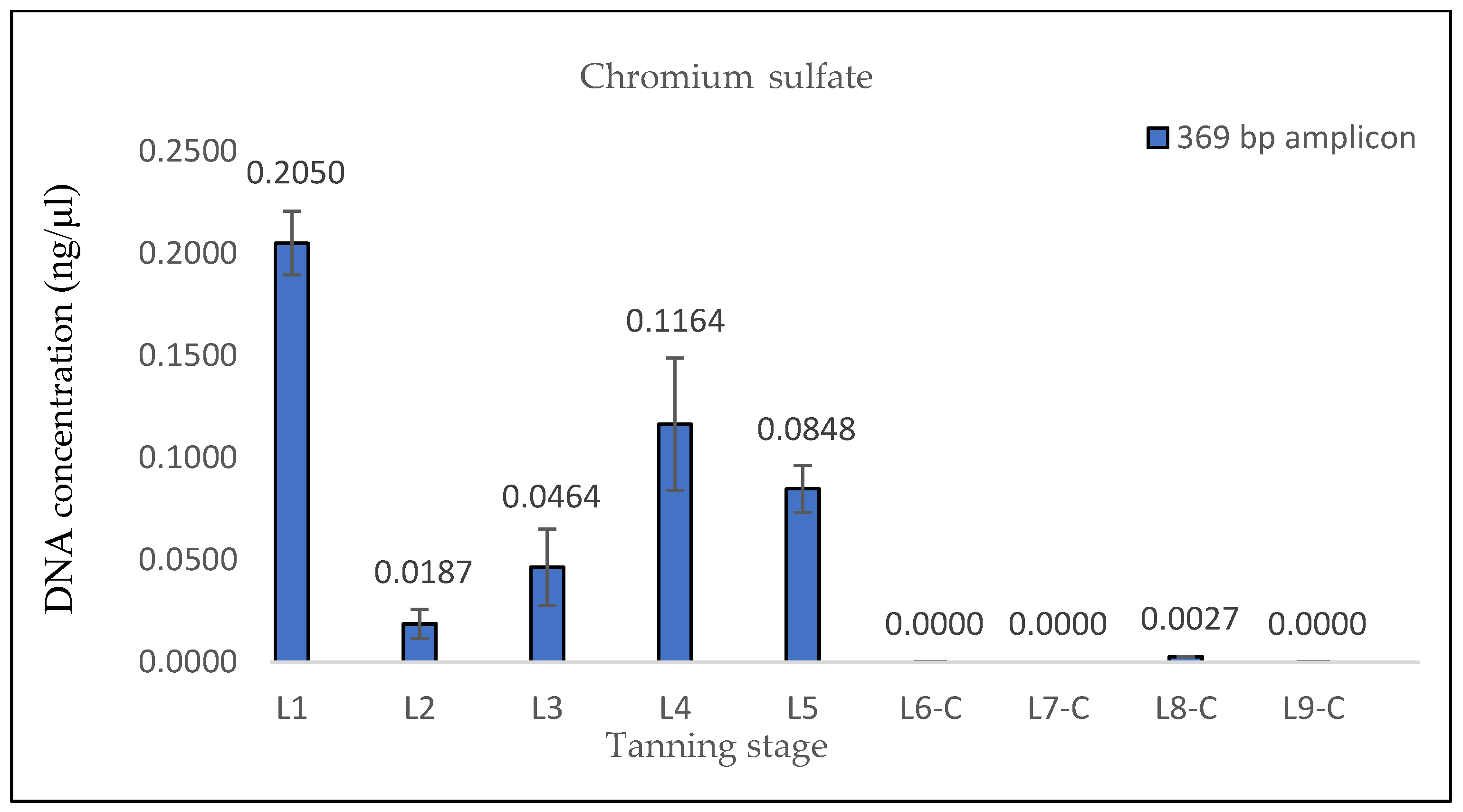
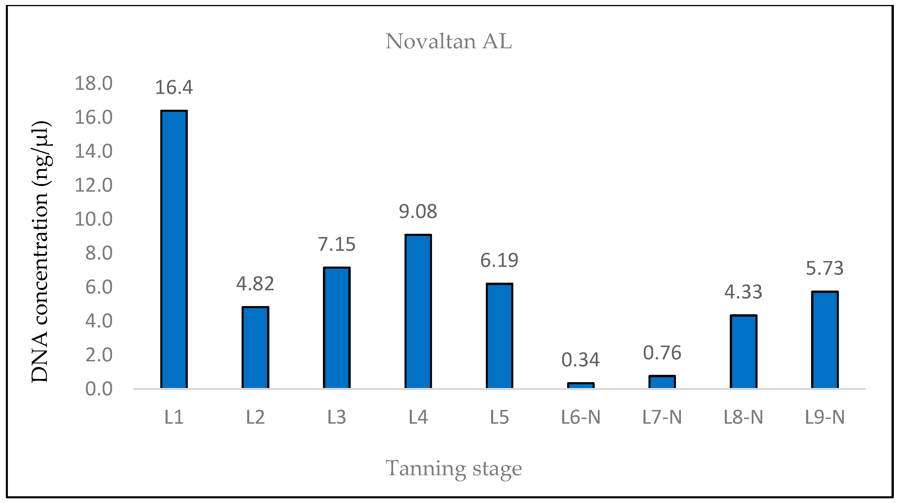
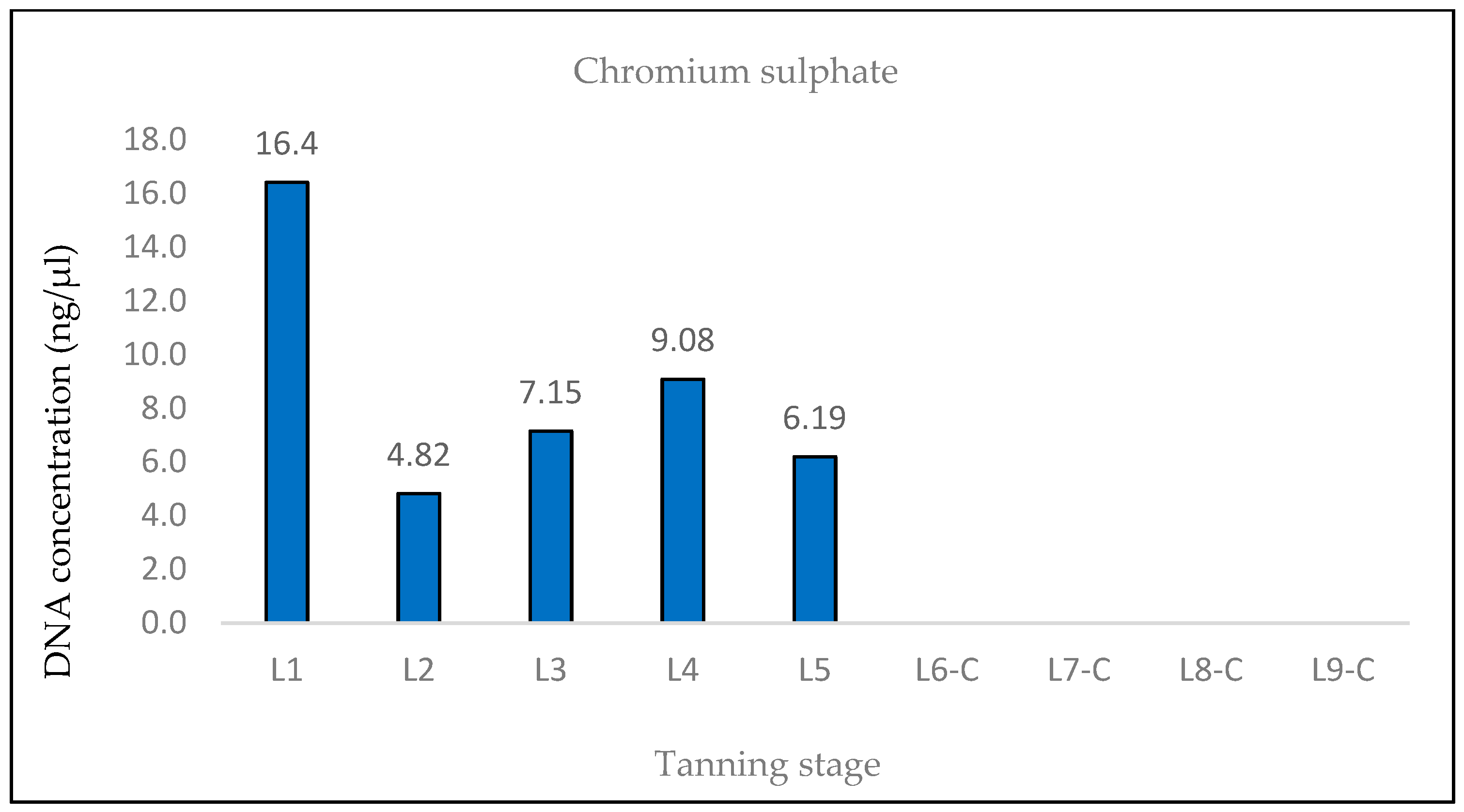
3.1. DNA Quantification
3.2. DNA Typing
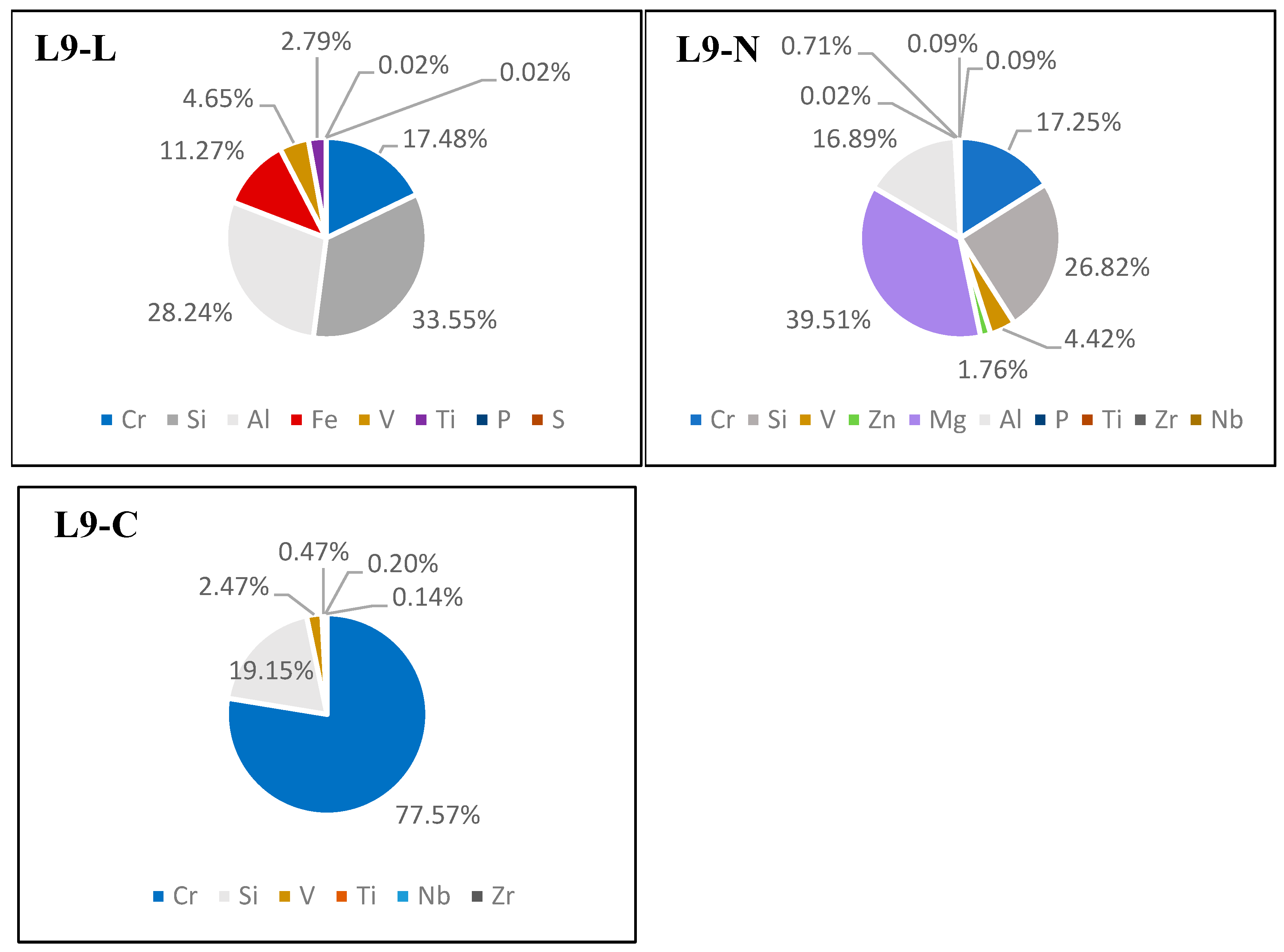
3.3. Chromium Detection in Hide Samples
4. Discussion
5. Conclusions
- The determination of the elemental composition of the tanned dermal tissue (e.g., via the XRF method) and the quantification of the chromium content.
- The acquisition of biological samples (via punch biopsy) from various distinct locations on the dermal tissue for DNA extraction.
- The utilization of the optimized extraction protocol outlined earlier for DNA extraction.
- The purification of the DNA isolates to eliminate copurified PCR inhibitors.
- The quantification of DNA utilizing quantitative PCR (qPCR) with two amplification targets, a shorter fragment (up to 150 bp) and a longer fragment (up to 400 bp), followed by the determination of the degradation index.
- An assessment of the quantification results and the selection of appropriate methodologies for subsequent analyses.
Supplementary Materials
Author Contributions
Funding
Institutional Review Board Statement
Informed Consent Statement
Data Availability Statement
Acknowledgments
Conflicts of Interest
References
- Scheffers, B.R.; Oliveira, B.R.; Lamb, I.; Edwards, D.P. Global wildlife across the tree of life. Science 2019, 366, 71–76. [Google Scholar] [CrossRef]
- Maxwell, S.L.; Fuller, R.A.; Brooks, T.M.; Watson, J.E.M. Biodiversity: The Ravages of Guns, Nets and Bulldozers. Nature 2016, 536, 143–145. [Google Scholar] [CrossRef] [PubMed]
- CITES. CITES Appendices I, II, and III. Available online: https://cites.org/eng/app/appendices.php (accessed on 16 October 2023).
- World Bank Group. Analysis of International Funding to Tackle Illegal Wildlife Trade. Available online: https://documents.worldbank.org/en/publication/documents-reports/documentdetail/695451479221164739/analysis-of-international-funding-to-tackle-illegal-wildlife-trade (accessed on 16 October 2023).
- Smart, U.; Cihlar, J.C.; Budowle, B. International Wildlife Trafficking: A Perspective on the Challenges and Potential Forensic Genetics Solutions. Forensic Sci. Int. Genet. 2021, 54, 102551. [Google Scholar] [CrossRef] [PubMed]
- UNODC. World Wildlife Crime Report—Trafficing in Protected Species. Available online: https://www.unodc.org/unodc/en/data-and-analysis/wildlife.html (accessed on 16 October 2023).
- Khedkar, G.; Khedkar, C.; Prakash, B.; Khedkar, A.; Haymer, D. DNA Barcode Based Identification of a Suspected Tiger Skin: A Case to Resolve Mimicry. Forensic Sci. Int. Rep. 2019, 1, 100027. [Google Scholar] [CrossRef]
- Vankova, L.; Vanek, D. DNA-Based Identification of Big Cats and Traditional Chinese Medicine Artifacts in the Czech Republic. Forensic Sci. Int. Genet. Suppl. Ser. 2022, 8, 122–124. [Google Scholar] [CrossRef]
- Vanek, D.; Ehler, E.; Vanková, L. Technical Note: Development of DNA Quantitation and STR Typing Systems for Panthera Tigris Species Determination and Individual Identification in Forensic Casework. Eur. J. Environ. Sci. 2021, 11, 113–118. [Google Scholar] [CrossRef]
- Rajani, C.V.; Patki, H.S.; Simanta, P.; Surjith, K.; Deepa, P.M.; Pradeep, M. Histomorphological Differentiation of the Skin of Leopard (Panthera pardus), Leopard Cat (Prionailurus bengalensis), Bengal Tiger (Panthera tigris), and Golden Jackal (Canis aureus). Vet. World 2020, 13, 827–832. [Google Scholar] [CrossRef]
- Votrubova, J.; Rihova, P.; Saskova, L.; Vanek, D. Operation Tiger’s Eye: DNA Testing of Traditional Chinese Medicine Artifacts in the Czech Republic. Forensic Sci. Int. Genet. Suppl. Ser. 2017, 6, e143–e144. [Google Scholar] [CrossRef]
- Kite, M.; Thompson, R. Conservation of Leather and Related Materials; Routledge, Taylor & Francis Group: London, UK, 2011; ISBN 978-0-7506-4881-3. [Google Scholar]
- Koehler, G.; Hobson, K.A. Effects of Tanning on the Stable Isotopic Compositions of Hair. Forensic Sci. Int. 2018, 292, 78–82. [Google Scholar] [CrossRef]
- Dubey, R.; Verma, P.; Kumar, S. Cr (III) Genotoxicity and Oxidative Stress: An Occupational Health Risk for Leather Tannery Workers of South Asian Developing Countries. Toxicol. Ind. Health 2022, 38, 112–126. [Google Scholar] [CrossRef]
- Gouda, S.; Kerry, R.G.; Das, A.; Chauhan, N.S. Wildlife Forensics: A Boon for Species Identification and Conservation Implications. Forensic Sci. Int. 2020, 317, 110530. [Google Scholar] [CrossRef] [PubMed]
- Vuissoz, A.; Worobey, M.; Odegaard, N.; Bunce, M.; Machado, C.A.; Lynnerup, N.; Peacock, E.E.; Gilbert, M.T.P. The Survival of PCR-Amplifiable DNA in Cow Leather. J. Archaeol. Sci. 2007, 34, 823–829. [Google Scholar] [CrossRef]
- Ražić, S.E.; Kopjar, N.; Kašuba, V.; Skenderi, Z.; Akalović, J.; Hrenović, J. Evaluation of DNA-Damaging Effects Induced by Different Tanning Agents Used in the Processing of Natural Leather—Pilot Study on HepG2 Cell Line. Molecules 2022, 27, 7030. [Google Scholar] [CrossRef]
- Hoffmann, O.I.; Kerekes, A.; Lipták, N.; Hiripi, L.; Bodo, S.; Szaloki, G.; Klein, S.; Ivics, Z.; Kues, W.A.; Bosze, Z. Transposon-Based Reporter Marking Provides Functional Evidence for Intercellular Bridges in the Male Germline of Rabbits. PLoS ONE 2016, 11, e0154489. [Google Scholar] [CrossRef] [PubMed]
- Dawoud Al-Bader, M.; Ali Al-Sarraf, H. Housekeeping Gene Expression during Fetal Brain Development in the Rat—Validation by Semi-Quantitative RT-PCR. Dev. Brain Res. 2005, 156, 38–45. [Google Scholar] [CrossRef] [PubMed]
- Vernarecci, S.; Ottaviani, E.; Agostino, A.; Mei, E.; Calandro, L.; Montagna, P. Quantifiler® Trio Kit and Forensic Samples Management: A Matter of Degradation. Forensic Sci. Int. Genet. 2015, 16, 77–85. [Google Scholar] [CrossRef]
- Hedmark, E.; Ellegren, H. Microsatellite Genotyping of DNA Isolated from Claws Left on Tanned Carnivore Hides. Int. J. Legal. Med. 2005, 119, 370–373. [Google Scholar] [CrossRef]
- Onem, E.; Yorgancioglu, A.; Karavana, H.A.; Yilmaz, O. Comparison of Different Tanning Agents on the Stabilization of Collagen via Differential Scanning Calorimetry. J. Therm. Anal. Calorim. 2017, 129, 615–622. [Google Scholar] [CrossRef]
- Gould, E.M.; Taylor, M.A.; Holmes, S.J. A More Consistent Method for Extracting and Amplifying DNA from Bee Wings. Apidologie 2011, 42, 721–727. [Google Scholar] [CrossRef]
- Vider, J.; Croaker, A.; Cox, A.J.; Raymond, E.; Rogers, R.; Adamson, S.; Doyle, M.; O’Brien, B.; Cripps, A.W.; West, N.P. Comparison of Skin Biopsy Sample Processing and Storage Methods on High Dimensional Immune Gene Expression Using the Nanostring NCounter System. Diagn. Pathol. 2020, 15, 57. [Google Scholar] [CrossRef]
- Trivedi, C.B.; Keuschnig, C.; Larose, C.; Rissi, D.V.; Mourot, R.; Bradley, J.A.; Winkel, M.; Benning, L.G. DNA/RNA Preservation in Glacial Snow and Ice Samples. Front. Microbiol. 2022, 13, 894893. [Google Scholar] [CrossRef] [PubMed]
- Gill, P.; Bleka, Ø.; Fonneløp, A.E. Limitations of QPCR to Estimate DNA Quantity: An RFU Method to Facilitate Inter-Laboratory Comparisons for Activity Level, and General Applicability. Forensic Sci. Int. Genet. 2022, 61, 2–7. [Google Scholar] [CrossRef] [PubMed]
- Zou, Z.-T.; Uphyrkina, O.V.; Fomenko, P.; Luo, S.-J. The Development and Application of a Multiplex Short Tandem Repeat (STR) System for Identifying Subspecies, Individuals and Sex in Tigers. Integr. Zool. 2015, 10, 376–388. [Google Scholar] [CrossRef] [PubMed]
- Bright, J.-A.; Taylor, D.; Curran, J.M.; Buckleton, J.S. Degradation of Forensic DNA Profiles. Aust. J. Forensic Sci. 2013, 45, 445–449. [Google Scholar] [CrossRef]
- Schulze Johann, K.; Bauer, H.; Wiegand, P.; Pfeiffer, H.; Vennemann, M. Detecting DNA Damage in Stored Blood Samples. Forensic Sci. Med. Pathol. 2022, 19, 50–59. [Google Scholar] [CrossRef]
- Pouliot, B.P.; Mass, J.; Kaplan, L. Using Xrf for the Identification of Chrome Tanning in Leather. In Proceedings of the 43rd Annual Meeting in Miami, Miami, FL, USA, 13–16 May 2015; p. 2015. [Google Scholar]


| Sample Label | Tanning Stage |
|---|---|
| L1 | Untanned skin |
| L2 | Degreasing solution |
| L3 | Pickling solution I |
| L4 | Fleshing and second degreasing bath |
| L5 | Pickling solution II |
| L6-L | Tanning solution—Lutan FN |
| L6-N | Tanning solution—Novaltan AL |
| L6-C | Tanning solution—chromium sulfate |
| L7-L | Tanning solution—Lutan FN with the addition of Eulan SPA 01 |
| L7-N | Tanning solution—Novaltan AL with the addition of Eulan SPA 01 |
| L7-C | Tanning solution—chromium sulfate with the addition of Eulan SPA 01 |
| L8-L | Leather oiling (Lutan FN tanned skin) |
| L8-N | Leather oiling (Novaltan AL tanned skin) |
| L8-C | Leather oiling (chromium sulfate tanned skin) |
| L9-L | Tumbling—Milling (Lutan FN tanned skin) |
| L9-N | Tumbling—Milling (Novaltan AL tanned skin) |
| L9-C | Tumbling—Milling (chromium sulfate tanned skin) |
| Solution | Composition |
|---|---|
| Degreasing solution I | water (30 °C) + Supralan 67 1 mL/L + Supralan 809 1 mL/L + disinfectant Sanitol 0.5 mL/L + deodorizer 0.25 mL/L + 9 g NaCl/L |
| Degreasing solution II | water (38 °C) + Supralan 67 2 mL/L + Supralan 809 3 mL/L + NaCl 80 g/L |
| Pickling solution I | water (room temperature) + NaCl 120 g/L + formic acid 8 g/L |
| Pickling solution II | water (room temperature) + NaCl 80 g/L + formic acid 6 g/L |
| Tanning solution—Lutan FN | water (room temperature) + NaCl 80 g/L + Lutan FN 8 g/L + Prinol M31 3 g/L + Pelgrassol SF 4 mL/L + Eulan SPA 01 2 mL/L |
| Tanning solution—Novaltan AL | water (room temperature) + NaCl 80 g/L + Novaltan Al 8 g/L + Prinol M31 3 g/L + Pelgrassol SF 4 mL/L + Eulan SPA 01 2 mL/L |
| Tanning solution—chromium sulphate | water (room temperature) + NaCl 80 g/L + chromium sulfate 12 g/L + Prinol M31 3 g/L + Pelgrassol SF 4 mL/L + Eulan SPA 01 2 mL/L |
| Primer | Sequence 5′ → 3′ | Amplicon Length | Reference |
|---|---|---|---|
| 28S-F | GTTGTTGCCATGGTAATCCTGCTCAGT | 133 bp | [18] |
| 28S-R | TCTGACTTAGAGGCGTTCAGTCATAAT | ||
| CY-F | CAACCCCACCGTGTTCTTCG | 369 bp | [19] |
| CY-R | TTGCCATCCAGCCACTCAGTC |
| Master Mix | 15 μL Reaction | Final Concentration |
|---|---|---|
| iQ SYBR Green Supermix | 7.5 µL | / |
| Primer forward (10 µM) | 0.3 µL | 0.3 µM |
| Primer reverse (10 µM) | 0.3 µL | 0.3 µM |
| Template DNA | 1 µL | different |
| H2O | 5.9 µL | / |
| qPCR Program | Melting Curve Analysis | ||||
|---|---|---|---|---|---|
| Step | Temperature | Time | Step | Temperature | Time |
| 1 | 95 °C | 3 min | 1 | 95 °C | 15 s |
| 2 | 95 °C | 15 s | 2 | 60 °C | 15 s |
| 3 | 60 °C | 1 min | 3 | 95 °C | 15 s |
| Number of cycles: 40 | |||||
| Degradation Index | |||
|---|---|---|---|
| Tanning Stage | Lutan FN | Novaltan AL | Chromium Sulphate |
| L1 | 6.8 | 6.8 | 6.8 |
| L2 | 101.4 | 101.4 | 101.4 |
| L3 | 38.1 | 38.1 | 38.1 |
| L4 | 17.2 | 17.2 | 17.2 |
| L5 | 32.7 | 32.7 | 32.7 |
| L6 | 7004.3 | 2933.6 | non-determinable |
| L7 | 10,564.2 | 5180.4 | non-determinable |
| L8 | 66,268.1 | 3587.0 | non-determinable |
| L9 | 2930.1 | 19,890.3 | non-determinable |
Disclaimer/Publisher’s Note: The statements, opinions and data contained in all publications are solely those of the individual author(s) and contributor(s) and not of MDPI and/or the editor(s). MDPI and/or the editor(s) disclaim responsibility for any injury to people or property resulting from any ideas, methods, instructions or products referred to in the content. |
© 2024 by the authors. Licensee MDPI, Basel, Switzerland. This article is an open access article distributed under the terms and conditions of the Creative Commons Attribution (CC BY) license (https://creativecommons.org/licenses/by/4.0/).
Share and Cite
Hebenstreitova, K.; Salaba, O.; Trubac, J.; Kufnerova, J.; Vanek, D. The Influence of Tanning Chemical Agents on DNA Degradation: A Robust Procedure for the Analysis of Tanned Animal Hide—A Pilot Study. Life 2024, 14, 147. https://doi.org/10.3390/life14010147
Hebenstreitova K, Salaba O, Trubac J, Kufnerova J, Vanek D. The Influence of Tanning Chemical Agents on DNA Degradation: A Robust Procedure for the Analysis of Tanned Animal Hide—A Pilot Study. Life. 2024; 14(1):147. https://doi.org/10.3390/life14010147
Chicago/Turabian StyleHebenstreitova, Kristyna, Ondrej Salaba, Jakub Trubac, Jitka Kufnerova, and Daniel Vanek. 2024. "The Influence of Tanning Chemical Agents on DNA Degradation: A Robust Procedure for the Analysis of Tanned Animal Hide—A Pilot Study" Life 14, no. 1: 147. https://doi.org/10.3390/life14010147
APA StyleHebenstreitova, K., Salaba, O., Trubac, J., Kufnerova, J., & Vanek, D. (2024). The Influence of Tanning Chemical Agents on DNA Degradation: A Robust Procedure for the Analysis of Tanned Animal Hide—A Pilot Study. Life, 14(1), 147. https://doi.org/10.3390/life14010147