Commentary

Nitric Oxide (NO) and Duchenne Muscular Dystrophy: NO Way to Go?

1
Institute for Health and Sport, Victoria University, Melbourne 8001, Victoria, Australia
2
Australian Institute for Musculoskeletal Science, St Albans 3021, Victoria, Australia
3
Institut de Myologie, Sorbonne University, INSERM UMRS974 Paris, France
*
Author to whom correspondence should be addressed.
Antioxidants 2020, 9(12), 1268; https://doi.org/10.3390/antiox9121268
Submission received: 26 November 2020 / Accepted: 11 December 2020 / Published: 13 December 2020
(This article belongs to the Special Issue Oxidative-Nitrative Stress in Human Health and Disease)

Abstract

:
The discordance between pre-clinical success and clinical failure of treatment options for Duchenne Muscular Dystrophy (DMD) is significant. The termination of clinical trials investigating the phosphodiesterase inhibitors, sildenafil and tadalafil (which prolong the second messenger molecule of nitric oxide (NO) signaling), are prime examples of this. Both attenuated key dystrophic features in the mdx mouse model of DMD yet failed to modulate primary outcomes in clinical settings. We have previously attempted to modulate NO signaling via chronic nitrate supplementation of the mdx mouse but failed to demonstrate beneficial modulation of key dystrophic features (i.e., metabolism). Instead, we observed increased muscle damage and nitrosative stress which exacerbated MD. Here, we highlight that acute nitrite treatment of human DMD myoblasts is also detrimental and suggest strategies for moving forward with NO replacement therapy in DMD.

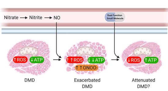
Graphical Abstract

1. Introduction

The failure of therapeutic candidates to translate from pre-clinical animal work to clinical trial success is unfortunately a common occurrence. This is particularly true for the muscle wasting condition, Duchenne Muscular Dystrophy (DMD), a rare and incurable disease that is fatal in all cases. Gene and exon skipping therapies, which partially restore the ablated dystrophin protein back into the muscle membrane, are the only curative options. However, therapeutic use is limited to those with specific dystrophin gene mutations (for exon skipping) and long-term safety and efficacy is yet to be established (for both therapies). Thus, new treatment options are crucial, particularly since standard-of-care corticosteroid practice is associated with side-effects which limits clinical utility.

2. Main

Re-purposing drugs is of particular benefit for rare diseases such as DMD as it can expedite new therapeutic options since it leverages off previous safety and tolerability trials in other diseases. One example of re-purposed drugs for the treatment of DMD are the phosphodiesterase (PDE) type 5 inhibitors, sildenafil and tadalafil, both commonly prescribed for erectile dysfunction [1]. PDE inhibition enhances blood flow by increasing the half-life of cyclic guanosine 3′,5′-monophosphate (cGMP), the second messenger of nitric oxide (NO) [1]. Pre-clinically, these PDE5 inhibitors prevented skeletal muscle fatigue, ischemia and damage in the dystrophin-deficient mdx mouse indicating that manipulation of the NO pathway is beneficial to dystrophic muscles [2,3,4,5,6,7]. Clinically, however, both have failed to modify primary endpoint measures, being delayed loss of ambulation (tadalafil; NCT01865084) [8] and improved cardiac function (sildenafil; NCT01168908) [9] in DMD patients, resulting in the termination of both trials. Similarly, sildenafil had no effect in Becker Muscular Dystrophy patients [9,10]. While there are no individualized data reported for the tadalafil trial (NCT01865084), there was evidence of participant-specific responsivity in the sildenafil trial where 4/14 participants (29%) experienced worsening of cardiac function with sildenafil versus 1/8 (12.9%) on placebo and the other 10/14 showed no response [9]. These data suggest that other factors interplay to orchestrate the biological effects of enhanced NO signaling in DMD.
In skeletal muscle, NO, produced by neuronal nitric oxide synthase (nNOS), is a critical signaling molecule that modulates key physiological processes including blood flow, contraction, glucose uptake and metabolism, among others [7]. In dystrophic muscles, nNOS dissociates from the sarcolemma resulting in its targeted degradation [11,12,13] and consequently, a diminished capacity for NO generation [7,14,15]. Reduced NO bioavailability impairs many processes including glucose metabolism [16]. This contributes to the global metabolic dysfunction (and reduced adenosine triphosphate (ATP) production) observed in dystrophic muscles [17,18,19], which we postulate is a key driver of dystrophic pathology since metabolic efficiency is essential for cell life [20]. This led us to question whether normalizing NO levels could have therapeutic potential. Since stimulating nNOS (i.e., with l-arginine supplementation [21,22]) to enhance endogenous NO generation is limited by the lower native nNOS protein content in dystrophic muscles, we sought to increase NO availability through nNOS-independent means. The nitrate-nitrite-NO pathway is an nNOS-independent mechanism that expands the NO pool [23]. Commensal bacteria reduce nitrate to nitrite (NIT), which is enzymatically converted in the blood/tissues to NO [23]. Nitrate supplementation improves mitochondrial efficiency and ATP production in healthy individuals [24], key parameters, which if improved, should theoretically mitigate the dystrophic condition.
In contrast to our hypothesis, in Timpani et al. we demonstrated that 8-week long treatment of dystrophic mdx mice with 1 mM nitrate was deleterious rather than therapeutic [25]. Despite nitrate increasing citrate synthase activity (a marker of mitochondrial density), it exacerbated mitochondrial uncoupling, muscle damage and nitrotyrosine staining (peroxynitrite marker) in dystrophic muscle [25]. The reaction of NO with superoxide results in the highly reactive nitrogen species, peroxynitrite, which damages proteins. While nitrate supplementation also increased peroxynitrite content in healthy control muscles (82% vs. 2775% increase in untreated vs. nitrate supplemented muscles, respectively), there was no evidence of concomitant muscle damage as was seen in dystrophic muscles [25]. This strain-dependent effect reflects the well-documented oxidative stress in dystrophic mdx muscles [26,27] and highlights that uncontrolled NO generation within a pro-oxidant environment exacerbates muscle damage. It is interesting to speculate whether variations in dietary antioxidant intake or endogenous antioxidant responsivity could explain why some patients experienced worsening of cardiomyopathy while others did not in the sildenafil trial [9].
We postulated that the detrimental effect of nitrate supplementation in the mdx mouse might be due to the chronic treatment period used. To test this hypothesis, here we cultured immortalized human DMD (fascia-lata of a 10 year old male with an exon 52 deletion in the dystrophin gene) and healthy control (CON; paraspinal muscle of a 12 year old female) myoblasts as described previously by us [28] (supplementary information) with 1 mM NIT (nitrate cannot be converted to NIT in cell models) for 24 h. Cells were grown in medium containing low glucose 199 media (1:5, Gibco 11150059; purchased from ThermoFisher Scientific (Scoresby, Australia)), high glucose Dulbecco’s modified eagle medium (DMEM; 4:5, Gibco 10566016), fetuin (25 µg/mL, Sigma Aldrich (Sydney, Australia) F2379), human epidermal growth factor (5 ng/mL; Sigma Aldrich E9644), basic human fibroblast growth factor (0.5 ng/mL, Sigma Aldrich F0291), insulin (5 µg/mL, Sigma Aldrich I5500), dexamethasone (0.2 µg/mL, Sigma Aldrich D4902), gentamycin (50 µg/mL, Gibco 15750060) and fetal bovine serum (20%, Bovogen Biologicals (East Keilor, Australia)). Cells were seeded at a confluency of ~10% in 75 cm2 flasks and passaged every 3 days and experiments were performed on undifferentiated myoblasts. Following NIT treatment, mitochondrial viability was determined as described previously [29] using 5000 cells per well and a positive control of FCCP and antimycin A (3 µM each) to dissipate the inner mitochondrial membrane potential (ΔΨ). Mitochondrial respiratory function (including spare respiratory capacity, ATP production and coupling efficiency) and extracellular acidification rate (ECAR), a measure of anaerobic glycolysis, were determined using the Seahorse Bioscience XF24 Analyzer as described in [28] with some modifications. On gelatin-coated (0.5%) Seahorse XF24 cell culture V7 microplates, CON (25,000 cells per well) and DMD myoblasts (15,000 cells per well) were seeded and treated with NIT. 24 h later, media was replaced with pre-warmed assay buffer (DMEM XF assay media (Seahorse Bioscience (North Billerica, MA, USA), 102365-10), 25 mM glucose and 1 mM sodium pyruvate; pH 7.4). To a pre-hydrated XF24 Sensor Cartridge, 50 µL of oligomycin (final concentration of 3 µM), 55 µL of FCCP (final concentration of 3 µM) and 60 µL of antimycin A and rotenone (final assay concentration of 1 µM and 2 µM, respectively) was added to ports A, B and C respectively. Finally, mitochondrial superoxide production was measured as described in [28] using antimycin A (3 µM) as a positive control to induce superoxide generation. Two-way ANOVA was used to detect between genotype and treatment differences. When a main effect/interaction was detected, unpaired T-tests were used to identify between group differences using SPSS (version 21; Armonk, NY, USA). An α value of 0.05 was considered significant.
Despite acute NIT treatment increasing mitochondrial viability (p < 0.01 DMD NIT vs. DMD untreated (UNTREAT); Figure 1A) and spare respiratory capacity (p < 0.05 DMD NIT vs. DMD UNTREAT; Figure 1B), this did not translate to improvements in other key metabolic parameters. In fact, acute NIT treatment exacerbated the repression of ATP production (p < 0.05 DMD NIT vs. DMD UNTREAT; Figure 1C) and mitochondrial uncoupling (p < 0.001 DMD NIT vs. DMD UNSUPP; Figure 1D) already evident in DMD myoblasts. While mitochondrial uncoupling could be considered protective against oxidative stress, in DMD myoblasts it only caused severe impact to ATP synthesis without any attenuation of mitochondrial superoxide, which was 83% higher in NIT-treated compared to UNTREAT dystrophic myoblasts (p < 0.05 DMD NIT vs. DMD UNTREAT; Figure 1E). Interestingly, though, mitochondrial superoxide content decreased with extended NIT treatment of 3 and 7 days in DMD myoblasts (by 66% and 93%, respectively compared to levels detected at 24 h; data not shown). Ostensibly, these data suggested to us induction of the endogenous antioxidant response to oxidative stress sufficient to attenuate detectable superoxide, which is a time-dependent event. However, when these data are considered in context of our data from nitrate-treated mdx mice, the attenuation of detectable superoxide seems more likely due to the vigorous formation of peroxynitrite. Our data demonstrate that amplifying NO bioavailability in an acute manner worsens the metabolic crisis in DMD cells and drives oxidative and nitrosative stress.
Augmentation of the spare respiratory capacity reflects the known metabolic modulatory effects of NO on glycolytic flux, observed here through NIT-stimulated ECAR (p < 0.05 DMD NIT vs. DMD UNTREAT; Figure 1F,G). Activity of phosphofructokinase (PFK), the rate-limiting enzyme of glycolysis, is reduced in DMD patients [30] which appears to be caused through loss of allosteric control by nNOS/NO [16]. Our observation suggests that in DMD myoblasts, PFK is activated by NIT-derived NO, which stimulates glycolytic flux and pyruvate shuttling into the mitochondria for the stimulation and expansion of the mitochondria tricarboxylic acid (TCA/Krebs) cycle. The outcome is a higher proton gradient across the inner mitochondrial membrane i.e., increased spare respiratory capacity. Paradoxically, this did not result in greater ATP production but rather in proton leak (most likely through uncoupling proteins) which consumes the proton gradient without ATP yield. Thus, the NO-driven compensatory stimulation of glycolytic flux cannot overcome the mitochondrial dysfunction to re-balance energy homeostasis in DMD myoblasts.
While our research suggests that manipulation of NO (particularly by the nitrate-NIT-NO pathway) is pernicious in dystrophic muscles, there does appear to be some merit in pursuing this treatment avenue. We saw more centronucleated fibers in nitrate-treated dystrophic muscles which suggests augmentation of muscle regeneration [25]. It stands to reason that if we could exploit this beneficial aspect of NO donor therapy while concomitantly buffering superoxide to prevent the oxidative/nitrosative damage elicited, then the true capacity for NO donor therapeutics would be known. For example, when sildenafil is administered in conjunction with vitamin E (a known free oxygen radical scavenger) in diabetic rats, vitamin E enhances the therapeutic effect of sildenafil [31]. If such a molecule were available, it could restore mitochondrial ATP production and energy homeostasis to lessen damage in the first place, but also facilitate regeneration to halt disease progression. Alternatively, other endogenous NO-generating enzymes such as endothelial NOS (eNOS) could be exploited to circumvent the loss of nNOS-mediated NO signaling and naturally elevate NO signaling within the vicinity of skeletal muscle tissue at beneficial physiological concentrations. For example, resveratrol treatment has been shown to increase eNOS expression [32] and skeletal muscle capillarization and oxygenation through eNOS-dependent NO signaling [33], and in mdx mice this leads to improved endurance [34] (Figure 2).

3. Conclusions

Here, we present the contraindicative nature of enhancing NO bioavailability as a treatment for DMD. Our previous findings in nitrate-treated mdx mice indicate that chronic NO (via nitrate) supplementation promotes nitrosative stress and muscle damage while here, acute NO (via NIT) supplementation of immortalized myoblasts derived from a DMD patient amplifies metabolic perturbations (with a flow on escalation of nitrosative stress likely). Taken with the failed clinical trials using PDE5 inhibitors to prolong NO signaling, it seems that clinical success through NO manipulation is unlikely. However, rather than abandon NO as a treatment option for DMD, modifying our approach may be the key. We suggest that a combination therapy or dual function small molecule, which increases the NO pool while addressing the oxidative stress (i.e., antioxidative capacity), may prove successful for the treatment of DMD (Figure 2). This should be tested in a human-based cell culture system (such as immortalized myoblasts/tubes derived from patients) in the first instance, which may improve translational outcomes over testing only in non-genetically and/or -phenotypically homologous animal models.

Author Contributions

Conceptualization, E.R. and C.A.T.; methodology, C.A.T., K.M., G.B.-B. and E.R.; validation, C.A.T., K.M. and G.B.-B.; formal analysis, C.A.T.; investigation, C.A.T.; resources, K.M., G.B.-B. and E.R.; data curation, C.A.T. and E.R., writing—original draft preparation, C.A.T. and E.R.; writing—review and editing, C.A.T., K.M., G.B.-B. and E.R.; visualization, C.A.T. and E.R.; supervision, E.R.; project administration, C.A.T. and E.R.; funding acquisition, E.R. All authors have read and agreed to the published version of the manuscript.

Funding

This research received no external funding.

Conflicts of Interest

The authors declare no conflict of interest relevant to this manuscript.

References

  1. Baillie, G.S.; Tejeda, G.S.; Kelly, M.P. Therapeutic targeting of 3′,5′-cyclic nucleotide phosphodiesterases: Inhibition and beyond. Nat. Rev. Drug Discov. 2019, 18, 770–796. [Google Scholar] [CrossRef]
  2. Nio, Y.; Tanaka, M.; Hirozane, Y.; Muraki, Y.; Okawara, M.; Hazama, M.; Matsuo, T. Phosphodiesterase 4 inhibitor and phosphodiesterase 5 inhibitor combination therapy has antifibrotic and anti-inflammatory effects in mdx mice with Duchenne muscular dystrophy. FASEB J. 2017, 31, 5307–5320. [Google Scholar] [CrossRef] [Green Version]
  3. De Arcangelis, V.; Strimpakos, G.; Gabanella, F.; Corbi, N.; Luvisetto, S.; Magrelli, A.; Onori, A.; Passananti, C.; Pisani, C.; Rome, S.; et al. Pathways Implicated in Tadalafil Amelioration of Duchenne Muscular Dystrophy. J. Cell Physiol. 2016, 231, 224–232. [Google Scholar] [CrossRef]
  4. Percival, J.M.; Whitehead, N.P.; Adams, M.E.; Adamo, C.M.; Beavo, J.A.; Froehner, S.C. Sildenafil reduces respiratory muscle weakness and fibrosis in the mdx mouse model of Duchenne muscular dystrophy. J. Pathol. 2012, 228, 77–87. [Google Scholar] [CrossRef] [PubMed] [Green Version]
  5. Adamo, C.M.; Dai, D.F.; Percival, J.M.; Minami, E.; Willis, M.S.; Patrucco, E.; Froehner, S.C.; Beavo, J.A. Sildenafil reverses cardiac dysfunction in the mdx mouse model of Duchenne muscular dystrophy. Proc. Natl. Acad. Sci. USA 2010, 107, 19079–19083. [Google Scholar] [CrossRef] [PubMed] [Green Version]
  6. Batra, A.; Vohra, R.S.; Chrzanowski, S.M.; Hammers, D.W.; Lott, D.J.; Vandenborne, K.; Walter, G.A.; Forbes, S.C. Effects of PDE5 inhibition on dystrophic muscle following an acute bout of downhill running and endurance training. J. Appl. Physiol. 2019, 126, 1737–1745. [Google Scholar] [CrossRef] [PubMed]
  7. Timpani, C.A.; Hayes, A.; Rybalka, E. Therapeutic strategies to address neuronal nitric oxide synthase deficiency and the loss of nitric oxide bioavailability in Duchenne Muscular Dystrophy. Orphanet J. Rare Dis. 2017, 12, 100. [Google Scholar] [CrossRef] [PubMed] [Green Version]
  8. Victor, R.G.; Sweeney, H.L.; Finkel, R.; McDonald, C.M.; Byrne, B.; Eagle, M.; Goemans, N.; Vandenborne, K.; Dubrovsky, A.L.; Topaloglu, H. A phase 3 randomized placebo-controlled trial of tadalafil for Duchenne muscular dystrophy. Neurology 2017, 89, 1811–1820. [Google Scholar] [CrossRef] [PubMed] [Green Version]
  9. Leung, D.G.; Herzka, D.A.; Thompson, W.R.; He, B.; Bibat, G.; Tennekoon, G.; Russell, S.D.; Schuleri, K.H.; Lardo, A.C.; Kass, D.A. Sildenafil does not improve cardiomyopathy in Duchenne/Becker muscular dystrophy. Ann. Neurol. 2014, 76, 541–549. [Google Scholar] [CrossRef]
  10. Witting, N.; Kruuse, C.; Nyhuus, B.; Prahm, K.P.; Citirak, G.; Lundgaard, S.J.; von Huth, S.; Vejlstrup, N.; Lindberg, U.; Krag, T.O.; et al. Effect of sildenafil on skeletal and cardiac muscle in Becker muscular dystrophy. Ann. Neurol. 2014, 76, 550–557. [Google Scholar] [CrossRef]
  11. Brenman, J.E.; Chao, D.S.; Xia, H.; Aldape, K.; Bredt, D.S. Nitric oxide synthase complexed with dystrophin and absent from skeletal muscle sarcolemma in Duchenne muscular dystrophy. Cell 1995, 82, 743–752. [Google Scholar] [CrossRef] [Green Version]
  12. Sander, M.; Chavoshan, B.; Harris, S.A.; Iannaccone, S.T.; Stull, J.T.; Thomas, G.D.; Victor, R.G. Functional muscle ischemia in neuronal nitric oxide synthase-deficient skeletal muscle of children with Duchenne muscular dystrophy. Proc. Natl. Acad. Sci. USA 2000, 97, 13818–13823. [Google Scholar] [CrossRef] [PubMed] [Green Version]
  13. Chang, W.-J.; Iannaccone, S.T.; Lau, K.S.; Masters, B.; McCabe, T.J.; McMillan, K.; Padre, R.C.; Spencer, M.J.; Tidball, J.G.; Stull, J.T. Neuronal nitric oxide synthase and dystrophin-deficient muscular dystrophy. Proc. Natl. Acad. Sci. USA 1996, 93, 9142–9147. [Google Scholar] [CrossRef] [PubMed] [Green Version]
  14. Gücüyener, K.; Ergenekon, E.; Erbas, D.; Pinarli, G.; Serdaroğlu, A. The serum nitric oxide levels in patients with Duchenne muscular dystrophy. Brain Dev. 2000, 22, 181–183. [Google Scholar] [CrossRef]
  15. Kasai, T.; Abeyama, K.; Hashiguchi, T.; Fukunaga, H.; Osame, M.; Maruyama, K. Decreased total nitric oxide production in patients with Duchenne muscular dystrophy. J. Biomed. Sci. 2004, 11, 534–537. [Google Scholar] [CrossRef] [PubMed]
  16. Wehling-Henricks, M.; Oltmann, M.; Rinaldi, C.; Myung, K.H.; Tidball, J.G. Loss of positive allosteric interactions between neuronal nitric oxide synthase and phosphofructokinase contributes to defects in glycolysis and increased fatigability in muscular dystrophy. Hum. Mol. Genet. 2009, 18, 3439–3451. [Google Scholar] [CrossRef] [Green Version]
  17. Austin, L.; De Niese, M.; McGregor, A.; Arthur, H.; Gurusinghe, A.; Gould, M. Potential oxyradical damage and energy status in individual muscle fibres from degenerating muscle diseases. Neuromuscul. Disord. 1992, 2, 27–33. [Google Scholar] [CrossRef]
  18. Cole, M.; Rafael, J.; Taylor, D.; Lodi, R.; Davies, K.; Styles, P. A quantitative study of bioenergetics in skeletal muscle lacking utrophin and dystrophin. Neuromuscul. Disord. 2002, 12, 247–257. [Google Scholar] [CrossRef]
  19. Rybalka, E.; Timpani, C.A.; Cooke, M.B.; Williams, A.D.; Hayes, A. Defects in mitochondrial ATP synthesis in dystrophin-deficient mdx skeletal muscles may be caused by complex I insufficiency. PLoS ONE 2014, 9, e115763. [Google Scholar] [CrossRef] [Green Version]
  20. Timpani, C.A.; Hayes, A.; Rybalka, E. Revisiting the dystrophin-ATP connection: How half a century of research still implicates mitochondrial dysfunction in Duchenne Muscular Dystrophy aetiology. Med. Hypothesis 2015, 85, 1021–1033. [Google Scholar] [CrossRef]
  21. Barton, E.R.; Morris, L.; Kawana, M.; Bish, L.T.; Toursel, T. Systemic administration of L-arginine benefits mdx skeletal muscle function. Muscle Nerve Off. J. Am. Assoc. Electrodiagn. Med. 2005, 32, 751–760. [Google Scholar] [CrossRef] [PubMed]
  22. Voisin, V.; Sébrié, C.; Matecki, S.; Yu, H.; Gillet, B.; Ramonatxo, M.; Israël, M.; De la Porte, S. L-arginine improves dystrophic phenotype in mdx mice. Neurobiol. Dis. 2005, 20, 123–130. [Google Scholar] [CrossRef] [PubMed]
  23. Kapil, V.; Khambata, R.S.; Jones, D.A.; Rathod, K.; Primus, C.; Massimo, G.; Fukuto, J.M.; Ahluwalia, A. The Noncanonical Pathway for In Vivo Nitric Oxide Generation: The Nitrate-Nitrite-Nitric Oxide Pathway. Pharmacol. Rev. 2020, 72, 692–766. [Google Scholar] [CrossRef] [PubMed]
  24. Larsen, F.J.; Schiffer, T.A.; Borniquel, S.; Sahlin, K.; Ekblom, B.; Lundberg, J.O.; Weitzberg, E. Dietary inorganic nitrate improves mitochondrial efficiency in humans. Cell Metab. 2011, 13, 149–159. [Google Scholar] [CrossRef] [Green Version]
  25. Timpani, C.A.; Trewin, A.J.; Stojanovska, V.; Robinson, A.; Goodman, C.A.; Nurgali, K.; Betik, A.C.; Stepto, N.; Hayes, A.; McConell, G.K. Attempting to compensate for reduced neuronal nitric oxide synthase protein with nitrate supplementation cannot overcome metabolic dysfunction but rather has detrimental effects in dystrophin-deficient mdx muscle. Neurotherapeutics 2017, 14, 429–446. [Google Scholar] [CrossRef] [Green Version]
  26. Rando, T.A.; Disatnik, M.-H.; Yu, Y.; Franco, A. Muscle cells from mdx mice have an increased susceptibility to oxidative stress. Neuromuscul. Disord. 1998, 8, 14–21. [Google Scholar] [CrossRef]
  27. Disatnik, M.-H.; Dhawan, J.; Yu, Y.; Beal, M.F.; Whirl, M.M.; Franco, A.A.; Rando, T.A. Evidence of oxidative stress in mdx mouse muscle: Studies of the pre-necrotic state. J. Neurol. Sci. 1998, 161, 77–84. [Google Scholar] [CrossRef]
  28. Timpani, C.A.; Goodman, C.A.; Stathis, C.G.; White, J.D.; Mamchaoui, K.; Butler-Browne, G.; Gueven, N.; Hayes, A.; Rybalka, E. Adenylosuccinic acid therapy ameliorates murine Duchenne Muscular Dystrophy. Sci. Rep. 2020, 10, 1125. [Google Scholar] [CrossRef] [Green Version]
  29. Rybalka, E.; Timpani, C.A.; Cheregi, B.D.; Sorensen, J.C.; Nurgali, K.; Hayes, A. Chemotherapeutic agents induce mitochondrial superoxide production and toxicity but do not alter respiration in skeletal muscle in vitro. Mitochondrion 2018, 42, 33–49. [Google Scholar] [CrossRef]
  30. Chi, M.M.Y.; Hintz, C.S.; McKee, D.; Felder, S.; Grant, N.; Kaiser, K.K.; Lowry, O.H. Effect of Duchenne muscular dystrophy on enzymes of energy metabolism in individual muscle fibers. Metabolism 1987, 36, 761–767. [Google Scholar] [CrossRef]
  31. De Young, L.; Yu, D.; Freeman, D.; Brock, G.B. Effect of PDE5 inhibition combined with free oxygen radical scavenger therapy on erectile function in a diabetic animal model. Int. J. Impot. Res. 2003, 15, 347–354. [Google Scholar] [CrossRef] [PubMed] [Green Version]
  32. Wallerath, T.; Deckert, G.; Ternes, T.; Anderson, H.; Li, H.; Witte, K.; Förstermann, U. Resveratrol, a polyphenolic phytoalexin present in red wine, enhances expression and activity of endothelial nitric oxide synthase. Circulation 2002, 106, 1652–1658. [Google Scholar] [CrossRef] [PubMed] [Green Version]
  33. Toniolo, L.; Formoso, L.; Torelli, L.; Crea, E.; Bergamo, A.; Sava, G.; Giacomello, E. Long-term resveratrol treatment improves the capillarization in the skeletal muscles of ageing C57BL/6J mice. Int. J. Food Sci. Nutr. 2020, 10, 1–8. [Google Scholar] [CrossRef] [PubMed]
  34. Sitzia, C.; Farini, A.; Colleoni, F.; Fortunato, F.; Razini, P.; Erratico, S.; Tavelli, A.; Fabrizi, F.; Belicchi, M.; Meregalli, M.; et al. Improvement of endurance of DMD animal model using natural polyphenols. BioMed Res. Int. 2015, 2015, 680615. [Google Scholar] [CrossRef] [PubMed]
Figure 1. Mitochondrial viability, functional indices and superoxide production and extracellular acidification rate (ECAR) of untreated (UNTREAT) and nitrite (NIT) treated control (CON) and Duchenne Muscular Dystrophy (DMD) myoblasts. Cells were treated for 24 h with 1 mM NIT. Mitochondrial viability was reduced in DMD myoblasts compared to CON (p < 0.0001), but NIT supplementation increased this parameter in DMD myoblasts (p < 0.01; A). Spare respiratory capacity, adenosine triphosphate (ATP) production and coupling efficiency (mitochondrial functional indices) were determined using the Seahorse Bioscience XF24 Analyzer with data presented per 5000 myoblasts due to different plating densities of CON and DMD myoblasts. While NIT increased the spare respiratory capacity in DMD myoblasts (p < 0.05; B), NIT further reduced ATP production (p < 0.05; C) and coupling efficiency (p < 0.0001; D) in DMD myoblasts. ATP production and coupling efficiency are already reduced in DMD myoblasts compared to CON (p < 0.0001). Mitochondrial superoxide production was higher in DMD myoblasts compared to CON (p < 0.01; E) and NIT treatment increased it in both CON and DMD myoblasts (p < 0.05). Extracellular acidification rate (ECAR), a metric of glycolytic flux, was measured using the Seahorse Bioscience XF24 Analyzer concomitantly with mitochondrial function. Basal ECAR (F) and maximal ECAR (G) were significantly higher in DMD myoblasts compared to CON (p < 0.01 and p < 0.0001 respectively) and NIT further stimulated ECAR in DMD myoblasts only (p < 0.05). Results are presented as mean ± standard error of the mean. Mitochondrial viability and superoxide assays: n = 4 per group; Seahorse assays: n = 7–8 CON UNTREAT, n = 6 CON NIT, n = 5 DMD UNTREAT, n = 5 DMD NIT.
Antioxidants 09 01268 g001
Figure 2. Proposed approach for nitric oxide (NO) therapy in Duchenne Muscular Dystrophy (DMD). (A) Dystrophic muscle is characterized by mitochondria which produce elevated reactive oxygen species (ROS) and reduced adenosine triphosphate (ATP). Along with other pathological processes, this culminates in muscle damage and wasting which impairs function. (B) We have previously demonstrated that the use of chronic nitrate supplementation to increase the NO pool is detrimental in dystrophic murine muscle. The superfluous ROS reacts with the NO to produce peroxynitrite (ONOO) which amplifies muscle damage. As such, manipulation of the nitrate-nitrite-NO pathway and NO donor/signal amplifier drugs are unfeasible as a treatment for DMD as they currently stand. (C) Since we observed that the increased muscle damage in dystrophic murine muscle was accompanied by markers of higher regenerative capacity (i.e., centronucleated muscle fibers), we postulate that a combination therapy/dual function small molecule which increases NO bioavailability while addressing the oxidative stress may be beneficial rather than damaging. (D) Alternatively, increasing endogenous NO signaling e.g., through endothelial NOS (eNOS) with compounds such as resveratrol could circumvent the loss of nNOS-mediated NO signaling and restore physiologically relevant NO levels to induce benefits such as enhanced capillarization and muscle oxygenation. Image created with BioRender.com.
Antioxidants 09 01268 g002
Publisher’s Note: MDPI stays neutral with regard to jurisdictional claims in published maps and institutional affiliations.