3.1. Hydrogel Synthesis and Temperature-Responsive Swelling
The ionic monomers and the drugs readily dissolved in DMSO and mixed with the other components of the hydrogels. After polymerization, transparent and consistent networks were obtained. An intensive washing step was then carried out in order to remove DMSO, unreacted substances and the drug molecules used as templates during synthesis; the cleaning was monitored by recording the absorbance of the washing solutions.
It is well known that copolymerization of NIPA with ionic monomers can alter the temperature threshold that induces the phase transition [
21,
22]. Thus, the first step was to test if the clean networks evidenced temperature-responsiveness in water in the common range of temperatures of biomedical applications,
i.e., from 4 °C for storage of labile substances to 37 °C for mimicking the physiological conditions. Two sets of experiments were run in parallel: some dried hydrogels were immersed first in cold water (4 °C) while other set was initially placed in warm water (37 °C).
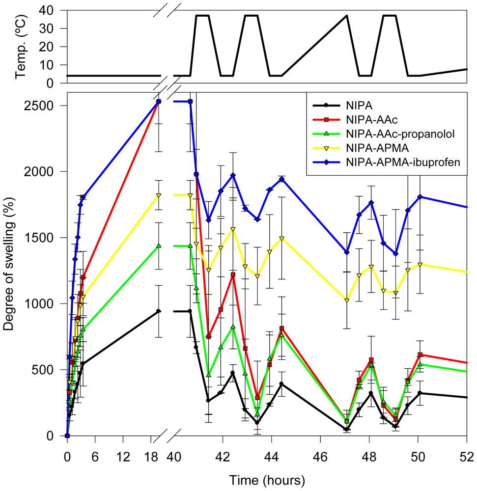
Figure 1 shows the changes in degree of swelling of the first set of hydrogels. At 4 °C (much below the LCST, lower critical solubility temperature) all networks behave as superabsorbent. Nevertheless, a remarkable influence of the comonomer and the template drug added during synthesis was noticed. The swelling at 4 °C ranked in the order: NIPA < NIPA-AAc-Propranolol < NIPA-APMA < NIPA-AAc < NIPA-APMA-Ibuprofen. Polyampholyte NIPA-APMA-AAc (data not shown) behaved as NIPA network, which indicates that internal cationic/anionic pair formation (salt bridges) occurred and that both types of monomers have been incorporated in a similar proportion. The presence of propranolol and ibuprofen in the polymerization mixture caused different effects in the swelling degree. It has been reported that cationic drugs can strongly interact with AAc [
27]. In the monomeric solutions without propranolol, AAc tends to interact with neighbour AAc through hydrogen bonds. Propranolol-AAc complexes can prevent the formation of AAc clusters and thus each AAc-mer might be more separate each other when polymerized in the presence of propranolol. Thus, when the drug is removed during the washing step, the repulsions among ionized AAc may be less intense than if AAc were closer in the space. As a consequence, the degree of swelling is also lower. Ibuprofen-APMA interactions are expected to be weaker in the polymerization mixture since the monomer was used in its salt form, namely hydrochloride, and the drug is also protonated. Thus, synthesis in the presence of ibuprofen may cause a different rearrangement of APMA monomers than when no drug is present.
Figure 1.
Temperature-responsiveness of the swelling of networks initially placed in cold water (4 °C) and that underwent cyclic changes in temperature in the 4–37 °C range.
Figure 1.
Temperature-responsiveness of the swelling of networks initially placed in cold water (4 °C) and that underwent cyclic changes in temperature in the 4–37 °C range.
Once fully swollen at 4 °C, the hydrogels were cyclically heated (37 °C) and cooled down (4 °C) to evaluate the temperature-sensitiveness and the rate and reproducibility of the volume phase transitions (
Figure 1). At 37 °C, a fast shrinking was observed for all hydrogels. Re-swelling also occurred rapidly when transferred to 4 °C medium. Those hydrogels prepared with NIPA solely or plus AAc with or without propranolol almost reached the collapse in less than 15 min at 37 °C (swelling below 100%), indicating that the hydrophobic interactions among the isopropyl groups of NIPA are the main responsible for the shrinking of the networks. Hydrogels bearing APMA did not shrink as much as the others in spite of the proportion of AAc and APMA used to synthesize the networks was the same. This finding is explained by the different degree of ionization of both monomers. It is well known that the
pKa of AAc raises as the monomers get close to each other [
20,
21] and, as a consequence, only partial ionization of the AAc mers is expected in the networks. Therefore, the contribution to the hydrophilicity of the network at 37 °C is minor. By contrast, APMA may remain as hydrochloride and thus positively charged (
pKa 8.3 in literature [
28] and 10.2 experimentally determined). The presence of the ionized groups in the networks led to swelling values in the shrunken state still above 1000%.
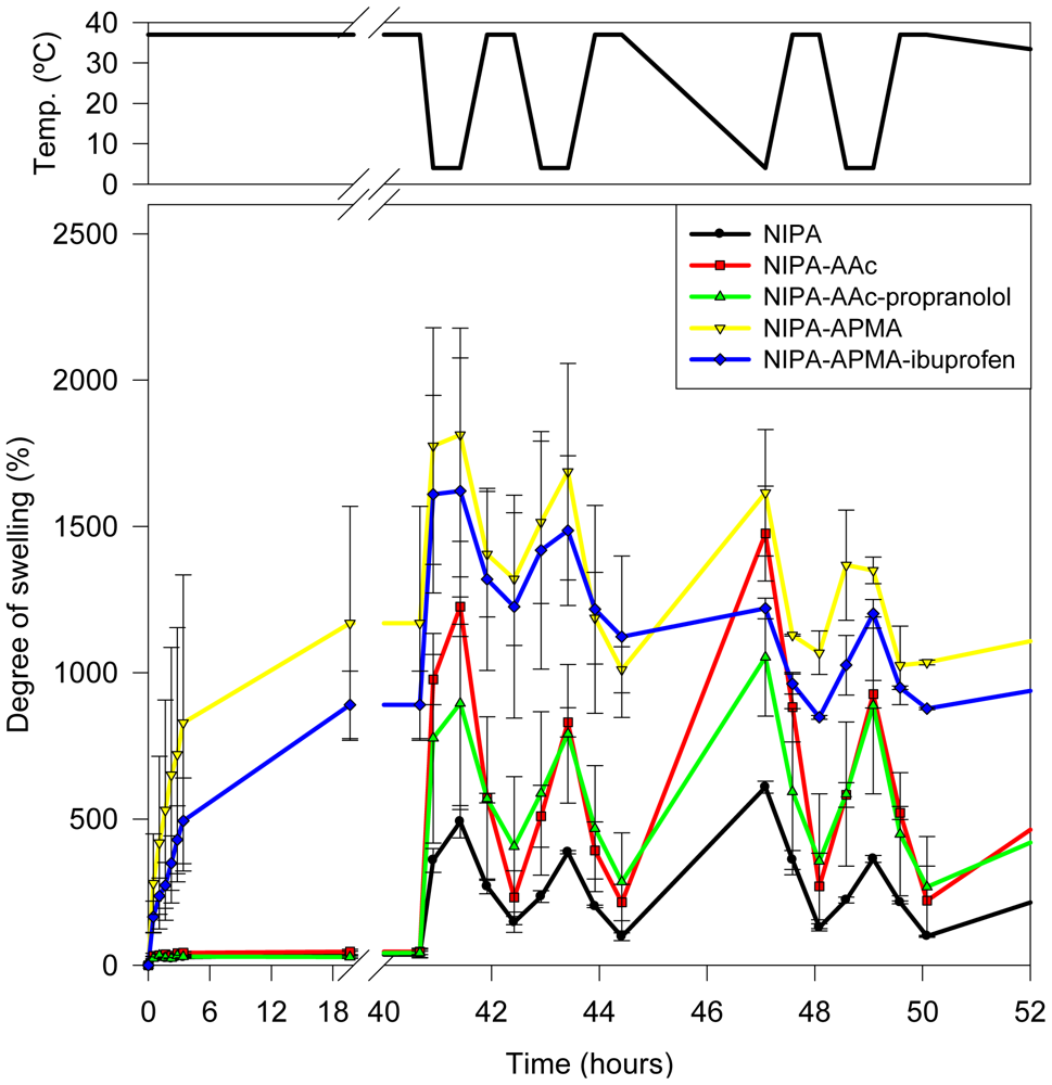
Similar temperature-induced swelling changes were observed when the hydrogels were initially placed in water at 37 °C (
Figure 2). Dried hydrogels prepared with NIPA solely or plus AAc with or without propranolol did not increase the volume when placed in the medium at 37 °C. By contrast, hydrogels bearing APMA swelled to ca. 1000%. When the temperature decreased to 4 °C, all networks swelled to a larger extent (
Figure 2) following a pattern similar to that shown in
Figure 1 after successive changes in temperature. Therefore, the networks behave as intelligent ones with rapid temperature-responsiveness and reproducible values of swelling and shrinking in water during several cycles.
Figure 2.
Temperature-responsiveness of the swelling of networks initially placed in water at 37 °C and that underwent cyclic changes in temperature in the 4–37 °C range.
Figure 2.
Temperature-responsiveness of the swelling of networks initially placed in water at 37 °C and that underwent cyclic changes in temperature in the 4–37 °C range.
3.2. Phase Transition Temperature in Salt and Drug Solutions
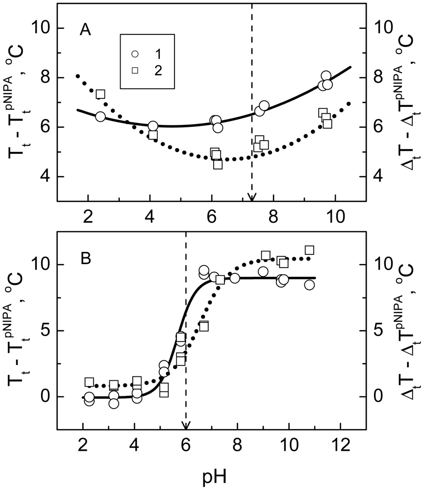
The thermodynamics of the phase transition (namely transition temperature and width) of the networks in media of various pH and salt and template concentrations was evaluated in order to gain an insight into the behavior of the hydrogels in environments that resemble the physiological conditions better than just water. It was of interest to compare some features of the collapse of the polyampholyte NIPA-APMA-AAc gel with those of the charged NIPA-AAc gel that has the same NIPA/AAc ratio and the same cross-linking density. The pH-dependences of the transition temperature and width of the polyampholyte and charged gels are shown in
Figure 3. Values of these parameters are calculated relative to the corresponding parameters of the PNIPA hydrogel. The values of the transition parameters of the polyampholyte gel exceed those of the reference PNIPA gel within the whole pH range. In other words, the polyampholyte gel undergoes the transition at the higher temperature and over wider temperature interval as compared with the PNIPA gel of the same cross-linking density. Besides, both transition parameters of the polyampholyte gel pass through a minimum upon changing pH. The minimum is particularly clearly seen in case of the transition width. From a general point of view one can expect that the polyampholyte gel should have an isoelectric point (pI), where the net charge of its network is zero. Upon shifting pH to the left and right from the pI, the network acquires a positive or negative net charge respectively. We have roughly estimated the pI value of the polyampholyte gel in the approximation of the proton binding to the independent sites (groups APMA and AAc) [
29], using an apparent copolymer composition of the gel and the values of the dissociation constants (
pKa 10.2 and
pKa 4.3, respectively) [
30] of these groups. The value pI 7.3 was obtained. It is marked in
Figure 3A by a dashed arrow. It is seen that the minima of the dependences of the transition parameters of the polyampholyte gel are located approximately in the vicinity of the hydrogel pI. It is known that an important factor affecting the collapse energetics and structure of the polyampholyte gels is the formation of salt bridges between the oppositely charged groups of the gel network [
24]. A maximal number of such bridges exists in the pI, where the number of the oppositely charged groups is maximal. Thus, it can be concluded that the presence of the salt bridges plays in favor of the collapse of the polyampholyte gel upon heating and makes its structure more cooperative (decreasing temperature and width of the transition at pI). At the same time, when comparing the polyampholyte gel with the reference PNIPA gel we should note that the inclusion of the salt bridges into the PNIPA network impedes its collapse and leads to a definite fragmentation of its structure. It is believed that these effects are related to the frustration phenomenon,
i.e., imposition of the restrictions on the subchain conformations in result of the formation of salt bridges between some subchain links [
24].
Figure 3.
Increments of the transition temperature (1 continuous line, left ordinate axis) and width (2, dotted line, right ordinate axis)
vs. pH for the polyampholyte
N-isopropylacrylamide (NIPA)-APMA-AAc (
A) and the charged NIPA-AAc (
B) hydrogels. The dashed arrows indicate (
A) the pI value of the polyampholyte gel calculated from its apparent copolymer composition and the dissociation constants of AAc (
pKa 4.3) [
30] and APMA (
pKa 10.2, determined experimentally), and (
B) the half-dissociation pH value of poly(AAc) [
30].
The pH-dependences of the transition parameters of the charged NIPA-AAc gel seem to be similar to the dissociation curve of the polyacrylic acid. These are the sigmoid curves with an inflection point in the vicinity of pH 6 [
30]. At pH 2, where the AAc dissociation is essentially suppressed, the transition parameters of the NIPA-AAc gel do not differ notably from the parameters of the reference PNIPA gel. With increasing pH the network of the NIPA-AAc gel acquires a larger charge, the transition temperature increases, and the transition is broadened. These changes are approximately exhausted at pH > 9. A probable cause of these changes in the transition parameters of the charged NIPA-AAc gel is most likely an electrostatic repulsion which impedes the rapprochement of subchains and perturbs the cooperative character of their interaction on the way to the collapsed state.
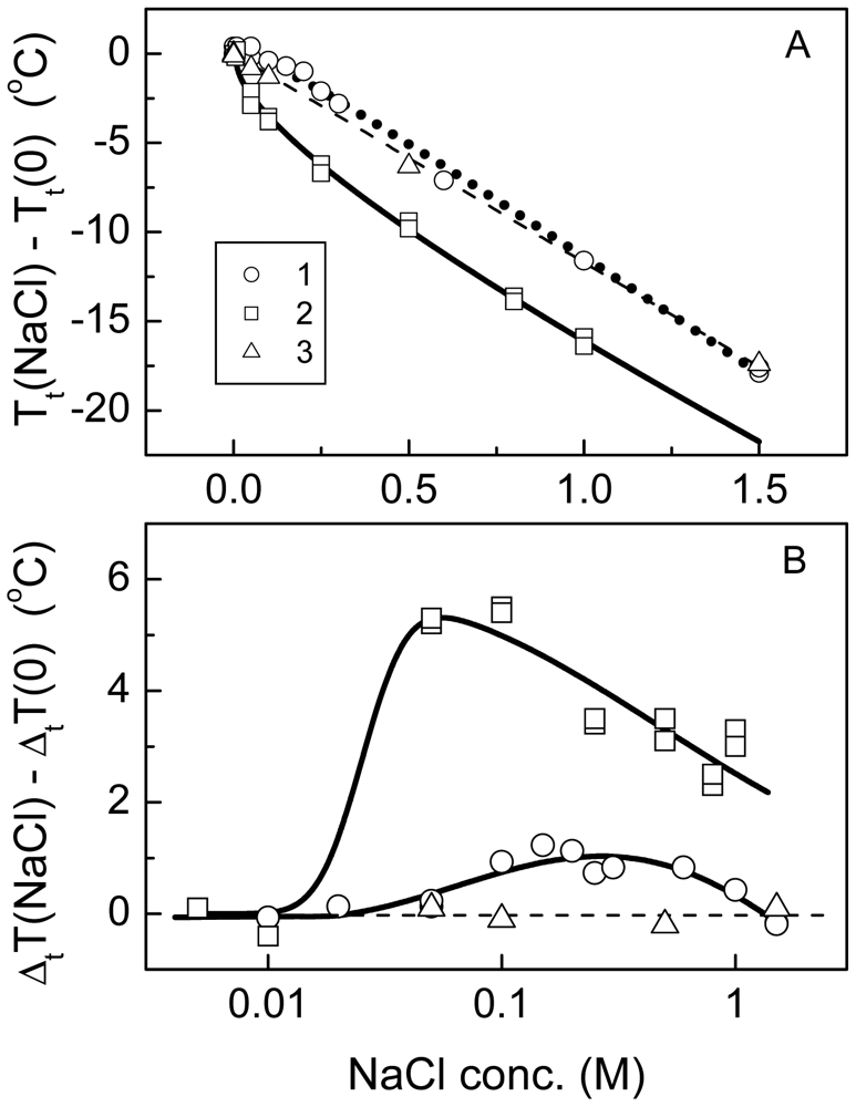
A strong effect of sodium chloride on the transition temperature and width of the NIPA-APMA-AAc, NIPA-AAc and NIPA hydrogels was observed (
Figure 4). The transition temperature decreased with increasing NaCl concentration (
Figure 4A). The dependences of the transition temperature on salt concentration,
Tt(
Cs), for the polyampholyte and charged gels were curvilinear at the initial sections and then tend to be linear at higher salt concentrations, while that for the neutral NIPA gel was strictly linear over all salt concentration range studied. In the case of the polyampholyte and charged gels, these dependences can be approximated by the equation:
while for the NIPA network, the Sechenov type equation should be used:
The parameters
Ke and
Ks take into account the electrostatics screening and the lyotropic effects on the transition free energy, respectively. The approximation of the data presented in
Figure 4A gives
Ke = −8.8 ± 0.8 K·M
−0.5 for the NIPA-AAc hydrogel, and
Ke = 3.0 ± 0.5 K·M
−0.5 for the polyampholyte gel. The negative value of
Ke in the case of the NIPA-AAc hydrogel indicates a decrease in the free energy of the collapsed state of the network due to salt screening of electrostatic repulsions. Alternatively, the salt screening of electrostatic interaction among the opposite charges of the network in the case of the polyampholyte gel gives rise to an increase in the free energy of the collapsed state. Thus the
Ke parameter of the polyampholyte gel is positive. Note, however, that thescreening effect on
Tt is expressed for the polyampholyte gel significantly less than for the chargedgel. It shows probably that salt bridges in the collapsed state of the polyampholyte gel are relatively stable and do not dissociate at low salt concentrations. The lyotropic constants
Ks of thepolyampholyte and NIPA-AAc hydrogels (−14.4 ± 0.5 and −7.3 ± 0.9 K·M
−1, respectively) are rather close to those of the neutral NIPA gel (−11.7 ± 0.1 K·M
−1). This seems to disclose that for all hydrogels the salt effect on
Tt at relatively high salt concentrations is mainly connected with the hydrophobic NIPA component of the gels. The negative value of
Ks is a consequence of the preferential increase in the free energy of the swollen state due to the salting-out phenomenon.
Figure 4.
Changes in the transition temperature (
A) and width (
B)
vs. NaCl concentration for the polyampholyte NIPA-APMA-AAc (1), NIPA-AAc (2) and NIPA (3) hydrogels: A—solid and dotted lines correspond to
equation (1); dashed line corresponds to
equation (2).
Figure 4.
Changes in the transition temperature (
A) and width (
B)
vs. NaCl concentration for the polyampholyte NIPA-APMA-AAc (1), NIPA-AAc (2) and NIPA (3) hydrogels: A—solid and dotted lines correspond to
equation (1); dashed line corresponds to
equation (2).
The effects of NaCl concentration on the transition width of the polyampholyte and NIPA-AAc hydrogels were more complex (
Figure 4B). The dependences of the transition width on NaCl concentration for both gels pass through a maximum, particularly evident for NIPA-AAc hydrogel, while no dependence was recorded for the neutral NIPA gel. It is particularly interesting that the maximum occurred in the range of the physiological salt concentration, namely at or slightly below 0.153 M (
i.e., 0.9% NaCl isotonic medium). The transition width is a merit of the system cooperativity: the higher is the system cooperativity, the narrower is the transition. The presence of different domains in a given network leads to the broadening of the transition [
22]. Alternatively, the transition must become narrower in result of coalescence of the domains. Thus, the represented dependences of the transition width on salt concentration for the polyampholyte and charged gels indicate changes in their domain structure as NaCl concentration raises. In the case of NIPA-AAc hydrogel, the disintegration into domains at very low salt concentration (less than 0.05 M) could be attributed to an increase in electrostatic repulsion between the gel subchains due to a trivial decrease in the
pKa of carboxyl groups, resulting in the increase in their ionization degree. In the case of the polyampholyte gel, a reason for the apparition of domains at low salt concentrations can be a partial dissociation of the salt bridges. On the other hand, coalescence of domains observed for both gels at relatively high salt concentrations could be caused by salting-out enhancement of hydrophobic interaction of the network subchains.
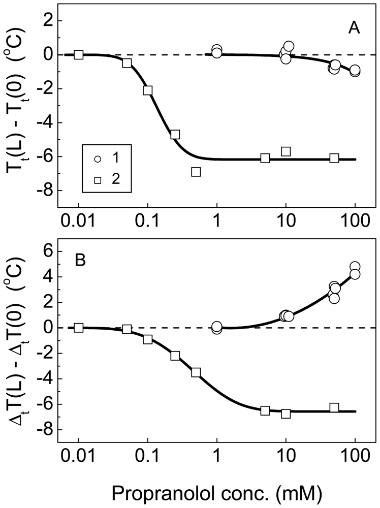
For hydrogels intended to behave as drug delivery systems, it is of relevance to know to what extent the binding of oppositely charged drugs can alter the phase transitions and consequently modify the swelling (and ultimately the mesh size) and drug diffusion through the networks. Influence of the binding of the positively charged amphiphilic ligand, propranolol, on the collapse of the polyampholyte and NIPA-AAc hydrogels is shown in
Figure 5. When propranolol concentration increased, the transition temperature of NIPA-AAc hydrogel decreased at first (more than 6 °C) and then reached a constant value (
Figure 5A). The transition temperature of the polyampholyte gel tends to decrease only at rather high concentrations of the ligand. Note that the changes in the transition temperature of the polyampholyte gel induced by the drug binding are significantly smaller than those of the charged gel. The different transition behavior of the polyampholyte and the NIPA-AAc hydrogel in the presence of propranolol is particularly evident from the plot of the transition width on ligand concentration (
Figure 5B). The transition width of the NIPA-AAc hydrogel diminished with increasing ligand concentration following a pattern that resembled that of the transition temperature. This means that the binding of propranolol and the subsequent neutralization of the AAc charges results in a weakening of the electrostatic repulsion of the network subchains and consequently leads to the coalescence of the gel domains. For the polyampholyte gel, the opposite tendency is observed. The transition width increases upon the increase in the ligand concentration. Propranolol can competitively displace APMA from the interaction with AAc groups, which results in a partial neutralization of negative charges of the network by the drug and the release of free APMA with non-neutralized positive charges. This results in a net positive charge of the network subchains. Repulsion forces arising between the subchains cause apparition of the domains. Thus, the comparison of the transition behavior of the polyampholyte and NIPA-AAc hydrogel discloses significant thermodynamic and structural differences. Mainly, the polyampholyte gel is less sensitive to changes in environmental variables such as pH, salt concentration and the presence of charged ligands. Such a stability of the network structure is a consequence of structural frustrations, that is, internal salt bridges between oppositely charged groups fixing some or other subchain conformations as additional cross-links.
Figure 5.
Dependence of the transition temperature (A) and width (B) of the polyampholyte NIPA-APMA-AAc (1) and charged NIPA-AAc (2) gels on propranolol concentration.
Figure 5.
Dependence of the transition temperature (A) and width (B) of the polyampholyte NIPA-APMA-AAc (1) and charged NIPA-AAc (2) gels on propranolol concentration.
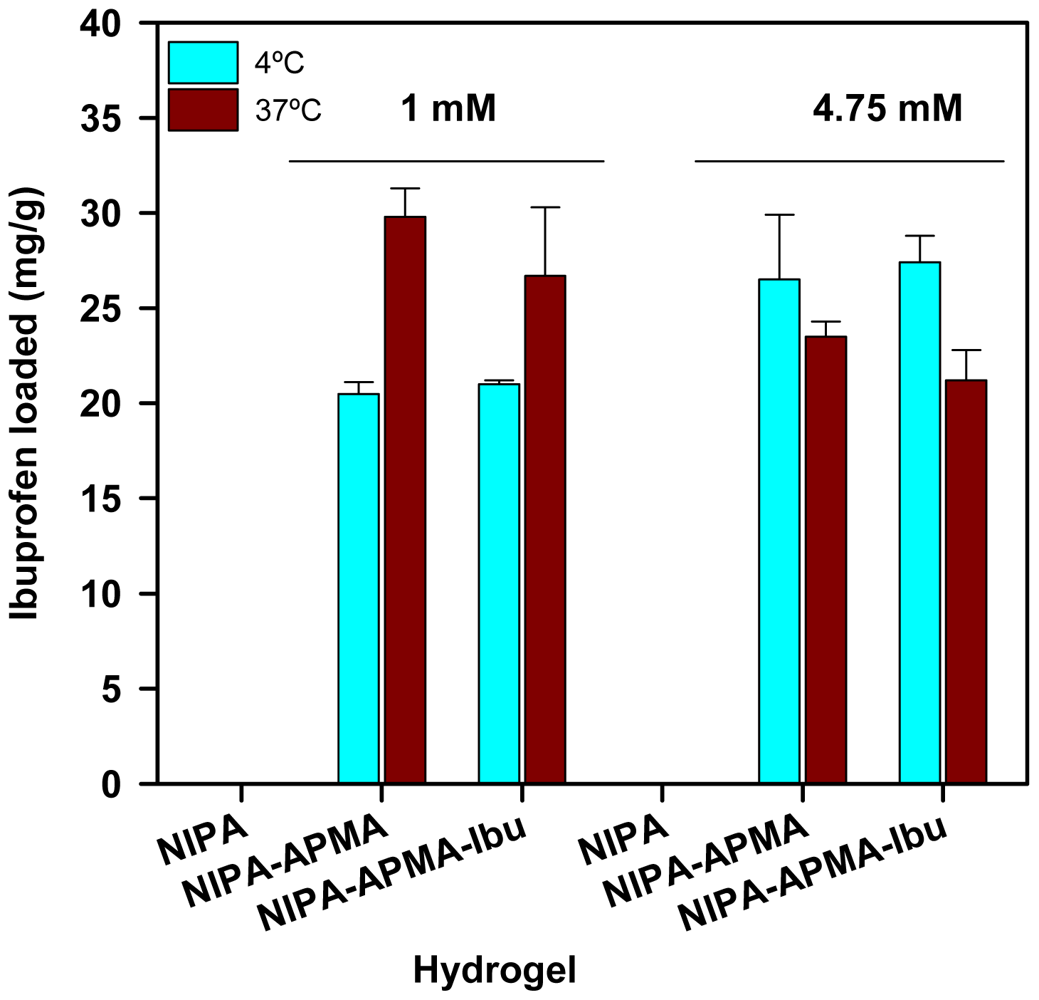
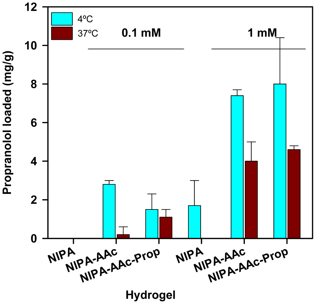
3.3. Drug Uptake
The ability of the hydrogels to uptake propranolol and ibuprofen when immersed in drug aqueous solutions was quantified at both 4 and 37 °C and at two different drug concentrations. Both drugs were tested at 1 mM, which was previously seen by HS-DSC (
Figure 5) that caused major changes in the network domains. Propranolol was also tested at lower concentration (0.1 mM) due to solubility limitations, while ibuprofen at higher concentration (4.75 mM) since the therapeutic doses are greater, but not exceeding its critical micellar concentration to avoid interferences due to the molecular aggregation [
31]. Drug loading reached the equilibrium in less than 24 h. PNIPA solely hydrogels did not load a significant amount of any drug, while copolymerization with APMA or AAc remarkably enhanced the affinity for ibuprofen (
Figure 6) and propranolol (
Figure 7).
Figure 6.
Ibuprofen loaded (mg/g) by NIPA (non-perceptible values) and NIPA-APMA hydrogels when immersed in 1 or 4.75 mM drug aqueous solutions at 4 °C or 37 °C.
Figure 6.
Ibuprofen loaded (mg/g) by NIPA (non-perceptible values) and NIPA-APMA hydrogels when immersed in 1 or 4.75 mM drug aqueous solutions at 4 °C or 37 °C.
Figure 7.
Propranolol loaded (mg/g) by NIPA and NIPA-AAc hydrogels when immersed in 0.1 and 1 mM drug aqueous solutions at 4 °C or 37 °C.
Figure 7.
Propranolol loaded (mg/g) by NIPA and NIPA-AAc hydrogels when immersed in 0.1 and 1 mM drug aqueous solutions at 4 °C or 37 °C.
The amounts of ibuprofen loaded by NIPA-APMA networks were remarkably high and similar for both drug concentrations tested, suggesting saturation of the binding capability already in 1 mM ibuprofen medium. Although minor, effects of temperature on loading in 1 and 4.5 mM drug solutions were observed. The uptake increased with ibuprofen concentration at 4 °C but slightly decreased at 37 °C. This small difference can be attributed to the somewhat smaller degree of swelling at 37 °C (
Figure 1 and
Figure 2). Nevertheless, at both temperatures NIPA-APMA networks swelled up to a large extent and, therefore, the effect of network mesh size on ibuprofen diffusion should be minor. It is interesting to note that the presence of the ligand during polymerization (codes NIPA-APMA-ibu in
Figure 6) led to a lower uptake of ibuprofen at 37 °C and for the two concentrations tested, compared to the network prepared in the absence of the drug (non-imprinted one). A similar effect has been previously found with drug-imprinted networks when loaded in drug solutions that saturate the binding ability and has been attributed to the fact that the drug causes a rearrangement of the functional monomers (APMA in our case) to gather to form binding sites with high affinity for the ligand [
32]. As more monomers are involved in the binding of each drug molecules to the imprinted network, less drug molecules are adsorbed. This hypothesis is also supported by the fact that the differences in drug uptake between networks synthesized in the presence and absence of ibuprofen are only noticed when the networks are shrunken, namely in a conformational state similar to that upon synthesis. At 4 °C, the networks are more swollen and the APMA mers become far apart acting each one as a binding site capable to establish electrostatic interactions with one ibuprofen molecule. Thus, the differences between the imprinted and non-imprinted networks are minimized.
In the case of propranolol, the effects of drug concentration and temperature on the amount of drug loaded by NIPA and NIPA-AAc hydrogels were notable (
Figure 7). The higher the concentration, the greater the amount loaded. Even NIPA network showed a small uptake when immersed in 1 mM propranolol concentration at 4 °C, which indicates that the drug can establish non-specific interactions with PNIPA chains; however, the collapsed state at 37 °C hinders drug uptake. Copolymerization with AAc notably enhanced drug loading owing to the contribution of the ionic interactions. Increase in drug concentration from 0.1 to 1 mM lead to ca. 3-fold increase in the amount loaded. Nevertheless, the lower amount of propranolol loaded compared to that achieved in the ibuprofen tests suggest that the binding capacity is not fulfilled. Under these conditions, the higher affinity of the networks synthesized in the presence of the drug (imprinted ones) is seen as an increase in the propranolol uptake at 37 °C, namely the collapsed state as upon polymerization. The arrangement of AAc mers to gather to form suitable binding sites for the drug is evidenced when the networks are at the shrunken state and the few drug molecules in the solution (diluted solution) can interact with the most perfectly formed binding sites. Such an affinity overcomes to some extent the hindrance to diffusion caused by the decrease in mesh size at 37 °C. Differences in the degree of swelling between imprinted and non-imprinted networks also corroborate this hypothesis.
3.4. Drug Release
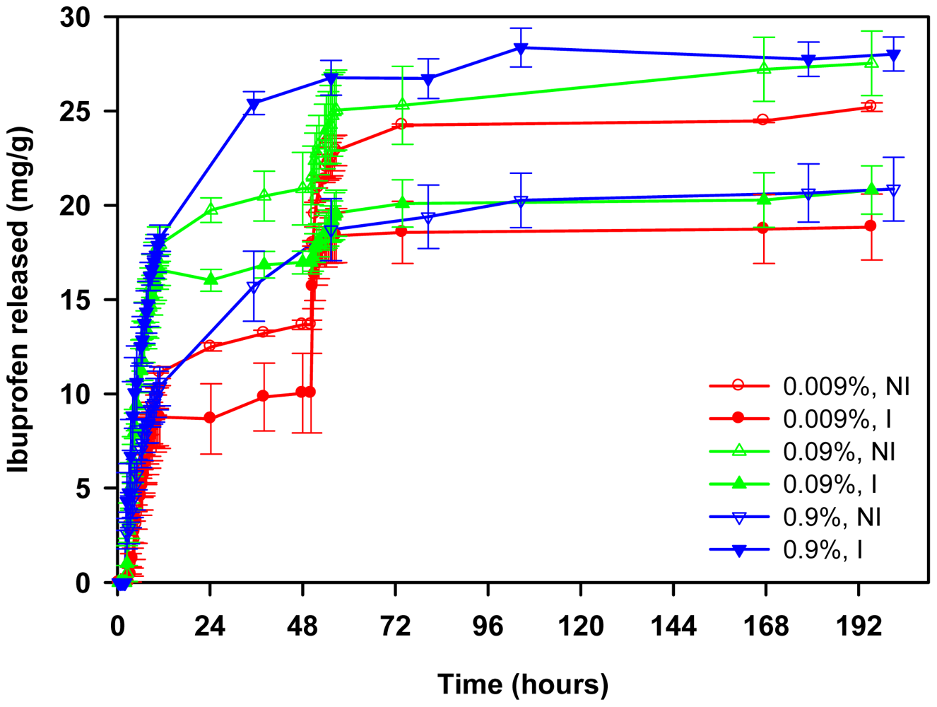
Release tests were carried out in water for the first two hours and, then, the hydrogels were transferred to NaCl solutions of various concentrations, namely 0.009, 0.09 or 0.9%. In this way, information about the role of the ionic interactions can be obtained and the performance of the hydrogels under physiological mimicking conditions can be elucidated. NIPA-APMA networks did not release ibuprofen at all when immersed in water for 2 h (
Figure 8), highlighting that the ionic interactions are so strong that in the absence of competitive ions no release occurs. In the case of propranolol, only NIPA network showed a burst release (100% released in few minutes) due to weak, non-specific interactions with the drug, while NIPA-AAc networks just led to a minor release (
Figure 9). Replacement of water for saline medium triggered the release and a marked effect of NaCl concentration on both amount released and release rate was observed.
Figure 8.
Ibuprofen release profiles in water (first 2 h; no release) and in NaCl solutions (subsequent time) at 37 °C from NIPA-APMA hydrogels loaded in 1 mM ibuprofen solution. After 48 h, the 0.009 or 0.09% NaCl solutions were replaced by 0.9% NaCl. Hydrogels synthesized in the presence of ibuprofen (imprinted) are indicated as “I”.
Figure 9.
Propranolol release profiles in water (first 2 h) and in NaCl solutions (subsequent time) at 37 °C from NIPA-AAc hydrogels previously loaded by immersion in 1 mM propranolol solution at 4 or 37 °C. Hydrogels synthesized in the presence of propranolol (imprinted) are indicated as “I”.
Figure 9.
Propranolol release profiles in water (first 2 h) and in NaCl solutions (subsequent time) at 37 °C from NIPA-AAc hydrogels previously loaded by immersion in 1 mM propranolol solution at 4 or 37 °C. Hydrogels synthesized in the presence of propranolol (imprinted) are indicated as “I”.
Drug release profiles fitted well to the Higuchi's equation (R
2 > 0.98):
where M
t and M
∞ represent the amount of drug released at time
t and the total amount loaded, respectively. The values of the release rate
kH are shown in
Figure 10. A detailed analysis of the release process in media of NaCl concentration ranging from 0.009% to 0.9% revealed that non-imprinted NIPA-APMA hydrogels released ibuprofen faster when the ionic strength was intermediate (namely, 0.09%). NaCl is needed to break the template-polymer ionic interactions, favoring the release of the drug, but at the same time the salt causes a salting out effect and makes the network to shrink (
Figure 10) and, consequently, drug diffusion becomes hindered [
33,34]. Therefore, equilibrium between both effects should result in the maximum release rate. Interestingly, the imprinted networks provided lower release rates of ibuprofen at any NaCl concentration, which confirms that the affinity of the drug for the imprinted pockets is higher than for the non-imprinted network. In 0.09% NaCl the release from the imprinted networks could be triggered but it stopped after a few ibuprofen molecules were released.
Figure 10.
Changes in diameter (blue bars) underwent when non-imprinted (clear colors) and imprinted (dark colors) NIPA-APMA hydrogels loaded with ibuprofen were immersed in NaCl solutions of various concentrations at 37 °C and drug release rate recorded for ibuprofen (green bars).
Figure 10.
Changes in diameter (blue bars) underwent when non-imprinted (clear colors) and imprinted (dark colors) NIPA-APMA hydrogels loaded with ibuprofen were immersed in NaCl solutions of various concentrations at 37 °C and drug release rate recorded for ibuprofen (green bars).

 and
and 


