
Inorganic Nanomaterials Used in Anti-Cancer Therapies:Further Developments

 ,
, 
Abstract
:1. Introduction
2. Thermal Ablation
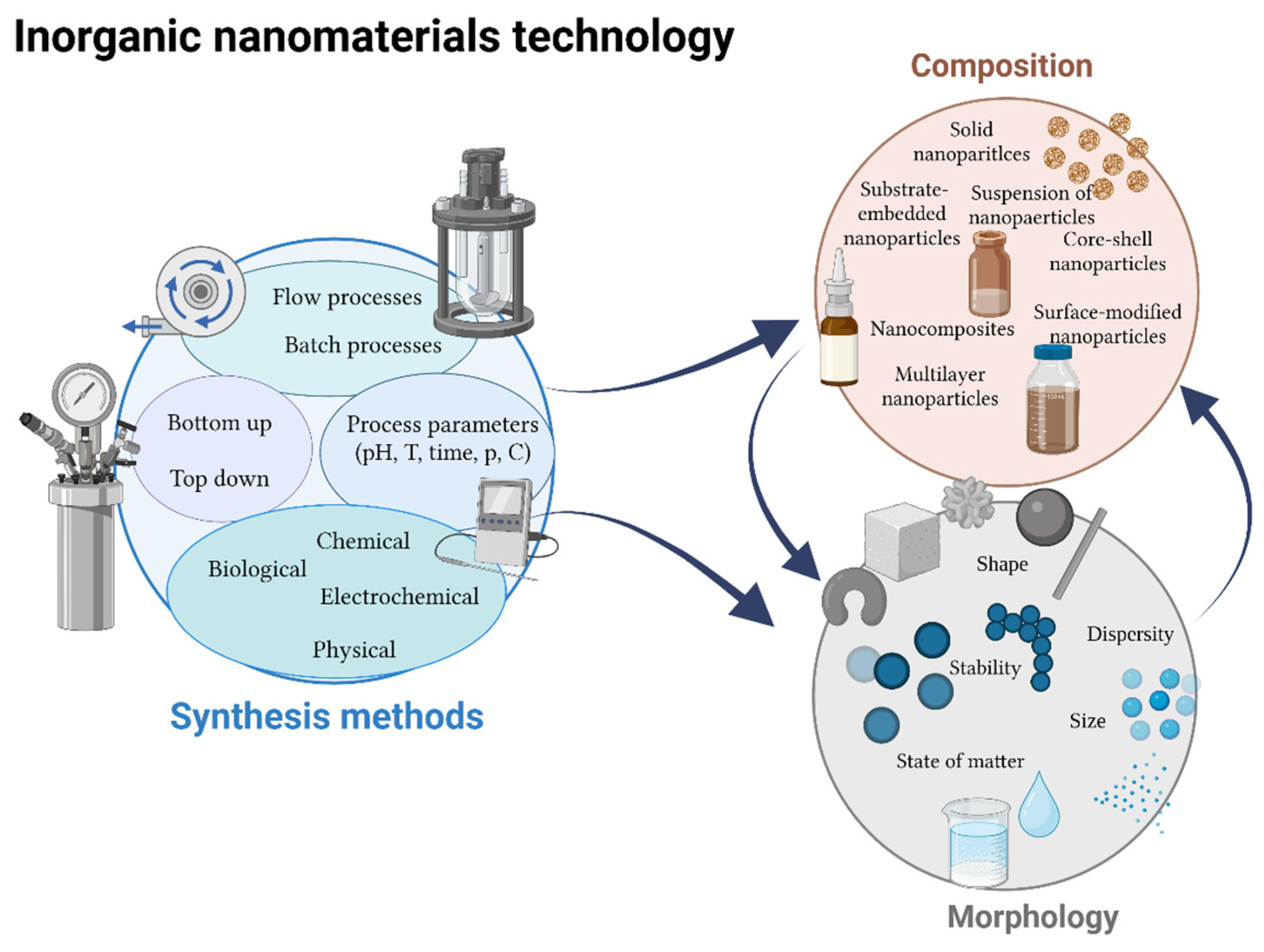
3. Nanotechnology
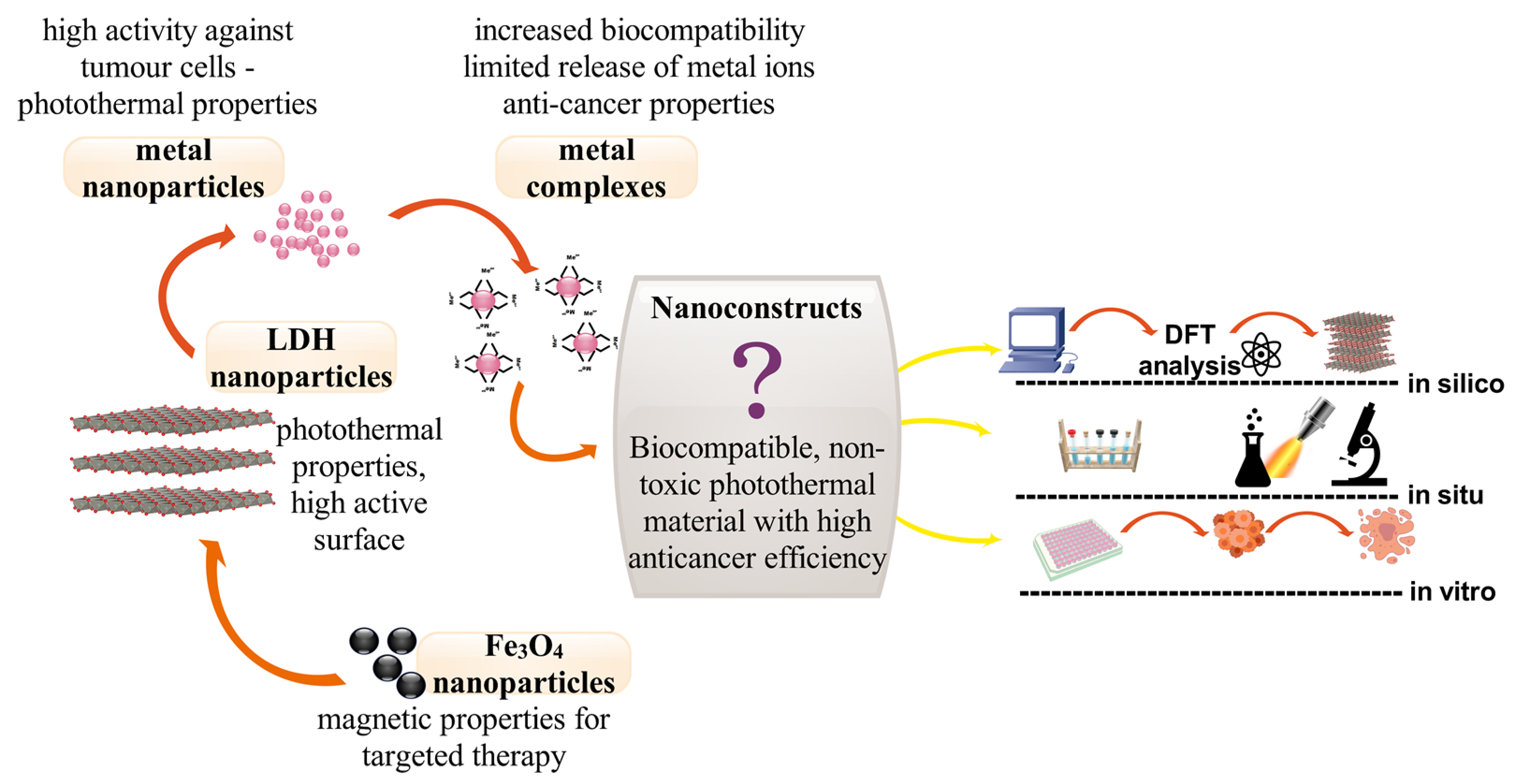
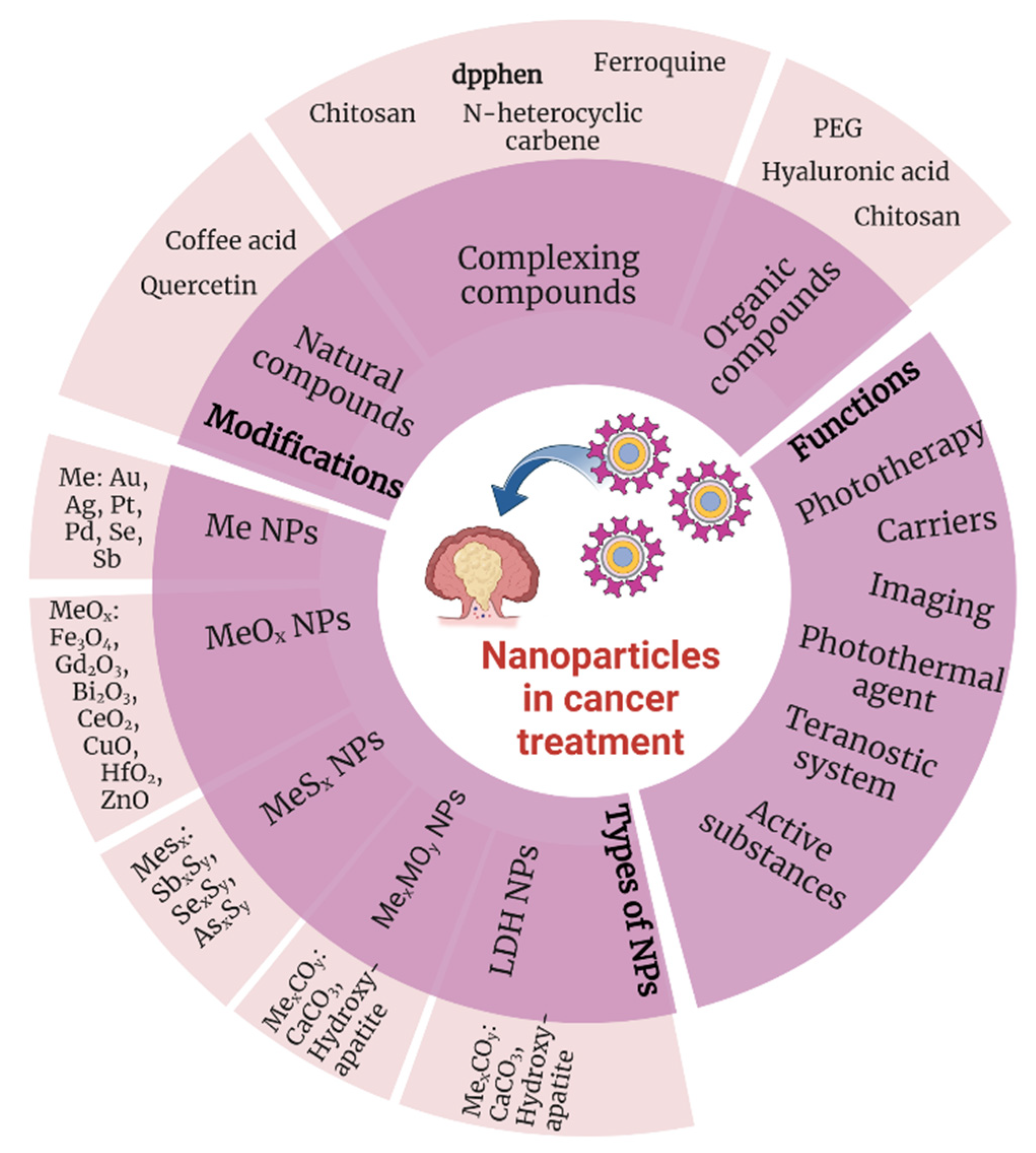
3.1. Metal Nanoparticles
3.2. Metal Oxide Nanoparticles
3.3. Layered Double Hydroxides (LDH)
4. Organic–Inorganic Complexes
5. Conclusions
Author Contributions
Funding
Institutional Review Board Statement
Informed Consent Statement
Data Availability Statement
Conflicts of Interest
References
- Hausman, D.M. What Is Cancer? Perspect. Biol. Med. 2019, 62, 778–784. [Google Scholar] [CrossRef]
- Hanna, D.H.; Saad, G.R. Induction of Mitochondria Mediated Apoptosis in Human Ovarian Cancer Cells by Folic Acid Coated Tin Oxide Nanoparticles. PLoS ONE 2021, 16, e0258115. [Google Scholar] [CrossRef]
- Jemal, A.; Ward, E.M.; Johnson, C.J.; Cronin, K.A.; Ma, J.; Ryerson, A.B.; Mariotto, A.; Lake, A.J.; Wilson, R.; Sherman, R.L.; et al. Annual Report to the Nation on the Status of Cancer, 1975–2014, Featuring Survival. J. Natl. Cancer Inst. 2017, 109, djx030. [Google Scholar] [CrossRef] [PubMed] [Green Version]
- Ferlay, J.; Colombet, M.; Soerjomataram, I.; Parkin, D.M.; Piñeros, M.; Znaor, A.; Bray, F. Cancer Statistics for the Year 2020: An Overview. Int. J. Cancer 2021, 149, 778–789. [Google Scholar] [CrossRef]
- Zhu, R.; Zhang, F.; Peng, Y.; Xie, T.; Wang, Y.; Lan, Y. Current Progress in Cancer Treatment Using Nanomaterials. Front. Oncol. 2022, 12, 3407. [Google Scholar] [CrossRef]
- Singh, R.; Sharma, A.; Saji, J.; Umapathi, A.; Kumar, S.; Daima, H.K. Smart Nanomaterials for Cancer Diagnosis and Treatment. Nano Converg. 2022, 9, 1–39. [Google Scholar] [CrossRef] [PubMed]
- Hilger, I.; Hiergeist, R.; Hergt, R.; Winnefeld, K.; Schubert, H.; Kaiser, W.A. Thermal Ablation of Tumors Using Magnetic Nanoparticles: An in Vivo Feasibility Study. Investig. Radiol. 2002, 37, 580–586. [Google Scholar] [CrossRef] [PubMed]
- Komarala, E.V.P.; Nigam, S.; Aslam, M.; Bahadur, D. In-Vitro Evaluation of Layered Double Hydroxide-Fe3O4 Magnetic Nanohybrids for Thermo-Chemotherapy. New J. Chem. 2016, 40, 423–433. [Google Scholar] [CrossRef]
- Costley, D.; Mc Ewan, C.; Fowley, C.; McHale, A.P.; Atchison, J.; Nomikou, N.; Callan, J.F. Treating Cancer with Sonodynamic Therapy: A Review. Int. J. Hyperth. 2015, 31, 107–117. [Google Scholar] [CrossRef] [PubMed]
- Takahashi, J.; Murakami, M.; Mori, T.; Iwahashi, H. Verification of Radiodynamic Therapy by Medical Linear Accelerator Using a Mouse Melanoma Tumor Model. Sci. Rep. 2018, 8, 2728. [Google Scholar] [CrossRef] [Green Version]
- Zhou, Z.; Song, J.; Nie, L.; Chen, X. Reactive Oxygen Species Generating Systems Meeting Challenges of Photodynamic Cancer Therapy. Chem. Soc. Rev. 2016, 45, 6597–6626. [Google Scholar] [CrossRef] [PubMed] [Green Version]
- Lin, H.; Chen, Y.; Shi, J. Nanoparticle-Triggered in Situ Catalytic Chemical Reactions for Tumour-Specific Therapy. Chem. Soc. Rev. 2018, 47, 1938–1958. [Google Scholar] [CrossRef] [PubMed]
- Ferrari, M. Cancer Nanotechnology: Opportunities and Challenges. Nat. Rev. Cancer 2005, 5, 161–171. [Google Scholar] [CrossRef] [PubMed]
- Xu, J.; Zhou, X.; Li, Y.; Tian, Y. Cancer Nanotechnology: Recent Trends and Developments in Strategies for Targeting Cancer Cells to Improve Cancer Imaging and Treatment. Curr. Drug Metab. 2017, 18, 266–279. [Google Scholar] [CrossRef]
- Zhang, L.; Fan, Y.; Yang, Z.; Yang, M.; Wong, C.-Y. NIR-II-Driven and Glutathione Depletion-Enhanced Hypoxia-Irrelevant Free Radical Nanogenerator for Combined Cancer Therapy. J. Nanobiotechnol. 2021, 19, 265. [Google Scholar] [CrossRef]
- Liu, X.; Yang, Y.; Ling, M.; Sun, R.; Zhu, M.; Chen, J.; Yu, M.; Peng, Z.; Yu, Z.; Liu, X. Near-Infrared II Light-Triggered Robust Carbon Radical Generation for Combined Photothermal and Thermodynamic Therapy of Hypoxic Tumors. Adv. Funct. Mater. 2021, 31, 2101709. [Google Scholar] [CrossRef]
- Liu, H.; Li, C.; Qian, Y.; Hu, L.; Fang, J.; Tong, W.; Nie, R.; Chen, Q.; Wang, H. Magnetic-Induced Graphene Quantum Dots for Imaging-Guided Photothermal Therapy in the Second near-Infrared Window. Biomaterials 2020, 232, 119700. [Google Scholar] [CrossRef]
- Tuchin, V.V.; Genina, E.A.; Tuchina, E.S.; Svetlakova, A.V.; Svenskaya, Y.I. Optical Clearing of Tissues: Issues of Antimicrobial Phototherapy and Drug Delivery. Adv. Drug Deliv. Rev. 2022, 180, 114037. [Google Scholar] [CrossRef]
- Khurana, A.; Tekula, S.; Saifi, M.A.; Venkatesh, P.; Godugu, C. Therapeutic Applications of Selenium Nanoparticles. Biomed. Pharmacother. 2019, 111, 802–812. [Google Scholar] [CrossRef]
- Yu, X.; Liu, X.; Yang, K.; Chen, X.; Li, W. Pnictogen Semimetal (Sb, Bi)-Based Nanomaterials for Cancer Imaging and Therapy: A Materials Perspective. ACS Nano 2021, 15, 2038–2067. [Google Scholar] [CrossRef]
- Yamada, M.; Foote, M.; Prow, T.W. Therapeutic Gold, Silver, and Platinum Nanoparticles. Wiley Interdiscip. Rev. Nanomed. Nanobiotechnol. 2015, 7, 428–445. [Google Scholar] [CrossRef]
- Chai, J.; Ma, Y.; Guo, T.; He, Y.; Wang, G.; Si, F.; Geng, J.; Qi, X.; Chang, G.; Ren, Z.; et al. Assembled Fe3O4 Nanoparticles on ZnAl LDH Nanosheets as a Biocompatible Drug Delivery Vehicle for PH-Responsive Drug Release and Enhanced Anticancer Activity. Appl. Clay Sci. 2022, 228, 106630. [Google Scholar] [CrossRef]
- Gu, T.; Wang, Y.; Lu, Y.; Cheng, L.; Feng, L.; Zhang, H.; Li, X.; Han, G.; Liu, Z. Platinum Nanoparticles to Enable Electrodynamic Therapy for Effective Cancer Treatment. Adv. Mater. 2019, 31, 1806803. [Google Scholar] [CrossRef] [PubMed]
- Benyettou, F.; Rezgui, R.; Ravaux, F.; Jaber, T.; Blumer, K.; Jouiad, M.; Motte, L.; Olsen, J.C.; Platas-Iglesias, C.; Magzoub, M.; et al. Synthesis of Silver Nanoparticles for the Dual Delivery of Doxorubicin and Alendronate to Cancer Cells. J. Mater. Chem. B 2015, 3, 7237–7245. [Google Scholar] [CrossRef] [PubMed]
- Srinivasan, S.Y.; Paknikar, K.M.; Gajbhiye, V.; Gajbhiye, K.R. Conjugated Polymer Nanoparticles as a Promising Tool for Anticancer Therapeutics. In Polymeric Nanoparticles as a Promising Tool for Anti-Cancer Therapeutics; Academic Press: Cambridge, MA, USA, 2019; pp. 257–280. [Google Scholar] [CrossRef]
- Sarkar, S.; Levi-Polyachenko, N. Conjugated Polymer Nano-Systems for Hyperthermia, Imaging and Drug Delivery. Adv. Drug Deliv. Rev. 2020, 163–164, 40–64. [Google Scholar] [CrossRef]
- Wang, Y.; Feng, L.; Wang, S.; Wang, Y.; Feng, L.; Wang, S. Conjugated Polymer Nanoparticles for Imaging, Cell Activity Regulation, and Therapy. Adv. Funct. Mater. 2019, 29, 1806818. [Google Scholar] [CrossRef]
- Chen, X.; Hussain, S.; Abbas, A.; Hao, Y.; Malik, A.H.; Tian, X.; Song, H.; Gao, R. Conjugated Polymer Nanoparticles and Their Nanohybrids as Smart Photoluminescent and Photoresponsive Material for Biosensing, Imaging, and Theranostics. Microchim. Acta 2022, 189, 1–33. [Google Scholar] [CrossRef]
- Fu, X.; Bai, H.; Lyu, F.; Liu, L.; Wang, S. Conjugated Polymer Nanomaterials for Phototherapy of Cancer. Chem. Res. Chin. Univ. 2020, 36, 237–242. [Google Scholar] [CrossRef]
- Dirheimer, L.; Pons, T.; Marchal, F.; Bezdetnaya, L. Quantum Dots Mediated Imaging and Phototherapy in Cancer Spheroid Models: State of the Art and Perspectives. Pharmaceutics 2022, 14, 2136. [Google Scholar] [CrossRef]
- Biswas, M.C.; Islam, M.T.; Nandy, P.K.; Hossain, M.M. Graphene Quantum Dots (GQDs) for Bioimaging and Drug Delivery Applications: A Review. ACS Mater. Lett. 2021, 3, 889–911. [Google Scholar] [CrossRef]
- Kumar, P.; Dhand, C.; Dwivedi, N.; Singh, S.; Khan, R.; Verma, S.; Singh, A.; Gupta, M.K.; Kumar, S.; Kumar, R.; et al. Graphene Quantum Dots: A Contemporary Perspective on Scope, Opportunities, and Sustainability. Renew. Sustain. Energy Rev. 2022, 157, 111993. [Google Scholar] [CrossRef]
- Li, B.; Zhao, S.; Huang, L.; Wang, Q.; Xiao, J.; Lan, M. Recent Advances and Prospects of Carbon Dots in Phototherapy. Chem. Eng. J. 2021, 408, 127245. [Google Scholar] [CrossRef]
- Triana, M.A.; Camargo, R.J.; Wu, S.-T.; Lanzafame, R.J.; Dong, Y. Quantum Dot Materials, Devices, and Their Applications in Photomedicine. In Quantum Materials, Devices, and Applications; Elsevier: Amsterdam, The Netherlands, 2023; pp. 155–200. [Google Scholar] [CrossRef]
- Xue, H.; Chen, K.; Zhou, Q.; Pan, D.; Zhang, Y.; Shen, Y. Antimony Selenide/Graphene Oxide Composite for Sensitive Photoelectrochemical Detection of DNA Methyltransferase Activity. J. Mater. Chem. B 2019, 7, 6789–6795. [Google Scholar] [CrossRef]
- Yu, X.; Li, A.; Zhao, C.; Yang, K.; Chen, X.; Li, W. Ultrasmall Semimetal Nanoparticles of Bismuth for Dual-Modal Computed Tomography/Photoacoustic Imaging and Synergistic Thermoradiotherapy. ACS Nano 2017, 11, 3990–4001. [Google Scholar] [CrossRef] [PubMed]
- Maiyo, F.; Singh, M. Selenium Nanoparticles: Potential in Cancer Gene and Drug Delivery. Nanomedicine 2017, 12, 1075–1089. [Google Scholar] [CrossRef]
- Liu, T.; Zeng, L.; Jiang, W.; Fu, Y.; Zheng, W.; Chen, T. Rational Design of Cancer-Targeted Selenium Nanoparticles to Antagonize Multidrug Resistance in Cancer Cells. Nanomedicine 2015, 11, 947–958. [Google Scholar] [CrossRef]
- Menon, S.K.S.D.; Santhiya, R.; Rajeshkumar, S.; Kumar, V. Selenium Nanoparticles: A Potent Chemotherapeutic Agent and an Elucidation of Its Mechanism. Colloids Surf. B Biointerfaces 2018, 170, 280–292. [Google Scholar] [CrossRef]
- Sun, X.; Sun, M.; Liu, M.; Yuan, B.; Gao, W.; Rao, W.; Liu, J. Shape Tunable Gallium Nanorods Mediated Tumor Enhanced Ablation through Near-Infrared Photothermal Therapy. Nanoscale 2019, 11, 2655–2667. [Google Scholar] [CrossRef]
- Shi Kam, N.W.; O’Connell, M.; Wisdom, J.A.; Dai, H.; Gray, H.B. Carbon Nanotubes as Multifunctional Biological Transporters and Near-Infrared Agents for Selective Cancer Cell Destruction on JSTOR. Proc. Natl. Acad. Sci. USA 2005, 102, 11600–11605. [Google Scholar] [CrossRef] [Green Version]
- Gannon, C.J.; Cherukuri, P.; Yakobson, B.I.; Cognet, L.; Kanzius, J.S.; Kittrell, C.; Weisman, R.B.; Pasquali, M.; Schmidt, H.K.; Smalley, R.E.; et al. Carbon Nanotube-Enhanced Thermal Destruction of Cancer Cells in a Noninvasive Radiofrequency Field. Cancer 2007, 110, 2654–2665. [Google Scholar] [CrossRef]
- Zhang, X.D.; Wu, D.; Shen, X.; Chen, J.; Sun, Y.M.; Liu, P.X.; Liang, X.J. Size-Dependent Radiosensitization of PEG-Coated Gold Nanoparticles for Cancer Radiation Therapy. Biomaterials 2012, 33, 6408–6419. [Google Scholar] [CrossRef] [PubMed] [Green Version]
- Guo, Y.; Zhang, Z.; Kim, D.H.; Li, W.; Nicolai, J.; Procissi, D.; Huan, Y.; Han, G.; Omary, R.A.; Larson, A.C. Photothermal Ablation of Pancreatic Cancer Cells with Hybrid Iron-Oxide Core Gold-Shell Nanoparticles. Int. J. Nanomed. 2013, 8, 3437–3446. [Google Scholar] [CrossRef] [PubMed] [Green Version]
- Liu, Y.; Crawford, B.M.; Vo-Dinh, T. Gold Nanoparticles-Mediated Photothermal Therapy and Immunotherapy. Immunotherapy 2018, 10, 1175–1188. [Google Scholar] [CrossRef]
- Moustafa, E.M.; Mohamed, M.A.; Thabet, N.M. Gallium Nanoparticle-Mediated Reduction of Brain Specific Serine Protease-4 in an Experimental Metastatic Cancer Model. Asian Pac. J. Cancer Prev. 2017, 18, 895. [Google Scholar] [CrossRef]
- Wang, X.; Yu, X.; Song, J.; Huang, W.; Xiang, Y.; Dai, X.; Zhang, H. Two-Dimensional Semiconducting Antimonene in Nanophotonic Applications—A Review. Chem. Eng. J. 2021, 406, 126876. [Google Scholar] [CrossRef]
- Markowska, A.; Kasprzak, B.; Jaszczyńska-Nowinka, K.; Lubin, J.; Markowska, J. Noble Metals in Oncology. Contemp. Oncol. 2015, 19, 271. [Google Scholar] [CrossRef] [Green Version]
- Manikandan, M.; Hasan, N.; Wu, H.F. Platinum Nanoparticles for the Photothermal Treatment of Neuro 2A Cancer Cells. Biomaterials 2013, 34, 5833–5842. [Google Scholar] [CrossRef]
- Hu, X.; Zhang, Y.; Ding, T.; Liu, J.; Zhao, H. Multifunctional Gold Nanoparticles: A Novel Nanomaterial for Various Medical Applications and Biological Activities. Front. Bioeng. Biotechnol. 2020, 8, 990. [Google Scholar] [CrossRef] [PubMed]
- Ahmad, N.; Sharma, A.K.; Sharma, S.; Khan, I.; Sharma, D.K.; Shamsi, A.; Santhosh Kumar, T.R.; Seervi, M. Biosynthesized Composites of Au-Ag Nanoparticles Using Trapa Peel Extract Induced ROS-Mediated P53 Independent Apoptosis in Cancer Cells. Drug Chem. Toxicol. 2018, 42, 43–53. [Google Scholar] [CrossRef]
- Hu, K.W.; Huang, C.C.; Hwu, J.R.; Su, W.C.; Shieh, D.B.; Yeh, C.S. A New Photothermal Therapeutic Agent: Core-Free Nanostructured Au x Ag1-x Dendrites. Chemistry 2008, 14, 2956–2964. [Google Scholar] [CrossRef]
- Heuer-Jungemann, A.; Feliu, N.; Bakaimi, I.; Hamaly, M.; Alkilany, A.; Chakraborty, I.; Masood, A.; Casula, M.F.; Kostopoulou, A.; Oh, E.; et al. The Role of Ligands in the Chemical Synthesis and Applications of Inorganic Nanoparticles. Chem. Rev. 2019, 119, 4819–4880. [Google Scholar] [CrossRef] [PubMed] [Green Version]
- Čubová, K.; Čuba, V. Synthesis of Inorganic Nanoparticles by Ionizing Radiation—A Review. Radiat. Phys. Chem. 2020, 169, 108774. [Google Scholar] [CrossRef]
- Jagdeo, K.R. Physical Methods for Synthesis of Nanoparticles. In Nanochemistry; CRC Press: Boca Raton, FL, USA, 2023; pp. 66–76. [Google Scholar] [CrossRef]
- Huang, X.; El-Sayed, M.A. Gold Nanoparticles: Optical Properties and Implementations in Cancer Diagnosis and Photothermal Therapy. J. Adv. Res. 2010, 1, 13–28. [Google Scholar] [CrossRef] [Green Version]
- Xie, X.; Liao, J.; Shao, X.; Li, Q.; Lin, Y. The Effect of Shape on Cellular Uptake of Gold Nanoparticles in the Forms of Stars, Rods, and Triangles. Sci. Rep. 2017, 7, 3827. [Google Scholar] [CrossRef] [PubMed] [Green Version]
- Zhuang, Y.; Liu, L.; Wu, X.; Tian, Y.; Zhou, X.; Xu, S.; Xie, Z.; Ma, Y. Size and Shape Effect of Gold Nanoparticles in “Far-Field” Surface Plasmon Resonance. Part. Part. Syst. Charact. 2019, 36, 1800077. [Google Scholar] [CrossRef] [Green Version]
- Amendola, V.; Pilot, R.; Frasconi, M.; Maragò, O.M.; Iatì, M.A. Surface Plasmon Resonance in Gold Nanoparticles: A Review. J. Phys. Condens. Matter 2017, 29, 203002. [Google Scholar] [CrossRef]
- Porcel, E.; Liehn, S.; Remita, H.; Usami, N.; Kobayashi, K.; Furusawa, Y.; Le Sech, C.; Lacombe, S. Platinum Nanoparticles: A Promising Material for Future Cancer Therapy? Nanotechnology 2010, 21, 085103. [Google Scholar] [CrossRef]
- Watanabe, A.; Kajita, M.; Kim, J.; Kanayama, A.; Takahashi, K.; Mashino, T.; Miyamoto, Y. In Vitro Free Radical Scavenging Activity of Platinum Nanoparticles. Nanotechnology 2009, 20, 455105. [Google Scholar] [CrossRef]
- Gunes, S.; He, Z.; van Acken, D.; Malone, R.; Cullen, P.J.; Curtin, J.F. Platinum Nanoparticles Inhibit Intracellular ROS Generation and Protect against Cold Atmospheric Plasma-Induced Cytotoxicity. Nanomedicine 2021, 36, 102436. [Google Scholar] [CrossRef]
- Yusof, F.; Akmal, N.; Ismail, S. Antioxidants Effects of Platinum Nanoparticles: A Potential Alternative Treatment to Lung Diseases. J. Appl. Pharm. Sci. 2015, 5, 140–145. [Google Scholar] [CrossRef] [Green Version]
- Pelka, J.; Gehrke, H.; Esselen, M.; Türk, M.; Crone, M.; Bräse, S.; Muller, T.; Blank, H.; Send, W.; Zibat, V.; et al. Cellular Uptake of Platinum Nanoparticles in Human Colon Carcinoma Cells and Their Impact on Cellular Redox Systems and DNA Integrity. Chem. Res. Toxicol. 2009, 22, 649–659. [Google Scholar] [CrossRef] [PubMed]
- Borm, P.J.A.; Robbins, D.; Haubold, S.; Kuhlbusch, T.; Fissan, H.; Donaldson, K.; Schins, R.; Stone, V.; Kreyling, W.; Lademann, J.; et al. The Potential Risks of Nanomaterials: A Review Carried out for ECETOC. Part. Fibre Toxicol. 2006, 3, 11. [Google Scholar] [CrossRef] [Green Version]
- Bendale, Y.; Bendale, V.; Paul, S. Evaluation of Cytotoxic Activity of Platinum Nanoparticles against Normal and Cancer Cells and Its Anticancer Potential through Induction of Apoptosis. Integr. Med. Res. 2017, 6, 141–148. [Google Scholar] [CrossRef]
- Gurunathan, S.; Kim, E.S.; Han, J.W.; Park, J.H.; Kim, J.H.; Grumezescu, A.M. Green Chemistry Approach for Synthesis of Effective Anticancer Palladium Nanoparticles. Molecules 2015, 20, 22476–22498. [Google Scholar] [CrossRef]
- Fontana, L.; Leso, V.; Marinaccio, A.; Cenacchi, G.; Papa, V.; Leopold, K.; Schindl, R.; Bocca, B.; Alimonti, A.; Iavicoli, I. The Effects of Palladium Nanoparticles on the Renal Function of Female Wistar Rats. Nanotoxicology 2014, 9, 843–851. [Google Scholar] [CrossRef] [PubMed]
- Petrarca, C.; Clemente, E.; di Giampaolo, L.; Mariani-Costantini, R.; Leopold, K.; Schindl, R.; Lotti, L.v.; Mangifesta, R.; Sabbioni, E.; Niu, Q.; et al. Palladium Nanoparticles Induce Disturbances in Cell Cycle Entry and Progression of Peripheral Blood Mononuclear Cells: Paramount Role of Ions. J. Immunol. Res. 2014, 2014, 295092. [Google Scholar] [CrossRef] [PubMed]
- Iavicoli, I.; Farina, M.; Fontana, L.; Lucchetti, D.; Leso, V.; Fanali, C.; Cufino, V.; Boninsegna, A.; Leopold, K.; Schindl, R.; et al. In Vitro Evaluation of the Potential Toxic Effects of Palladium Nanoparticles on Fibroblasts and Lung Epithelial Cells. Toxicol. In Vitro 2017, 42, 191–199. [Google Scholar] [CrossRef] [PubMed]
- Xu, L.; Wang, Y.Y.; Huang, J.; Chen, C.Y.; Wang, Z.X.; Xie, H. Silver Nanoparticles: Synthesis, Medical Applications and Biosafety. Theranostics 2020, 10, 8996. [Google Scholar] [CrossRef]
- Beyene, H.D.; Werkneh, A.A.; Bezabh, H.K.; Ambaye, T.G. Synthesis Paradigm and Applications of Silver Nanoparticles (AgNPs), a Review. Sustain. Mater. Technol. 2017, 13, 18–23. [Google Scholar] [CrossRef]
- Burdușel, A.C.; Gherasim, O.; Grumezescu, A.M.; Mogoantă, L.; Ficai, A.; Andronescu, E. Biomedical Applications of Silver Nanoparticles: An Up-to-Date Overview. Nanomaterials 2018, 8, 681. [Google Scholar] [CrossRef] [Green Version]
- Antony, J.J.; Sivalingam, P.; Chen, B. Toxicological Effects of Silver Nanoparticles. Env. Toxicol Pharm. 2015, 40, 729–732. [Google Scholar] [CrossRef] [PubMed]
- McShan, D.; Ray, P.C.; Yu, H. Molecular Toxicity Mechanism of Nanosilver. J. Food Drug Anal. 2014, 22, 116–127. [Google Scholar] [CrossRef] [PubMed] [Green Version]
- Pongrac, I.M.; Ahmed, L.B.; Mlinarić, H.; Jurašin, D.D.; Pavičić, I.; Marjanović Čermak, A.M.; Milić, M.; Gajović, S.; Vinković Vrček, I. Surface Coating Affects Uptake of Silver Nanoparticles in Neural Stem Cells. J. Trace Elem. Med. Biol. 2018, 50, 684–692. [Google Scholar] [CrossRef] [PubMed]
- Mattea, F.; Vedelago, J.; Malano, F.; Gomez, C.; Strumia, M.C.; Valente, M. Silver Nanoparticles in X-ray Biomedical Applications. Radiat. Phys. Chem. 2017, 130, 442–450. [Google Scholar] [CrossRef]
- Sharma, H.; Mishra, P.K.; Talegaonkar, S.; Vaidya, B. Metal Nanoparticles: A Theranostic Nanotool against Cancer. Drug Discov. Today 2015, 20, 1143–1151. [Google Scholar] [CrossRef]
- Cui, Y.; Yang, J.; Zhou, Q.; Liang, P.; Wang, Y.; Gao, X.; Wang, Y. Renal Clearable Ag Nanodots for in Vivo Computer Tomography Imaging and Photothermal Therapy. ACS Appl. Mater. Interfaces 2017, 9, 5900–5906. [Google Scholar] [CrossRef]
- Du, F.; Lou, J.; Jiang, R.; Fang, Z.; Zhao, X.; Niu, Y.; Zou, S.; Zhang, M.; Gong, A.; Wu, C. Hyaluronic Acid-Functionalized Bismuth Oxide Nanoparticles for Computed Tomography Imaging-Guided Radiotherapy of Tumor. Int. J. Nanomed. 2017, 12, 5973–5992. [Google Scholar] [CrossRef] [Green Version]
- Bogusz, K.; Tehei, M.; Stewart, C.; Mcdonald, M.; Cardillo, D.; Lerch, M.; St´, S.; Corde, S.; Rosenfeld, A.; Hua, B.; et al. Synthesis of Potential Theranostic System Consisting of Methotrexate-Immobilized (3-Aminopropyl)Trimethoxysilane Coated a-Bi2O3 Nanoparticles for Cancer Treatment. RSC Adv. 2014, 4, 24412–24419. [Google Scholar] [CrossRef] [Green Version]
- Stewart, C.; Konstantinov, K.; McKinnon, S.; Guatelli, S.; Lerch, M.; Rosenfeld, A.; Tehei, M.; Corde, S. First Proof of Bismuth Oxide Nanoparticles as Efficient Radiosensitisers on Highly Radioresistant Cancer Cells. Available online: https://reader.elsevier.com/reader/sd/pii/S1120179716309577?token=9FD57E88B0AB130E3E7CCE1176954E2CCBB029CDA7227D188311654B0CB65BD0D9A8DE5E7A76FF6E7F5A4EE9D10C1EC5&originRegion=eu-west-1&originCreation=20211202112301 (accessed on 2 December 2021).
- Anandhakumar, S.; Mahalakshmi, V.; Raichur, A.M. Silver Nanoparticles Modified Nanocapsules for Ultrasonically Activated Drug Delivery. Mater. Sci. Eng. C 2012, 32, 2349–2355. [Google Scholar] [CrossRef]
- Kooti, M.; Sedeh, A.N.; Motamedi, H.; Rezatofighi, S.E. Magnetic Graphene Oxide Inlaid with Silver Nanoparticles as Antibacterial and Drug Delivery Composite. Appl. Microbiol. Biotechnol. 2018, 102, 3607–3621. [Google Scholar] [CrossRef]
- Gul, A.R.; Shaheen, F.; Rafique, R.; Bal, J.; Waseem, S.; Park, T.J. Grass-Mediated Biogenic Synthesis of Silver Nanoparticles and Their Drug Delivery Evaluation: A Biocompatible Anti-Cancer Therapy. Chem. Eng. J. 2021, 407, 127202. [Google Scholar] [CrossRef]
- Sakr, T.M.; Khowessah, O.M.; Motaleb, M.A.; Abd El-Bary, A.; El-Kolaly, M.T.; Swidan, M.M. I-131 Doping of Silver Nanoparticles Platform for Tumor Theranosis Guided Drug Delivery. Eur. J. Pharm. Sci. 2018, 122, 239–245. [Google Scholar] [CrossRef] [PubMed]
- Zeng, F.; Xu, D.; Zhan, C.; Liang, C.; Zhao, W.; Zhang, J.; Feng, H.; Ma, X. Surfactant-Free Synthesis of Graphene Oxide Coated Silver Nanoparticles for Sers Biosensing and Intracellular Drug Delivery. ACS Appl. Nano Mater. 2018, 1, 2748–2753. [Google Scholar] [CrossRef]
- Bian, K.; Zhang, X.; Liu, K.; Yin, T.; Liu, H.; Niu, K.; Cao, W.; Gao, D. Peptide-Directed Hierarchical Mineralized Silver Nanocages for Anti-Tumor Photothermal Therapy. ACS Sustain. Chem. Eng. 2018, 6, 7574–7588. [Google Scholar] [CrossRef]
- Fan, B.; Guo, H.; Shi, J.; Shi, C.; Jia, Y.; Wang, H.; Chen, D.; Yang, Y.; Lu, H.; Xu, H.; et al. Facile One-Pot Preparation of Silver/Reduced Graphene Oxide Nanocomposite for Cancer Photodynamic and Photothermal Therapy. J. Nanosci. Nanotechnol. 2016, 16, 7049–7054. [Google Scholar] [CrossRef]
- Viswanathan, S.; Palaniyandi, T.; Shanmugam, R.; Karunakaran, S.; Pandi, M.; Wahab, M.R.A.; Baskar, G.; Rajendran, B.K.; Sivaji, A.; Moovendhan, M. Synthesis, Characterization, Cytotoxicity, and Antimicrobial Studies of Green Synthesized Silver Nanoparticles Using Red Seaweed Champia Parvula. Biomass Convers. Biorefin. 2023, 1, 1–14. [Google Scholar] [CrossRef]
- Yesilot, S.; Bayram, D.; Özgöçmen, M.; Toğay, V.A. Apoptotic Effects of Phlomis Armeniaca Mediated Biosynthesized Silver Nanoparticles in Monolayer (2D) and Spheroid (3D) Cultures of Human Breast Cancer Cell Lines. 3 Biotech 2023, 13, 4. [Google Scholar] [CrossRef]
- Majeed, S.; Saravanan, M.; Danish, M.; Zakariya, N.A.; Ibrahim, M.N.M.; Rizvi, E.H.; un NisaAndrabi, S.; Barabadi, H.; Mohanta, Y.K.; Mostafavi, E. Bioengineering of Green-Synthesized TAT Peptide-Functionalized Silver Nanoparticles for Apoptotic Cell-Death Mediated Therapy of Breast Adenocarcinoma. Talanta 2023, 253, 124026. [Google Scholar] [CrossRef]
- Ganesh Kumar, A.; Pugazhenthi, E.; Sankarganesh, P.; Muthusamy, C.; Rajasekaran, M.; Lokesh, E.; Khusro, A.; Kavya, G. Cleome Rutidosperma Leaf Extract Mediated Biosynthesis of Silver Nanoparticles and Anti-Candidal, Anti-Biofilm, Anti-Cancer, and Molecular Docking Analysis. Biomass Convers. Biorefin. 2023, 1, 1–13. [Google Scholar] [CrossRef]
- Kusumaningsih, T.; Prasetyo, W.E.; Istiqomah, A.; Firdaus, M.; Wibowo, F.R. Sustainable Synthesis of Silver Nanoparticles with Enhanced Anticancer, Antibacterial, and Antioxidant Properties Mediated by Dimeric 2,4-Diacetyl Phloroglucinol: Experimental and Computational Insights. Surf. Interfaces 2023, 36, 102545. [Google Scholar] [CrossRef]
- Hanna, D.H.; El-Mazaly, M.H.; Mohamed, R.R. Synthesis of Biodegradable Antimicrobial PH-Sensitive Silver Nanocomposites Reliant on Chitosan and Carrageenan Derivatives for 5-Fluorouracil Drug Delivery toward HCT116 Cancer Cells. Int. J. Biol. Macromol. 2023, 231, 123364. [Google Scholar] [CrossRef] [PubMed]
- Tambunlertchai, S.; Geary, S.M.; Naguib, Y.W.; Salem, A.K. Investigating Silver Nanoparticles and Resiquimod as a Local Melanoma Treatment. Eur. J. Pharm. Biopharm. 2023, 183, 1–12. [Google Scholar] [CrossRef] [PubMed]
- Chahardoli, A.; Mavaei, M.; Shokoohinia, Y.; Fattahi, A. Galbanic Acid, a Sesquiterpene Coumarin as a Novel Candidate for the Biosynthesis of Silver Nanoparticles: In Vitro Hemocompatibility, Antiproliferative, Antibacterial, Antioxidant, and Anti-Inflammatory Properties. Adv. Powder Technol. 2023, 34, 103928. [Google Scholar] [CrossRef]
- González-Pedroza, M.G.; Benítez, A.R.T.; Navarro-Marchal, S.A.; Martínez-Martínez, E.; Marchal, J.A.; Boulaiz, H.; Morales-Luckie, R.A. Biogeneration of Silver Nanoparticles from Cuphea Procumbens for Biomedical and Environmental Applications. Sci. Rep. 2023, 13, 790. [Google Scholar] [CrossRef] [PubMed]
- Singh, C.; Anand, S.K.; Upadhyay, R.; Pandey, N.; Kumar, P.; Singh, D.; Tiwari, P.; Saini, R.; Tiwari, K.N.; Mishra, S.K.; et al. Green Synthesis of Silver Nanoparticles by Root Extract of Premna integrifolia L. and Evaluation of Its Cytotoxic and Antibacterial Activity. Mater. Chem. Phys. 2023, 297, 127413. [Google Scholar] [CrossRef]
- Chauhan, V.; Dhiman, V.K.; Mahajan, G.; Pandey, A.; Kanwar, S.S. Synthesis and Characterization of Silver Nanoparticles Developed Using a Novel Lipopeptide(s) Biosurfactant and Evaluating Its Antimicrobial and Cytotoxic Efficacy. Process Biochem. 2023, 124, 51–62. [Google Scholar] [CrossRef]
- Rana, K.; Kumar Pandey, S.; Chauhan, S.; Preet, S. Anticancer Therapeutic Potential of 5-Fluorouracil and Nisin Co-Loaded Chitosan Coated Silver Nanoparticles against Murine Skin Cancer. Int. J. Pharm. 2022, 620, 121744. [Google Scholar] [CrossRef]
- Narasimha, V.R.; Latha, T.S.; Pallu, R.; Panati, K.; Narala, V.R. Anticancer Activities of Biogenic Silver Nanoparticles Targeting Apoptosis and Inflammatory Pathways in Colon Cancer Cells. J. Clust. Sci. 2022, 33, 2215–2231. [Google Scholar] [CrossRef]
- Marghani, B.H.; Fehaid, A.; Ateya, A.I.; Ezz, M.A.; Saleh, R.M. Photothermal Therapeutic Potency of Plasmonic Silver Nanoparticles for Apoptosis and Anti-Angiogenesis in Testosterone Induced Benign Prostate Hyperplasia in Rats. Life Sci. 2022, 291, 120240. [Google Scholar] [CrossRef]
- Huang, Y.; He, L.; Liu, W.; Fan, C.; Zheng, W.; Wong, Y.S.; Chen, T. Selective Cellular Uptake and Induction of Apoptosis of Cancer-Targeted Selenium Nanoparticles. Biomaterials 2013, 34, 7106–7116. [Google Scholar] [CrossRef]
- Ramamurthy, C.H.; Sampath, K.S.; Arunkumar, P.; Suresh Kumar, M.; Sujatha, V.; Premkumar, K.; Thirunavukkarasu, C. Green Synthesis and Characterization of Selenium Nanoparticles and Its Augmented Cytotoxicity with Doxorubicin on Cancer Cells. Bioprocess Biosyst. Eng. 2013, 36, 1131–1139. [Google Scholar] [CrossRef] [PubMed]
- Toubhans, B.; Gazze, S.A.; Bissardon, C.; Bohic, S.; Gourlan, A.T.; Gonzalez, D.; Charlet, L.; Conlan, R.S.; Francis, L.W. Selenium Nanoparticles Trigger Alterations in Ovarian Cancer Cell Biomechanics. Nanomedicine 2020, 29, 102258. [Google Scholar] [CrossRef] [PubMed]
- Rengan, A.K.; Bukhari, A.B.; Pradhan, A.; Malhotra, R.; Banerjee, R.; Srivastava, R.; De, A. In Vivo Analysis of Biodegradable Liposome Gold Nanoparticles as Efficient Agents for Photothermal Therapy of Cancer. Nano Lett. 2015, 15, 842–848. [Google Scholar] [CrossRef] [PubMed]
- Craciun, B.F.; Clima, L.; Bostiog, D.I.; Silion, M.; Calin, M.; Peptanariu, D.; Pinteala, M. Multilayer Gold Nanoparticles as Non-Viral Vectors for Targeting MCF-7 Cancer Cells. Biomater. Adv. 2023, 144, 213201. [Google Scholar] [CrossRef] [PubMed]
- Wei, G.; He, W.; Bai, Y.; Yu, H. Design and Evaluation of a Novel Kaolin-Chitosan/Gold Nanocomposite for the Treatment of Human Lung Cancer. Inorg. Chem. Commun. 2022, 4, 109474. [Google Scholar] [CrossRef]
- Lorenzoni, S.; Cerra, S.; Angulo-Elizari, E.; Salamone, T.A.; Battocchio, C.; Marsotto, M.; Scaramuzzo, F.A.; Sanmartín, C.; Plano, D.; Fratoddi, I. Organoselenium Compounds as Functionalizing Agents for Gold Nanoparticles in Cancer Therapy. Colloids Surf. B Biointerfaces 2022, 219, 112828. [Google Scholar] [CrossRef]
- Kip, B.; Tunc, C.U.; Aydin, O. Triple-Combination Therapy Assisted with Ultrasound-Active Gold Nanoparticles and Ultrasound Therapy against 3D Cisplatin-Resistant Ovarian Cancer Model. Ultrason. Sonochem. 2022, 82, 105903. [Google Scholar] [CrossRef]
- Yu, S.; Zhang, J.; Liu, S.; Ma, Z.; Sun, H.; Liu, Z.; Wang, L. Self-Assembly Synthesis of Flower-like Gold Nanoparticles for Photothermal Treatment of Cancer. Colloids Surf. A Phys. Eng. Asp. 2022, 647, 129163. [Google Scholar] [CrossRef]
- Luan, S.; Xie, R.; Yang, Y.; Xiao, X.; Zhou, J.; Li, X.; Fang, P.; Zeng, X.; Yu, X.; Chen, M.; et al. Acid-Responsive Aggregated Gold Nanoparticles for Radiosensitization and Synergistic Chemoradiotherapy in the Treatment of Esophageal Cancer. Small 2022, 18, 2200115. [Google Scholar] [CrossRef]
- Rezaeian, A.; Amini, S.M.; Najafabadi, M.R.H.; Farsangi, Z.J.; Samadian, H. Plasmonic Hyperthermia or Radiofrequency Electric Field Hyperthermia of Cancerous Cells through Green-Synthesized Curcumin-Coated Gold Nanoparticles. Lasers Med. Sci. 2022, 37, 1333–1341. [Google Scholar] [CrossRef]
- Zhang, D.; Wu, T.; Qin, X.; Qiao, Q.; Shang, L.; Song, Q.; Yang, C.; Zhang, Z. Intracellularly Generated Immunological Gold Nanoparticles for Combinatorial Photothermal Therapy and Immunotherapy against Tumor. Nano Lett. 2019, 19, 6635–6646. [Google Scholar] [CrossRef] [PubMed]
- Mendes, R.; Pedrosa, P.; Lima, J.C.; Fernandes, A.R.; Baptista, P.V. Photothermal Enhancement of Chemotherapy in Breast Cancer by Visible Irradiation of Gold Nanoparticles. Sci. Rep. 2017, 7, 10872. [Google Scholar] [CrossRef] [PubMed]
- Cheng, X.; Sun, R.; Yin, L.; Chai, Z.; Shi, H.; Gao, M. Light-Triggered Assembly of Gold Nanoparticles for Photothermal Therapy and Photoacoustic Imaging of Tumors In Vivo. Adv. Mater. 2017, 29, 1604894. [Google Scholar] [CrossRef] [PubMed]
- Park, S.; Lee, W.J.; Park, S.; Choi, D.; Kim, S.; Park, N. Reversibly PH-Responsive Gold Nanoparticles and Their Applications for Photothermal Cancer Therapy. Sci. Rep. 2019, 9, 20180. [Google Scholar] [CrossRef] [Green Version]
- Kim, H.S.; Lee, D.Y. Near-Infrared-Responsive Cancer Photothermal and Photodynamic Therapy Using Gold Nanoparticles. Polymers 2018, 10, 961. [Google Scholar] [CrossRef] [Green Version]
- Sun, M.; Liu, F.; Zhu, Y.; Wang, W.; Hu, J.; Liu, J.; Dai, Z.; Wang, K.; Wei, Y.; Bai, J.; et al. Salt-Induced Aggregation of Gold Nanoparticles for Photoacoustic Imaging and Photothermal Therapy of Cancer. Nanoscale 2016, 8, 4452–4457. [Google Scholar] [CrossRef] [Green Version]
- Sztandera, K.; Gorzkiewicz, M.; Klajnert-Maculewicz, B. Gold Nanoparticles in Cancer Treatment. Mol. Pharm. 2019, 16, 1–23. [Google Scholar] [CrossRef]
- Abadeer, N.S.; Murphy, C.J. Recent Progress in Cancer Thermal Therapy Using Gold Nanoparticles. J. Phys. Chem. C 2016, 120, 4691–4716. [Google Scholar] [CrossRef]
- Her, S.; Jaffray, D.A.; Allen, C. Gold Nanoparticles for Applications in Cancer Radiotherapy: Mechanisms and Recent Advancements. Adv. Drug Deliv. Rev. 2017, 109, 84–101. [Google Scholar] [CrossRef]
- Khan, A.K.; Rashid, R.; Murtaza, G.; Zahra, A. Gold Nanoparticles: Synthesis and Applications in Drug Delivery. Trop. J. Pharm. Res. 2014, 13, 1169–1177. [Google Scholar] [CrossRef]
- Kong, F.-Y.; Zhang, J.-W.; Li, R.-F.; Wang, Z.-X.; Wang, W.-J.; Wang, W. Unique Roles of Gold Nanoparticles in Drug Delivery, Targeting and Imaging Applications. Molecules 2017, 22, 1445. [Google Scholar] [CrossRef] [Green Version]
- Varlamova, E.G.; Goltyaev, M.V.; Mal’tseva, V.N.; Turovsky, E.A.; Sarimov, R.M.; Simakin, A.V.; Gudkov, S.V. Mechanisms of the Cytotoxic Effect of Selenium Nanoparticles in Different Human Cancer Cell Lines. Int. J. Mol. Sci. 2021, 22, 7798. [Google Scholar] [CrossRef] [PubMed]
- Garrido, C.; Simpson, C.A.; Dahl, N.P.; Bresee, J.; Whitehead, D.C.; Lindsey, E.A.; Harris, T.L.; Smith, C.A.; Carter, C.J.; Feldheim, D.L.; et al. Gold Nanoparticles to Improve HIV Drug Delivery. Future Med. Chem. 2015, 7, 1097–1107. [Google Scholar] [CrossRef]
- Hu, D.; Li, H.; Wang, B.; Ye, Z.; Lei, W.; Jia, F.; Jin, Q.; Ren, K.F.; Ji, J. Surface-Adaptive Gold Nanoparticles with Effective Adherence and Enhanced Photothermal Ablation of Methicillin-Resistant Staphylococcus Aureus Biofilm. ACS Nano 2017, 11, 9330–9339. [Google Scholar] [CrossRef]
- Lu, S.T.; Xu, D.; Liao, R.F.; Luo, J.Z.; Liu, Y.H.; Qi, Z.H.; Zhang, C.J.; Ye, N.L.; Wu, B.; Xu, H.B. Single-Component Bismuth Nanoparticles as a Theranostic Agent for Multimodal Imaging-Guided Glioma Therapy. Comput. Struct. Biotechnol. J. 2019, 17, 619–627. [Google Scholar] [CrossRef]
- Ren, X.; Yang, S.; Yu, N.; Sharjeel, A.; Jiang, Q.; Macharia, D.K.; Yan, H.; Lu, C.; Geng, P.; Chen, Z. Cell Membrane Camouflaged Bismuth Nanoparticles for Targeted Photothermal Therapy of Homotypic Tumors. J. Colloid Interface Sci. 2021, 591, 229–238. [Google Scholar] [CrossRef] [PubMed]
- Deng, J.; Xu, S.; Hu, W.; Xun, X.; Zheng, L.; Su, M. Tumor Targeted, Stealthy and Degradable Bismuth Nanoparticles for Enhanced X-Ray Radiation Therapy of Breast Cancer. Biomaterials 2018, 154, 24–33. [Google Scholar] [CrossRef] [PubMed]
- Yan, L.; Zhou, R.; Liu, X.; Wu, Y.; Xiang, H.; Cao, J.; Li, Y.; Yin, W.; Zu, Y.; Li, J.; et al. Suppressing the Radiation-Induced Corrosion of Bismuth Nanoparticles for Enhanced Synergistic Cancer Radiophototherapy. ACS Nano 2020, 14, 13016–13029. [Google Scholar] [CrossRef]
- Torrisi, L.; Silipigni, L.; Restuccia, N.; Cuzzocrea, S.; Cutroneo, M.; Barreca, F.; Fazio, B.; di Marco, G.; Guglielmino, S. Laser-Generated Bismuth Nanoparticles for Applications in Imaging and Radiotherapy. J. Phys. Chem. Solids 2018, 119, 62–70. [Google Scholar] [CrossRef]
- Kandil, E.I.; El-sonbaty, S.M.; Moawed, F.S.M.; Khedr, O.M.S. Anticancer Redox Activity of Gallium Nanoparticles Accompanied with Low Dose of Gamma Radiation in Female Mice. Tumor Biol. 2018, 40, 3. [Google Scholar] [CrossRef] [Green Version]
- Moawed, F.S.M.; El-Sonbaty, S.M.; Mansour, S.Z. Gallium Nanoparticles along with Low-Dose Gamma Radiation Modulate TGF-β/MMP-9 Expression in Hepatocellular Carcinogenesis in Rats. Tumor Biol. 2019, 41, 3. [Google Scholar] [CrossRef] [PubMed] [Green Version]
- Sánchez, J.; Cortés-Hernández, D.A.; Escobedo-Bocardo, J.C.; Jasso-Terán, R.A.; Zugasti-Cruz, A. Bioactive Magnetic Nanoparticles of Fe-Ga Synthesized by Sol-Gel for Their Potential Use in Hyperthermia Treatment. J. Mater. Sci. Mater. Med. 2014, 25, 2237–2242. [Google Scholar] [CrossRef] [PubMed]
- Gurunathan, S.; Qasim, M.; Park, C.H.; Arsalan Iqbal, M.; Yoo, H.; Ho Hwang, J.; Uhm, S.J.; Song, H.; Park, C.; Choi, Y.; et al. Cytotoxicity and Transcriptomic Analyses of Biogenic Palladium Nanoparticles in Human Ovarian Cancer Cells (SKOV3). Nanomaterials 2019, 9, 787. [Google Scholar] [CrossRef] [PubMed] [Green Version]
- Zhang, X.-F.; Yan, Q.; Shen, W.; Gurunathan, S. Trichostatin A Enhances the Apoptotic Potential of Palladium Nanoparticles in Human Cervical Cancer Cells. Int. J. Mol. Sci. 2016, 17, 1354. [Google Scholar] [CrossRef] [PubMed]
- Phan, T.T.V.; Hoang, G.; Nguyen, V.T.; Nguyen, T.P.; Kim, H.H.; Mondal, S.; Manivasagan, P.; Moorthy, M.S.; Lee, K.D.; Junghwan, O. Chitosan as a Stabilizer and Size-Control Agent for Synthesis of Porous Flower-Shaped Palladium Nanoparticles and Their Applications on Photo-Based Therapies. Carbohydr. Polym. 2019, 205, 340–352. [Google Scholar] [CrossRef]
- Ameri, A.; Shakibaie, M.; Rahimi, H.-R.; Adeli-Sardou, M.; Raeisi, M.; Najafi, A.; Forootanfar, H. Rapid and Facile Microwave-Assisted Synthesis of Palladium Nanoparticles and Evaluation of Their Antioxidant Properties and Cytotoxic Effects Against Fibroblast-Like (HSkMC) and Human Lung Carcinoma (A549) Cell Lines. Biol. Trace Elem. Res. 2011, 197, 132–140. [Google Scholar] [CrossRef] [PubMed]
- Vaghela, H.; Shah, R.; Pathan, A. Palladium Nanoparticles Mediated Through Bauhinia Variegata: Potent In Vitro Anticancer Activity Against MCF-7 Cell Lines and Antimicrobial Assay. Curr. Nanomater. 2019, 3, 168–177. [Google Scholar] [CrossRef]
- Rubio-Ruiz, B.; Pérez-López, A.; Bray, T.; Lee, M.; Serrels, A.; Prieto, M.; Arruebo, M.; Carragher, N.; Sebastián, V.; Unciti-Broceta, A. High-Precision Photothermal Ablation Using Biocompatible Palladium Nanoparticles and Laser Scanning Microscopy. Appl. Mater. Interfaces 2018, 10, 3341–3348. [Google Scholar] [CrossRef] [Green Version]
- Nellore, J.; Pauline, C.; Amarnath, K. Bacopa Monnieri Phytochemicals Mediated Synthesis of Platinum Nanoparticles and Its Neurorescue Effect on 1-Methyl 4-Phenyl 1,2,3,6 Tetrahydropyridine-Induced Experimental Parkinsonism in Zebrafish. J. Neurodegener. Dis. 2013, 2013, 972391. [Google Scholar] [CrossRef] [Green Version]
- Alshatwi, A.A.; Athinarayanan, J.; Subbarayan, V.P. Green Synthesis of Platinum Nanoparticles That Induce Cell Death and G2/M-Phase Cell Cycle Arrest in Human Cervical Cancer Cells. Engineering and Nano-Engineering Approaches for Medical Devices. J. Mater. Sci. Mater. Med. 2015, 26, 5330. [Google Scholar] [CrossRef]
- Baskaran, B.; Muthukumarasamy, A.; Chidambaram, S.; Sugumaran, A.; Ramachandran, K.; Manimuthu, T.R. Cytotoxic Potentials of Biologically Fabricated Platinum Nanoparticles from Streptomyces Sp. on MCF-7 Breast Cancer Cells. IET Nanobiotechnol. 2017, 11, 241–246. [Google Scholar] [CrossRef] [PubMed]
- Bendale, Y.; Bendale, V.; Paul, S.; Bhattacharyya, S.S. Green Synthesis, Characterization and Anticancer Potential of Platinum Nanoparticles Bioplatin. Zhong Xi Yi Jie He Xue Bao 2012, 10, 681–689. [Google Scholar] [CrossRef] [PubMed]
- Mironava, T.; Simon, M.; Rafailovich, M.H.; Rigas, B. Platinum Folate Nanoparticles Toxicity: Cancer vs. Normal Cells. Toxicol. In Vitro 2013, 27, 882–889. [Google Scholar] [CrossRef]
- Teow, Y.; Valiyaveettil, S. Active Targeting of Cancer Cells Using Folic Acid-Conjugated Platinum Nanoparticles. Nanoscale 2010, 2, 2607–2613. [Google Scholar] [CrossRef]
- Samadi, A.; Klingberg, H.; Jauffred, L.; Kjær, A.; Bendix, P.M.; Oddershede, L.B. Platinum Nanoparticles: A Non-Toxic, Effective and Thermally Stable Alternative Plasmonic Material for Cancer Therapy and Bioengineering. Nanoscale 2018, 10, 9097–9107. [Google Scholar] [CrossRef] [PubMed]
- Yang, X.; Wu, R.; Xu, N.; Li, X.; Dong, N.; Ling, G.; Liu, Y.; Zhang, P. Application and Prospect of Antimonene: A New Two-Dimensional Nanomaterial in Cancer Theranostics. J. Inorg. Biochem. 2020, 212, 111232. [Google Scholar] [CrossRef] [PubMed]
- Dibaba, S.T.; Caputo, R.; Xi, W.; Zhang, J.Z.; Wei, R.; Zhang, Q.; Zhang, J.; Ren, W.; Sun, L. NIR Light-Degradable Antimony Nanoparticle-Based Drug-Delivery Nanosystem for Synergistic Chemo-Photothermal Therapy in Vitro. ACS Appl. Mater. Interfaces 2019, 11, 48290–48299. [Google Scholar] [CrossRef]
- Tao, W.; Ji, X.; Xu, X.; Islam, M.A.; Li, Z.; Chen, S.; Saw, P.E.; Zhang, H.; Bharwani, Z.; Guo, Z.; et al. Antimonene Quantum Dots: Synthesis and Application as Near-Infrared Photothermal Agents for Effective Cancer Therapy. Angew Chem. Int. Ed. Engl. 2017, 56, 11896–11900. [Google Scholar] [CrossRef]
- Chen, Y.; Wang, M.; Zheng, K.; Ren, Y.; Xu, H.; Yu, Z.; Zhou, F.; Liu, C.; Qu, J.; Song, J. Antimony Nanopolyhedrons with Tunable Localized Surface Plasmon Resonances for Highly Effective Photoacoustic-Imaging-Guided Synergistic Photothermal/Immunotherapy. Adv. Mater. 2021, 33, 2100039. [Google Scholar] [CrossRef]
- Lu, G.; Lv, C.; Bao, W.; Li, F.; Zhang, F.; Zhang, L.; Wang, S.; Gao, X.; Zhao, D.; Wei, W.; et al. Antimonene with Two-Orders-of-Magnitude Improved Stability for High-Performance Cancer Theranostics. Chem. Sci. 2019, 10, 4847–4853. [Google Scholar] [CrossRef] [Green Version]
- Duo, Y.; Huang, Y.; Liang, W.; Yuan, R.; Li, Y.; Chen, T.; Zhang, H. Ultraeffective Cancer Therapy with an Antimonene-Based X-Ray Radiosensitizer. Adv. Funct. Mater. 2020, 30, 1906010. [Google Scholar] [CrossRef]
- Tao, W.; Ji, X.; Zhu, X.; Li, L.; Wang, J.; Zhang, Y.; Saw, P.E.; Li, W.; Kong, N.; Islam, M.A.; et al. Two-Dimensional Antimonene-Based Photonic Nanomedicine for Cancer Theranostics. Adv. Mater. 2018, 30, 1802061. [Google Scholar] [CrossRef] [PubMed]
- Yu, J.; Wang, X.H.; Feng, J.; Meng, X.; Bu, X.; Li, Y.; Zhang, N.; Wang, P. Antimonene Nanoflakes: Extraordinary Photoacoustic Performance for High-Contrast Imaging of Small Volume Tumors. Adv. Health Mater. 2019, 8, 1900378. [Google Scholar] [CrossRef]
- Bai, D.P.; Zhang, X.F.; Zhang, G.L.; Huang, Y.F.; Gurunathan, S. Zinc Oxide Nanoparticles Induce Apoptosis and Autophagy in Human Ovarian Cancer Cells. Int. J. Nanomed. 2017, 12, 6521–6535. [Google Scholar] [CrossRef] [Green Version]
- Alamer, A.; Ali, D.; Alarifi, S.; Alkahtane, A.; AL-Zharani, M.; Abdel-Daim, M.M.; Albasher, G.; Almeer, R.; Al-Sultan, N.K.; Almalik, A.; et al. Bismuth Oxide Nanoparticles Induce Oxidative Stress and Apoptosis in Human Breast Cancer Cells. Environ. Sci. Pollut. Res. 2021, 28, 7379–7389. [Google Scholar] [CrossRef]
- Ganguly, B.N.; Verma, V.; Chatterjee, D.; Satpati, B.; Debnath, S.; Saha, P. Study of Gallium Oxide Nanoparticles Conjugated with β-Cyclodextrin: An Application to Combat Cancer. ACS Appl. Mater. Interfaces 2016, 8, 17127–17137. [Google Scholar] [CrossRef] [PubMed]
- Shahabadi, N.; Razlansari, M.; Zhaleh, H. In Vitro Cytotoxicity Studies of Smart PH-Sensitive Lamivudine-Loaded CaAl-LDH Magnetic Nanoparticles against Mel-Rm and A-549 Cancer Cells. J. Biomol. Struct. Dyn. 2020, 40, 213–225. [Google Scholar] [CrossRef]
- Chang, D.; Lim, M.; Goos, J.A.C.M.; Qiao, R.; Ng, Y.Y.; Mansfeld, F.M.; Jackson, M.; Davis, T.P.; Kavallaris, M. Biologically Targeted Magnetic Hyperthermia: Potential and Limitations. Front. Pharmacol. 2018, 9, 831. [Google Scholar] [CrossRef] [Green Version]
- Zhao, X.F.; Wang, W.Y.; Li, X.D.; Li, S.P.; Song, F.G. Core-Shell Structure of Fe3O4@MTX-LDH/Au NPs for Cancer Therapy. Mater. Sci. Eng. C 2018, 89, 422–428. [Google Scholar] [CrossRef]
- Suleman, M.; Riaz, S. In Silico Study of Hyperthermia Treatment of Liver Cancer Using Core-Shell CoFe2O4@MnFe2O4 Magnetic Nanoparticles. J. Magn. Magn. Mater. 2020, 498, 166143. [Google Scholar] [CrossRef]
- Rajan, A.; Sahu, N.K. Review on Magnetic Nanoparticle-Mediated Hyperthermia for Cancer Therapy. J. Nanopart. Res. 2020, 22, 1–25. [Google Scholar] [CrossRef]
- Rajeshkumar, S.; Kumar, S.V.; Ramaiah, A.; Agarwal, H.; Lakshmi, T.; Roopan, S.M. Biosynthesis of Zinc Oxide Nanoparticles UsingMangifera Indica Leaves and Evaluation of Their Antioxidant and Cytotoxic Properties in Lung Cancer (A549) Cells. Enzym. Microb. Technol. 2018, 117, 91–95. [Google Scholar] [CrossRef]
- Zhang, T.; Du, E.; Liu, Y.; Cheng, J.; Zhang, Z.; Xu, Y.; Qi, S.; Chen, Y. Anticancer Effects of Zinc Oxide Nanoparticles Through Altering the Methylation Status of Histone on Bladder Cancer Cells. Int. J. Nanomed. 2020, 2020, 1457–1468. [Google Scholar] [CrossRef] [Green Version]
- Wang, Y.; Zhang, Y.; Guo, Y.; Lu, J.; Veeraraghavan, V.P.; Mohan, S.K.; Wang, C.; Yu, X. Synthesis of Zinc Oxide Nanoparticles from Marsdenia Tenacissima Inhibits the Cell Proliferation and Induces Apoptosis in Laryngeal Cancer Cells (Hep-2). J. Photochem. Photobiol. B 2019, 201, 111624. [Google Scholar] [CrossRef]
- Nazaripour, E.; Mousazadeh, F.; Doosti Moghadam, M.; Najafi, K.; Borhani, F.; Sarani, M.; Ghasemi, M.; Rahdar, A.; Iravani, S.; Khatami, M. Biosynthesis of Lead Oxide and Cerium Oxide Nanoparticles and Their Cytotoxic Activities against Colon Cancer Cell Line. Inorg. Chem. Commun. 2021, 131, 108800. [Google Scholar] [CrossRef]
- Miri, A.; Darroudi, M.; Sarani, M. Biosynthesis of Cerium Oxide Nanoparticles and Its Cytotoxicity Survey against Colon Cancer Cell Line. Appl. Organomet. Chem. 2020, 34, e5308. [Google Scholar] [CrossRef]
- Wason, M.S.; Lu, H.; Yu, L.; Lahiri, S.K.; Mukherjee, D.; Shen, C.; Das, S.; Seal, S.; Zhao, J. Cerium Oxide Nanoparticles Sensitize Pancreatic Cancer to Radiation Therapy through Oxidative Activation of the JNK Apoptotic Pathway. Cancers 2018, 10, 303. [Google Scholar] [CrossRef] [Green Version]
- Gao, Y.; Chen, K.; Ma, J.L.; Gao, F. Cerium Oxide Nanoparticles in Cancer. Onco Targets 2014, 7, 835. [Google Scholar] [CrossRef] [PubMed] [Green Version]
- Journal, A.I.; Goushbolagh, N.A.; Firouzjah, R.A.; Kourosh, E.; Gorji, M.; Khosravanipour, S.; Moradi, A.; Banaei, A.; Astani, M.; Najafi, M.; et al. Estimation of Radiation Dose-Reduction Factor for Cerium Oxide Nanoparticles in MRC-5 Human Lung Fibroblastic Cells and MCF-7 Breast-Cancer Cells. Artif. Cells Nanomed. Biotechnol. 2018, 46, S1215–S1225. [Google Scholar] [CrossRef] [Green Version]
- Saravanakumar, K.; Shanmugam, S.; Varukattu, N.B.; MubarakAli, D.; Kathiresan, K.; Wang, M.H. Biosynthesis and Characterization of Copper Oxide Nanoparticles from Indigenous Fungi and Its Effect of Photothermolysis on Human Lung Carcinoma. J. Photochem. Photobiol. B 2019, 190, 103–109. [Google Scholar] [CrossRef]
- Sankar, R.; Maheswari, R.; Karthik, S.; Shivashangari, K.S.; Ravikumar, V. Anticancer Activity of Ficus Religiosa Engineered Copper Oxide Nanoparticles. Mater. Sci. Eng. C Mater. Biol. Appl. 2014, 44, 234–239. [Google Scholar] [CrossRef]
- Gayathri, T.; Sundaram, N.M.; Kumar, R.A. Gadolinium Oxide Nanoparticles for Magnetic Resonance Imaging and Cancer Theranostics. J. Bionanosci. 2015, 9, 409–423. [Google Scholar] [CrossRef]
- Mortezazadeh, T.; Gholibegloo, E.; Khoobi, M.; Alam, N.R.; Haghgoo, S.; Mesbahi, A. In Vitro and in Vivo Characteristics of Doxorubicin-Loaded Cyclodextrine-Based Polyester Modified Gadolinium Oxide Nanoparticles: A Versatile Targeted Theranostic System for Tumour Chemotherapy and Molecular Resonance Imaging. J. Drug Target. 2020, 28, 533–546. [Google Scholar] [CrossRef] [PubMed]
- Faucher, L.; Guay-Bégin, A.A.; Lagueux, J.; Côté, M.F.; Petitclerc, É.; Fortin, M.A. Ultra-Small Gadolinium Oxide Nanoparticles to Image Brain Cancer Cells in Vivo with MRI. Contrast Media Mol. Imaging 2011, 6, 209–218. [Google Scholar] [CrossRef] [PubMed]
- Zhang, P.; Darmon, A.; Naeemunnisa, J.M.; Anesary, M.; Paris, S. Radiotherapy-Activated Hafnium Oxide Nanoparticles Produce Abscopal Effect in a Mouse Colorectal Cancer Model. Int. J. Nanomed. 2020, 2020, 3843–3850. [Google Scholar] [CrossRef]
- Bonvalot, S.; Rutkowski, P.L.; Thariat, J.; Carrère, S.; Ducassou, A.; Sunyach, M.P.; Agoston, P.; Hong, A.; Mervoyer, A.; Rastrelli, M.; et al. NBTXR3, a First-in-Class Radioenhancer Hafnium Oxide Nanoparticle, plus Radiotherapy versus Radiotherapy Alone in Patients with Locally Advanced Soft-Tissue Sarcoma (Act.In.Sarc): A Multicentre, Phase 2–3, Randomised, Controlled Trial. Lancet Oncol. 2019, 20, 1148–1159. [Google Scholar] [CrossRef]
- Zhang, P.; Marill, J.; Darmon, A.; Anesary, N.M.; Lu, B.; Paris, S. NBTXR3 Radiotherapy-Activated Functionalized Hafnium Oxide Nanoparticles Show Efficient Antitumor Effects Across a Large Panel of Human Cancer Models. Int. J. Nanomed. 2021, 2021, 2761–2773. [Google Scholar] [CrossRef]
- Li, Y.; Qi, Y.; Zhang, H.; Xia, Z.; Xie, T.; Li, W.; Zhong, D.; Zhu, H.; Zhou, M. Gram-Scale Synthesis of Highly Biocompatible and Intravenous Injectable Hafnium Oxide Nanocrystal with Enhanced Radiotherapy Efficacy for Cancer Theranostic. Biomaterials 2020, 226, 119538. [Google Scholar] [CrossRef]
- Ovsyannikov, V.A.; Zamoryanskaya, M.V.; Semencha, A.V.; Lycheva, K.A.; Kol’tsova, T.S.; Tolochko, O.V.; Blinov, L.N. Development of Bismuth Oxide-Based Nanopreparation for the Destruction of Malignant Neoplasms: Theoretical Prerequisites, Challenges, and Practical Approaches. Glass Phys. Chem. 2015, 41, 533–536. [Google Scholar] [CrossRef]
- Ahamed, M.; Akhtar, M.J.; Khan, M.A.M.; Alrokayan, S.A.; Alhadlaq, H.A. Oxidative Stress Mediated Cytotoxicity and Apoptosis Response of Bismuth Oxide (Bi2O3) Nanoparticles in Human Breast Cancer (MCF-7) Cells. Chemosphere 2019, 216, 823–831. [Google Scholar] [CrossRef]
- Pešić, M.; Podolski-Renić, A.; Stojković, S.; Matović, B.; Zmejkoski, D.; Kojić, V.; Bogdanović, G.; Pavićević, A.; Mojović, M.; Savić, A.; et al. Anti-Cancer Effects of Cerium Oxide Nanoparticles and Its Intracellular Redox Activity. Chem. Biol. Interact. 2015, 232, 85–93. [Google Scholar] [CrossRef]
- Renu, G.; Divya Rani, V.V.; Nair, S.V.; Subramanian, K.R.V.; Lakshmanan, V.K. Development of Cerium Oxide Nanoparticles and Its Cytotoxicity in Prostate Cancer Cells. Adv. Sci. Lett. 2012, 6, 17–25. [Google Scholar] [CrossRef]
- Jiang, Y.W.; Gao, G.; Jia, H.R.; Zhang, X.; Zhao, J.; Ma, N.; Liu, J.B.; Liu, P.; Wu, F.G. Copper Oxide Nanoparticles Induce Enhanced Radiosensitizing Effect via Destructive Autophagy. ACS Biomater. Sci. Eng. 2019, 5, 1569–1579. [Google Scholar] [CrossRef]
- Gnanavel, V.; Palanichamy, V.; Roopan, S.M. Biosynthesis and Characterization of Copper Oxide Nanoparticles and Its Anticancer Activity on Human Colon Cancer Cell Lines (HCT-116). J. Photochem. Photobiol. B 2017, 171, 133–138. [Google Scholar] [CrossRef] [PubMed]
- Elbialy, N.S.; Fathy, M.M.; Khalil, W.M. Doxorubicin Loaded Magnetic Gold Nanoparticles for in Vivo Targeted Drug Delivery. Int. J. Pharm. 2015, 490, 190–199. [Google Scholar] [CrossRef] [PubMed]
- Ho, S.L.; Choi, G.; Yue, H.; Kim, H.-K.; Jung, K.-H.; Park, J.A.; Kim, M.H.; Lee, Y.J.; Kim, J.Y.; Miao, X.; et al. In Vivo Neutron Capture Therapy of Cancer Using Ultrasmall Gadolinium Oxide Nanoparticles with Cancer-Targeting Ability. RSC Adv. 2020, 10, 865–874. [Google Scholar] [CrossRef] [Green Version]
- Ahamed, M.; Akhtar, M.J.; Majeed Khan, M.A.; Alhadlaq, H.A. Oxidative Stress Mediated Cytotoxicity of Tin (IV) Oxide (SnO2) Nanoparticles in Human Breast Cancer (MCF-7) Cells. Colloids Surf. B Biointerfaces 2018, 172, 152–160. [Google Scholar] [CrossRef]
- Behnam, M.A.; Emami, F.; Sobhani, Z.; Dehghanian, A.R. The Application of Titanium Dioxide (TiO2) Nanoparticles in the Photo-Thermal Therapy of Melanoma Cancer Model. Iran. J. Basic Med. Sci. 2018, 21, 1133. [Google Scholar] [CrossRef] [PubMed]
- Oh, J.M.; Choi, S.J.; Lee, G.E.; Han, S.H.; Choy, J.H. Inorganic Drug-Delivery Nanovehicle Conjugated with Cancer-Cell-Specific Ligand. Adv. Funct. Mater. 2009, 19, 1617–1624. [Google Scholar] [CrossRef]
- Qiu, M.; Duo, Y.; Liang, W.; Yang, Y.; Zhang, B.; Xie, Z.; Yang, X.; Wang, G.; Xie, N.; Nie, G.; et al. Nanopoxia: Targeting Cancer Hypoxia by Antimonene-Based Nanoplatform for Precision Cancer Therapy. Adv. Funct. Mater. 2021, 31, 2104607. [Google Scholar] [CrossRef]
- Choi, G.; Choy, J.H. Recent Progress in Layered Double Hydroxides as a Cancer Theranostic Nanoplatform. Wiley Interdiscip. Rev. Nanomed. Nanobiotechnol. 2021, 13, e1679. [Google Scholar] [CrossRef]
- Liu, J.; Sun, L.; Li, L.; Zhang, R.; Xu, Z.P. Synergistic Cancer Photochemotherapy via Layered Double Hydroxide-Based Trimodal Nanomedicine at Very Low Therapeutic Doses. ACS Appl. Mater. Interfaces 2021, 13, 7115–7126. [Google Scholar] [CrossRef] [PubMed]
- Li, L.; Qian, Y.; Sun, L.; Han, F.Y.; Zhang, R.; Wang, P.Y.; Xu, Z.P. Albumin-Stabilized Layered Double Hydroxide Nanoparticles Synergized Combination Chemotherapy for Colorectal Cancer Treatment. Nanomedicine 2021, 34, 102369. [Google Scholar] [CrossRef] [PubMed]
- Shen, W.; Hu, T.; Liu, X.; Zha, J.; Meng, F.; Wu, Z.; Cui, Z.; Yang, Y.; Li, H.; Zhang, Q.; et al. Defect Engineering of Layered Double Hydroxide Nanosheets as Inorganic Photosensitizers for NIR-III Photodynamic Cancer Therapy. Nat. Commun. 2022, 13, 3384. [Google Scholar] [CrossRef] [PubMed]
- Wen, J.; Yang, K.; Huang, J.; Sun, S. Recent Advances in LDH-Based Nanosystems for Cancer Therapy. Mater. Des. 2021, 198, 109298. [Google Scholar] [CrossRef]
- Li, C.; Liang, R.; Tian, R.; Guan, S.; Yan, D.; Luo, J.; Wei, M.; Evans, D.G.; Duan, X. A Targeted Agent with Intercalation Structure for Cancer Near-Infrared Imaging and Photothermal Therapy. RSC Adv. 2016, 6, 16608–16614. [Google Scholar] [CrossRef]
- Oh, J.M.; Park, M.; Kim, S.T.; Jung, J.Y.; Kang, Y.G.; Choy, J.H. Efficient Delivery of Anticancer Drug MTX through MTX-LDH Nanohybrid System. J. Phys. Chem. Solids 2006, 67, 1024–1027. [Google Scholar] [CrossRef]
- Kim, J.Y.; Choi, S.J.; Oh, J.M.; Park, T.; Choy, J.H. Anticancer Drug-Inorganic Nanohybrid and Its Cellular Interaction. J. Nanosci. Nanotechnol. 2007, 7, 3700–3705. [Google Scholar] [CrossRef]
- Kiani, M.; Bagherzadeh, M.; Ghadiri, A.M.; Makvandi, P.; Rabiee, N. Multifunctional Green Synthesized Cu-Al Layered Double Hydroxide (LDH) Nanoparticles: Anti-Cancer and Antibacterial Activities. Sci. Rep. 2022, 12, 9461. [Google Scholar] [CrossRef]
- Bertrand, B.; Stefan, L.; Pirrotta, M.; Monchaud, D.; Bodio, E.; Richard, P.; le Gendre, P.; Warmerdam, E.; de Jager, M.H.; Groothuis, G.M.M.; et al. Caffeine-Based Gold(I) N-Heterocyclic Carbenes as Possible Anticancer Agents: Synthesis and Biological Properties. Inorg. Chem. 2014, 53, 2296–2303. [Google Scholar] [CrossRef]
- Chtchigrovsky, M.; Eloy, L.; Jullien, H.; Saker, L.; Ségal-Bendirdjian, E.; Poupon, J.; Bombard, S.; Cresteil, T.; Retailleau, P.; Marinetti, A. Antitumor Trans-N-Heterocyclic Carbene-Amine-Pt(II) Complexes: Synthesis of Dinuclear Species and Exploratory Investigations of DNA Binding and Cytotoxicity Mechanisms. J. Med. Chem. 2013, 56, 2074–2086. [Google Scholar] [CrossRef] [PubMed]
- El-Tabl, A.S.; Mohamed Abd El-Waheed, M.; Wahba, M.A.; Abd El-Halim Abou El-Fadl, N. Synthesis, Characterization, and Anticancer Activity of New Metal Complexes Derived from 2-Hydroxy-3-(Hydroxyimino)-4-Oxopentan-2-Ylidene)Benzohydrazide. Bioinorg. Chem. Appl. 2015, 2015, 126023. [Google Scholar] [CrossRef] [PubMed] [Green Version]
- Rana, B.K.; Nandy, A.; Bertolasi, V.; Bielawski, C.W.; Das Saha, K.; Dinda, J. Novel Gold(I)- and Gold(III)-N-Heterocyclic Carbene Complexes: Synthesis and Evaluation of Their Anticancer Properties. Organometallics 2014, 33, 2544–2548. [Google Scholar] [CrossRef]
- Hackenberg, F.; Müller-Bunz, H.; Smith, R.; Streciwilk, W.; Zhu, X.; Tacke, M. Novel Ruthenium(II) and Gold(I) NHC Complexes: Synthesis, Characterization, and Evaluation of Their Anticancer Properties. Organometallics 2013, 32, 5551–5560. [Google Scholar] [CrossRef]
- Xiao, H.; Song, H.; Zhang, Y.; Qi, R.; Wang, R.; Xie, Z.; Huang, Y.; Li, Y.; Wu, Y.; Jing, X. The Use of Polymeric Platinum(IV) Prodrugs to Deliver Multinuclear Platinum(II) Drugs with Reduced Systemic Toxicity and Enhanced Antitumor Efficacy. Biomaterials 2012, 33, 8657–8669. [Google Scholar] [CrossRef]
- Dik-Lung Ma, S.; Cai, Z.; David Wang, H.-M.; Zhong, H.-J.; Lu, L.; Leung, K.-H.; Wong, C.C.L.; Peng, C.; Yan, S.-C.; Ma, D.-L.; et al. As Featured in: An Iridium(III)-Based Irreversible Protein-Protein Interaction Inhibitor of BRD4 as a Potent Anticancer Agent. Chem. Sci. 2015, 6, 5400–5408. [Google Scholar] [CrossRef] [Green Version]
- Urig, S.; Fritz-Wolf, K.; Réau, R.; Herold-Mende, C.; Tóth, K.; Davioud-Charvet, E.; Becker, K. Undressing of Phosphine Gold(I) Complexes as Irreversible Inhibitors of Human Disulfide Reductases. Angew. Chem. Int. Ed. 2006, 45, 1881–1886. [Google Scholar] [CrossRef] [PubMed]
- Failes, T.W.; Cullinane, C.; Diakos, C.I.; Yamamoto, N.; Lyons, J.G.; Hambley, T.W. Studies of a Cobalt(III) Complex of the MMP Inhibitor Marimastat: A Potential Hypoxia-Activated Prodrug. Chem. A Eur. J. 2007, 13, 2974–2982. [Google Scholar] [CrossRef]
- James, P.; Neudörfl, J.; Eissmann, M.; Jesse, P.; Prokop, A.; Schmalz, H.G. Enantioselective Synthesis of Ferrocenyl Nucleoside Analogues with Apoptosis-Inducing Activity. Org. Lett. 2006, 8, 2763–2766. [Google Scholar] [CrossRef]
- Fernández-Gallardo, J.; Elie, B.T.; Sulzmaier, F.J.; Sanaú, M.; Ramos, J.W.; Contel, M. Organometallic Titanocene–Gold Compounds as Potential Chemotherapeutics in Renal Cancer. Study of Their Protein Inhibitory Properties. Organometallics 2014, 33, 6669. [Google Scholar] [CrossRef] [Green Version]
- Shokohi-Pour, Z.; Chiniforoshan, H.; Momtazi-Borojeni, A.A.; Notash, B. A Novel Schiff Base Derived from the Gabapentin Drug and Copper (II) Complex: Synthesis, Characterization, Interaction with DNA/Protein and Cytotoxic Activity. J. Photochem. Photobiol. B 2016, 162, 34–44. [Google Scholar] [CrossRef] [PubMed] [Green Version]
- Adhikari, H.S.; Yadav, P.N. Anticancer Activity of Chitosan, Chitosan Derivatives, and Their Mechanism of Action. Int. J. Biomater. 2018, 2018, 2952085. [Google Scholar] [CrossRef] [PubMed] [Green Version]


| NPs | Size/Shape of Nanoparticles | Function | Ref. |
|---|---|---|---|
| Ag | 25 ± 5 nm, spherical | Hybrid nanocapsules for drug delivery containing silver nanoparticles on the surface, enabling controlled drug release under ultrasound | [82] |
| Ag/GO | 15 nm (PXRD) | Synergistic antimicrobial effect of AgNPs-ciprofloxacin with reduced cytotoxicity and high stability | [83] |
| Ag | 37 ± 8 nm, spherical | Stable and non-toxic drug carrier | [84] |
| Ag | 20 ± 4 nm, spherical | Stable radionuclide carrier for radiotherapy captured by cancer cells with low toxicity to healthy cells | [85] |
| Ag/GO | ≈20 nm, spherical | Easy to functionalize hybrid drug nanocarrier that also enables SERS bioimaging | [86] |
| Ag | ≈120 nm, nanocages | Biocompatible nanocrystalline material for photothermal therapy | [87] |
| Ag | ≈6 nm, nanodots | Low toxic material with high X-ray attenuation for imaging and drug for photothermal therapy | [88] |
| Ag/RGO | ≈12 nm, spherical | Material for photothermal and photodynamic therapy | [79] |
| Ag | ≈79 nm, spherical | Material with antimicrobial, antioxidant, and anti-cancer activity | [89] |
| Ag | 38–63 nm, cubic/square | Material with anti-cancer effects | [90] |
| Ag | ≈27 nm, spherical | Functionalized material with anti-tumour activity and enhanced biocompatibility | [91] |
| Ag | 5–25 nm, oval and spherical | Material with antifungal and antitumour activity | [92] |
| Ag | ≈23 nm, spherical | DDAPG drug carrier with anti-cancer, antimicrobial and antioxidant activity, with enhanced bioactivity and biocompatibility | [93] |
| Ag | 21–25 nm, spherical | Functionalized 5-fluorouracil drug carrier, pH-sensitive with modulated release, with antitumour and antimicrobial activity | [94] |
| Ag | 11 nm, spherical | Drug carrier with reduced toxicity and anti-tumour effects | [95] |
| Ag | 142 ± 33 nm, spherical | Material with anti-cancer, antimicrobial, antioxidant, and anti-inflammatory activity with reduced toxicity | [96] |
| Ag | 2–24 nm, spherical | Material with anti-cancer and antimicrobial activity | [97] |
| Ag | ≈30 nm, spherical | Material with anti-cancer and antimicrobial activity | [98] |
| Ag | 42 ± 5 nm, spherical | Functionalized anti-cancer material with low cytotoxicity against healthy cells, antimicrobial activity | [99] |
| Ag | ≈72 nm, spherical | Functionalized drug carrier with anti-tumour activity to increase the effectiveness of the drug used | [100] |
| Ag | ≈20 nm, spherical | Functionalized material with anti-cancer activity | [101] |
| Ag | 50–90 nm, spherical | Functionalized material for photothermal therapy with enhanced antioxidant activity, anti-cancer activity, and increased biocompatibility, and low toxicity to healthy tissues (organs) | [102] |
| Ag | 37 nm, spherical shape | Drug carrier in anti-cancer therapy | [103] |
| Ag | 45 nm, spherical and oval particles | Cytotoxic activity (against Human hepatoblastoma cells (Hep G2)) and antibacterial activity | [37] |
| Ag-Chitosan | 72 nm, oligomeric chitosan coated silver nanoparticles | Drug carrier with anti-cancer therapeutic potential | [104] |
| Ag | 20 nm, spherical shape | Anti-cancer activity with dual inhibitory action on COX-2 and NF-jB expression | [38] |
| Ag-PVP | 50–90 nm, spherical shape | Photothermal therapy technique for benign prostate hyperplasia (BPH) | [105] |
| Au | 10–15 nm | Biodegradable material for photothermal therapy, embedded in liposomes | [106] |
| Au | 5–12 nm, spherical | Low toxicity and highly selective gene carrier for cancer therapies | [107] |
| Au | 10–20 nm, spherical | Element of a non-toxic and antioxidant antitumour composite (chemotherapeutic) | [108] |
| Au | <10 nm, spherical | Stable carrier possible for functionalization with organic selenium compounds, reducing cytotoxicity, and increasing selectivity and efficiency against cancer cells | [109] |
| Au | ≈100 nm, triangular flakes | Functionalized drug carrier active in the presence of ultrasound to enhance the effectiveness of cisplatin against cancer cells resistant to the drug | [110] |
| Au | ≈50 nm, nanoflowers | Photothermal therapy material embedded with polymyxin E (PE) with high photothermal conversion, antimicrobial activity, and low toxicity to healthy tissues | [111] |
| Au | ≈22 nm, spherical | Drug carrier (doxorubicin), increasing the effectiveness of radiotherapy and radiochemotherapy with increased accumulation in the acidic tumour environment | [112] |
| Au | 7 ± 4 nm, spherical | Functionalized material that induces hyperthermia under the influence of light or radiofrequency electric field with high biocompatibility and low cytotoxicity | [113] |
| Au | NPs encapsulated in two types of cell vesicles (~30 nm and ~4–6 nm membrane thickness) | Inducing an immune response against cancer cells via Au nanoparticles camouflaged with exocytotic vesicles derived from B16F10 cancer cells and CDs dendritic cells | [114] |
| Au | Spherical, 14 ± 3 nm modified with citrate; ~19 nm modified with PEG350 | Photothermal agent, enhancing cytotoxic effect of DOX drug in breast cancer treatment using PTT; effect confirmed against MCF-7 cells with λ = 530 nm 3.44 W/cm2 irradiation | [115] |
| Au | 20.5 ± 1.9 nm after modification | Photocrosslinking PEGylated and diazirine-decorated particles for enhanced PTT and photoacoustic tumour imaging, confirmed in female BALB/c mice, 2 mg/L with λ = 405 nm 1 W/cm2 irradiation | [116] |
| Au | 12 nm before modification | Cytochrome c-modified pH-responsive photothermal agent, activity confirmed on B16F10 and MDCK-GFP cells with λ = 660 nm 14 W/cm2 irradiation | [117] |
| Au | Various particle shapes and sizes in the range of 7 × 26–400 nm | Particles targeting anti-cancer activity using different mechanisms in PTT and PDT with photosensitizing properties | [118] |
| Au | 9.9–11.6 nm before modification | Photothermal agent using PBS or modified PEG for simultaneous PT/PA and PTT imaging of tumours, confirmed to work on C26 organisms with λ = 808 nm, 1 W/cm2 irradiation | [119] |
| Au | Different shapes and sizes of particles—an overview | Modified gold nanoparticles in anti-cancer therapy—PTT, RFA, drug transport, and modulation of angiogenesis | [120] |
| Au | Different shapes and sizes of particles—an overview | Modified gold nanoparticles in anti-cancer therapy—PTT, RFA | [121] |
| Au | 1.9–74 nm after various modifications | Nanoparticles as radiosensitizers, physical, chemical, and biological mechanisms | [122] |
| Au | Different sizes depending on the method of obtaining | Drug carriers, contrast agent in imaging and photosensitizer in PTT, substrate in SERS imaging | [123] |
| Au | Different shapes and sizes of particles—an overview | Modified carriers of drugs, antibiotics, genes, proteins, molecular nanoprobes for detection and monitoring of target molecules | [124] |
| Au | Gold nanoparticles embedded on liposomes, 100–120 nm, spherical shape | Killing cancer cells via photothermal therapy | [125] |
| Au | 1.8 ± 0.32 nm after modification | Increased targeting of HIV drug carriers, p-mercaptobenzoic acid modification, effect confirmed on PBMCs, HBMECs, and macrophages cells | [126] |
| Au | 14 nm before modification | Antimicrobial agent capable of surface self-adaptation, modified with a mixture of SAMs and fast responders to pH change, effect confirmed on MRSA bacterial biofilm | [127] |
| Bi | 105 nm after modification) | Photosensitizer, contrast and photoacoustic agent; stabilized DSPE-PEG2000 with proven activity against C6 cells (LC80 200 μg/mL with λ = 808 nm 1 W/cm2 irradiation) | [128] |
| Bi | ~42 ± 2 nm, after modification ~50 ± 2 nm | DSPE-PEG2000-stabilized photosensitizer directed and camouflaged by CT26 cell membranes deposited on a surface with proven activity against CT26 cells (LC99 100 μg/mL with λ = 808 nm 1 W/cm2 irradiation) | [129] |
| Bi | 40 nm, after modification 56 nm | Radiosensitizer directed and stabilized by folic acid-PEG2000-DSPE, camouflaged by RBCs cell membranes deposited on the surface with proven effect against 4T1 cells (LC80 100 μg/mL with 9 Gy X-ray irradiation) | [130] |
| Bi | ~10 nm, after modification ~300 nm | PVP-modified radiosensitizer with attached anti-corrosive graphene oxide with proven activity against 4T1 cells (IC44 2 mg/mL under NIR λ = 808 nm irradiation, IC59 2 mg/mL under X-ray irradiation, and IC90 2 mg/mL under NIR λ = 808 nm and X-ray irradiation) | [131] |
| Bi | 25 nm, spherical | Potential radiosensitizer, contrast agent with high biocompatibility (tested in mice) | [132] |
| Bi | 3.6 nm, after modification | Radio- and photosensitizing agent with contrast and photoacoustic properties, stabilized by DSPE-PEG2000, directed by LyP-1 peptide with confirmed activity against 4T1 cells (IC mg/mL on NIR λ = 1064 nm irradiation, IC mg/mL on 4 Gy X-ray irradiation, and IC mg/mL on NIR λ = 1064 nm and X-ray irradiation) | [133] |
| Ga | 8–20 nm | An anti-cancer agent based on gallium nanoparticles combined with gamma radiation. Efficacy was confirmed by a study in female mice that had solid Erlich cancer. Gallium nanoparticles were biologically synthesized using Lactobacillus helveticus cells. | [36] |
| Ga | 8–20 nm | A therapeutic agent in the form of gallium nanoparticles combined with low levels of gamma radiation was used to treat hepatocellular carcinoma induced by dietary nitrosamine in rats. A strain of Bacillus helveticus was used to synthesize GaNPs. | [134] |
| Ga | 5–7 nm | An agent that prevents hepatocellular carcinoma (HCC) from metastasizing to the brain by inhibiting BSSP4 mRNA expression, leading to suppression of multiple tumour growth factors. The study was conducted in rats. | [46] |
| Ga | GaNS: 220 nm GaNR: 255 nm LMNR: 237 nm Nanospheric, rod-like structures | A therapeutic agent in the form of liquid gallium nanoparticles, characterized by variable shape (from spherical to rod-shaped). During the synthesis, liquid metal sonication was used along with HS-PEG-HS to increase the stability of the system and biosafety in the bloodstream (spherical structure- GaNS). In addition, gallium- GANR nanorods and gallium- indium- LMNR alloy nanorods were synthesized. The nanoparticles have the ability to target tumours through specific binding between HA and overexpressed CD44 receptors on breast tumour membranes. | [40] |
| Fe/Ga | 15–20 nm | Agents with potential therapeutic properties for hard and soft tissue cancers via hyperthermia. Magnetic Fe-Ga nanoparticles were synthesized using sol-gel polycondensation reaction. | [135] |
| Pd | 10 nm 4 to 14 nm | Human Ovarian Cancer Cells (SKOV3). Potential agent for the treatment of ovarian cancer- SKOV3 cells. NPs were synthesized by treating palladium chloride with hesperidin. | [136] |
| Pd | Spherical in shape, 5–20 nm | IC50 300 nM for human breast cancer cells (MDA-MB-231) An agent with potential verpaeutic therapeutic properties, synthesized using saponin. Strong synergistic interactions have been demonstrated between PdNs, and trichostatin A (TSA) in cervical cancer cells. | [137] |
| Pd | 30–153 flower shaped, size dependent on addiction of chitosan | The agent has been used for in vitro photothermal therapy and in vitro near-infrared photoacoustic imaging. Porous flower-shaped palladium nanoparticles were synthesized using chitosan and vitamin C. | [138] |
| Pd | In MV process: spherical particles, 11–33 nm | Antioxidant and Cytotoxic Effects Against Fibroblast-Like (HSkMC) 320 μg/mL and Human Lung Carcinoma (A549) Cell Lines (7.2 ± 1.7 μg/mL) | [139] |
| Pd | Cubic structure, an average size about 2–9 nm | Anti-cancer Activity Against MCF-7 Cell Lines | [140] |
| Pd | Various shapes, 3.1–6.5 nm | Reduced inherent cytotoxicity and high photothermal conversion capacity in the presence of NIR irradiation | [141] |
| Pt | 55 nm, spherical | Photoacoustic/photothermal multimodal imaging at tumour sites | [142] |
| Pt/TPP | Flower shaped, 30–60 nm | Induction of cell death and G2/M-phase cell cycle arrest in human cervical cancer cells | [143] |
| Pt | Spherical shaped NPs with size ranges from 20 to 50 nm. | Cytoxicityactivity against MCF-7 cell line using PtNPs. | [144] |
| Se | Spherical shape, 25 nm, | Anti-cancer action and low toxicity to normal cells and its selectivity towards tumour cells | [145] |
| Sb | antimonene quantum dots coated with PEG, 2.8 nm, spherical shape | Notable NIR-induced tumour ablation ability | [146] |
| Sb | Sb nanopolyherdrons with PEG and 1-methyl-d-tryptophan, 65 nm, polyhedrons | A highly efficient photoacoustic-imaging-guided synergistic photothermal/immune-therapy of tumours in vivo | [147] |
| Sb | PEG coated antimoneny 4 nm, spherical nanosheets | Drug delivery, sensing, imaging, photothermal therapy and other cancer treatment fields | [49] |
| Sb | 34–42 nm, spherical shape | Synergistic chemo—photothermal therapy | [148] |
| Sb | 4 nm, spherical shape | NIR light-induced tumour ablation | [149] |
| Sb | Spherical particles, 1.6–2.9 nm | Infrared degradability of antimonene in tumour treatment | [66] |
| Sb | 34–42 nm, spherical | Photosensitizer, DOX drug carrier degradable under NIR, modified with PAA, at a concentration of 200 mg/mL irradiation, and as a result of the release of the drug killed 97% of HeLa cells | [150] |
| Sb | 2.8 nm quantum dots | PEG-modified photosensitizer; at a concentration of 200 mg/mL and the irradiation killed 90% of MCF-7 and HeLa cells) | [151] |
| Sb | Nanopolyhedrons | Drug/antibody carrier, photosensitizer, with photoacoustic properties modified by oleylamine, dodecylthiol, and DSPE-PEG2000 with activity confirmed against 4T1 cells LC85 62.5 µg/ml | [152] |
| Sb | 2D and 3D nanostructures | Drug/antibody carrier, photosensitizer, with photoacoustic properties modified by 4T1 cell membrane action confirmed against 4T1 cells | [153] |
| Sb | 2D nanostructures (237.1 ± 67.2 nm) and quantum spheres (164.3 ± 27.4) | Radiosensitizer that undergoes X-ray conversion to toxic Sb2O3, modified with PLGA with activity confirmed against A375 cells | [154] |
| Sb | 140 nm × 4 nm; after modification, 90 nm × 6 nm | DOX drug carrier, photosensitizer with photoacoustic properties, stabilized with DSPE-PEG3000 (achieved 43.3–43.7 °C after NIR λ = 808 nm, 0.5 W/cm2 irradiation, 41.8% conversion; with confirmed effect against MCF-7 cells-upon irradiation and drug release killed 91.5% of cells; in mouse study 98% inhibition of tumour growth) | [155] |
| Sb | 2.0 ± 0.6 nm; quantum dots, 150–200 nm after modification; spherical | Photosensitizer and drug carrier, stabilized and directed by modification of HS-PLGA-PEG-FA (obtained 40.0 °C after NIR irradiation λ = 808 nm, 1 W/cm2, 42.53% conversion; with confirmed activity against HeLa, MCF-7, HepG2, PC3 cells) | [156] |
| Sb | Nanoplatelets (52.52 × 20.24 × 15 nm;) | Contrast agent with photoacoustic properties and high photosensitizing potential (obtained 207.9 °C after NIR irradiation λ = 808 nm, 2 W/cm2, 42.36% conversion) | [49] |
| Se | 130 nm | DOX drug carrier directed by transferase and stabilized by chitosan with proven activity against MCF-7, HepG2, A375 (IC50 7.1 to 11.1 μm) | [157] |
| Se | 180 nm | RuPOP drug carrier directed folic acid with proven activity against HepG2 (IC50 0.33 ± 0.02 μm) and R-HepG2 (IC50 0.24 ± 0.02 μm) | [104] |
| Se | 50–150 Sb nm | DOX drug carrier, with proven effect against MCF7 cells, a significant amount of cell killing was observed after 6 and 36 h at 100 μg/mL and 25 μg/mL, respectively; there was a marked improvement in effect against DOX drug alone | [19] |
| Material | Nanoparticles Parameters | Nanoparticles Functions | Ref. |
|---|---|---|---|
| Bi2O3 | Spherical shape, 35 nm | Combination with photodytase for targetable phototherapy | [183] |
| Bi2O3 | Spherical particles, 97 nm | Examined dose-dependent cytotoxicity and 23 apoptosis response of Bi2O3 NPs in human breast cancer (MCF-7) cells | [184] |
| Bi2O3 | 43 nm-α-B2iO3, 37 nm-α-Bi2O3 -APTMS (3-aminopropyl)trimethoxysilane | Theranostic system (diagnosis and treatment in one) | [80] |
| Bi2O3 | Bi2O3 with hyaluronic acid, quasi-spherical shape, 45 ± 0.6 nm | Targeted computed tomography imaging and tumour radiosensitization | [79] |
| CeO2 | 15–20 nm | Cytotoxic effect against colorectal cancer cell line | [169] |
| CeO2 | Band-gap was 4.1 eV, spherical shape, 15–20 nm | Cytotoxic effect against colorectal cancer cell line | [170] |
| CeO2 | Calculated crystallites from Scherrer around 4 nm | Sensitization to radiotherapy | [185] |
| CeO2 | Spherical particles, 100–200 nm | Cytotoxic effect on prostate cancer cells | [186] |
| CuO | Spherical particles average 5.4 nm | Sensibiliser for radiation therapy | [187] |
| CuO | Nanoparticles can be seen in TEM images, but the measured diameter was about 1–2 μm | Antitumour activity on human colorectal cancer cell lines | [188] |
| CuO | 10 to 190 nm average 110 nm | Photothermal activity against lung cancer | [174] |
| Fe3O4-DOX | ~22 nm, spherical, Au-Fe3O4, DOX | Magnetic gold nanoparticles modified with iron oxide nanoparticles and functionalized with PEG for transporting the anti-cancer drug DOX | [189] |
| Gd2O3 | 3–4 nm | MRI imaging | [178] |
| Gd2O3 | Average size 1.8 nm | Electron capture anti-cancer therapy | [190] |
| Gd2O3 | 100 nm | Magnetic imaging and theranostics | [177] |
| GaO(OH) | Oval GaO(OH) with β-cyclodextrin NPs, 380–400 nm | A drug carrier | [159] |
| HfO2 | 65 nm | Anti-cancer effects Enhancing the effects of radiation therapy | [191] |
| SnO2 | Irregular NPs, 21.3 ± 11.4 nm | Cytotoxicity, ROS and H2O2 generation along with lipid peroxidation, SnO2 NPs weakened the antioxidant capacity of cells | [192] |
| SnO2 | Folic acid coated tin oxide nanoparticles (FA-SnO2 NPs), Spherical NPs, 157 nm, | Targeting human ovarian cancer cells with minimal side effects against normal cells | [2] |
| TiO2 | TiO2 -PEG Spherical particles, 10–25 nm, | Energy conversion to enhance induced heat efficacy in photo-thermal therapy; reduction of melanoma tumour size after PTT | [193] |
| ZnO | Spherical and hexagonal NPs, 45–60 nm | Antioxidant and cytotoxic properties in lung cancer (A549) cells | [166] |
| ZnO | Approximately 20 nm | Cytotoxic properties against human ovarian cancer cells | [158] |
| ZnO | Nanoparticles can be seen in TEM images, but the measured hydrodynamic diameter came out to 13.87 nm | Anti-cancer agents | [167] |
| ZnO | Marsdenia Tenacissima hexagonal, elongated and rod-like ZnO at different nanometre scales | Anti-cancer agent | [168] |
| Material | Nanoparticles Parameters | Nanoparticles Functions | Ref. |
|---|---|---|---|
| LDH | (Mg0.68Al0.32(OH)2(CO3)0.16·0.1H2O), hexagonal, 200 nm | Carrier of anti-cancer drugs and their targeting of lung cancer cells A549 | [198] |
| LDH | (Mg2Al(OH)6(CO3)0.5·0.1H2O), 100 ± 25 nm | Anti-cancer drug carriers with targeting function through chemical conjugation with the ligand folic acid (FA) | [195] |
| LDH | Mg2Al(OH)6(NO3)·0.1H2O | Carrier of the anti-cancer drug methotrexate (MTX) in the control of osteosarcoma cells Saos-2 and MG-63 | [201] |
| LDH | Mg0.68Al0.32(OH)2(NO3)0.32·0.1H2O | MTX anti-cancer drug carrier with low cytotoxicity tested on human breast adenocarcinoma MCF-7 cells | [202] |
| Cu-Al LDH | Shape of plates before modification | DOX anti-cancer drug carrier modified with Plantago ovata plant extract with antibacterial properties | [203] |
| Fe3O4/Zn-Al LDH | Large specific surface area of 121.7 m2/g | Carrier of anti-cancer drugs with controlled release and cytotoxic, magnetic properties, tested on human hepatocellular carcinoma cells HepG2 | [22] |
| Complex | Organic–Inorganic Compound | Metal-Ligand | IC 50 (Micromolar) | Ref. |
|---|---|---|---|---|
| NHC |  | Au | 5.2 (for A549) | [207] |
| NHC |  | Ru | 2,4-80 (for MCF7) | [208] |
| di-DACH-Pt(IV)-COOH |  | Pt | 36.4-51.6 (for HCT-8) | [209] |
| dpphen |  | Rh/Ir | 12,5 (for A375) | [210] |
| Phosphole 14 |  | Au | 5,4 (for NCH37) | [211] |
| 17 marimastat |  | Co | 0.9–1 (for MMP-9) | [212] |
| Eonate 21 |  | Fe | 0,9-16,0 (for MCF-7) | [213] |
| Titanocene |  | Ti/Au | 3,2-8,7 (for A498) | [214] |
| Schiff base |  | Cu | 28,4 (for U87) | [215] |
| Chitosan |  | Cu | 28,0 (for HCT15) | [216] |
Disclaimer/Publisher’s Note: The statements, opinions and data contained in all publications are solely those of the individual author(s) and contributor(s) and not of MDPI and/or the editor(s). MDPI and/or the editor(s) disclaim responsibility for any injury to people or property resulting from any ideas, methods, instructions or products referred to in the content. |
© 2023 by the authors. Licensee MDPI, Basel, Switzerland. This article is an open access article distributed under the terms and conditions of the Creative Commons Attribution (CC BY) license (https://creativecommons.org/licenses/by/4.0/).
Share and Cite
Długosz, O.; Matyjasik, W.; Hodacka, G.; Szostak, K.; Matysik, J.; Krawczyk, P.; Piasek, A.; Pulit-Prociak, J.; Banach, M. Inorganic Nanomaterials Used in Anti-Cancer Therapies:Further Developments. Nanomaterials 2023, 13, 1130. https://doi.org/10.3390/nano13061130
Długosz O, Matyjasik W, Hodacka G, Szostak K, Matysik J, Krawczyk P, Piasek A, Pulit-Prociak J, Banach M. Inorganic Nanomaterials Used in Anti-Cancer Therapies:Further Developments. Nanomaterials. 2023; 13(6):1130. https://doi.org/10.3390/nano13061130
Chicago/Turabian StyleDługosz, Olga, Wiktoria Matyjasik, Gabriela Hodacka, Krzysztof Szostak, Julia Matysik, Patrycja Krawczyk, Anna Piasek, Jolanta Pulit-Prociak, and Marcin Banach. 2023. "Inorganic Nanomaterials Used in Anti-Cancer Therapies:Further Developments" Nanomaterials 13, no. 6: 1130. https://doi.org/10.3390/nano13061130
APA StyleDługosz, O., Matyjasik, W., Hodacka, G., Szostak, K., Matysik, J., Krawczyk, P., Piasek, A., Pulit-Prociak, J., & Banach, M. (2023). Inorganic Nanomaterials Used in Anti-Cancer Therapies:Further Developments. Nanomaterials, 13(6), 1130. https://doi.org/10.3390/nano13061130