Article

Acquisition of Colistin Resistance Links Cell Membrane Thickness Alteration with a Point Mutation in the lpxD Gene in Acinetobacter baumannii

1
Department of Microbiology, Division of Basic Medical Science, National Organization for Drug Control and Research (NODCAR), 12553 Giza, Egypt
2
Department of Microbiology and Immunology, Faculty of Pharmacy, University of Cairo, 11562 Cairo, Egypt
*
Author to whom correspondence should be addressed.
Antibiotics 2020, 9(4), 164; https://doi.org/10.3390/antibiotics9040164
Submission received: 10 March 2020 / Revised: 1 April 2020 / Accepted: 2 April 2020 / Published: 6 April 2020
(This article belongs to the Special Issue Microbial Drug Resistance Genes)

Abstract

:
Acinetobacter baumannii is one of the most common causes of nosocomial infections in intensive care units. Its ability to acquire diverse mechanisms of resistance limits the therapeutic choices for its treatment. This especially concerns colistin, which has been reused recently as a last-resort drug against A. baumannii. Here, we explored the impact of gaining colistin resistance on the susceptibility of A. baumannii to other antibiotics and linked colistin resistance acquisition to a gene mutation in A. baumannii. The susceptibility of 95 A. baumannii isolates revealed that 89 isolates were multi-drug resistance (MDR), and nine isolates were resistant to colistin. Subsequently, three isolates, i.e., MS48, MS50, and MS64, exhibited different resistance patterns when colistin resistance was induced and gained resistance to almost all tested antibiotics. Upon TEM examination, morphological alterations were reported for all induced isolates and a colistin-resistant clinical isolate (MS34Col-R) compared to the parental sensitive strains. Finally, genetic alterations in PmrB and LpxACD were assessed, and a point mutation in LpxD was identified in the MS64Col-R and MS34Col-R mutants, corresponding to Lys117Glu substitution in the lipid-binding domain. Our findings shed light on the implications of using colistin in the treatment of A. baumannii, especially at sub-minimum inhibitory concentrations concentrations, since cross-resistance to other classes of antibiotics may emerge, beside the rapid acquisition of resistance against colistin itself due to distinct genetic events.

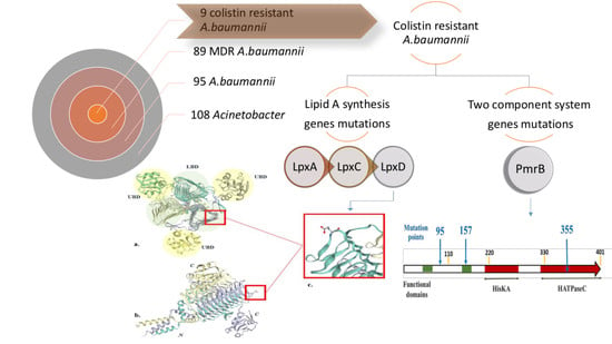
Graphical Abstract

1. Introduction

Acinetobacter baumannii is a highly versatile nosocomial Gram-negative pathogen that has become a critical challenge for common antibiotic treatments [1,2]. Moreover, since it causes catheter-correlated infections, urinary tract infections, pneumonia, wound infections, and bacteremia, its infection is associated with a great risk of hospital death [3].
Carbapenem-resistant A. baumannii (crab) was often recorded in hospitals, which forced physicians to reuse the old polymyxin antibiotic, colistin, regardless of its high systematic toxicity, owing to its favorable properties of rapid bacterial killing, a narrow spectrum of activity, and slow development of resistance [1,4]. Unfortunately, colistin resistance has been reported recently [1].
Colistin, a cationic peptide that disrupts Outer membrane(OM), was recognized as a last-resort treatment. Unfortunately, the abuse of colistin, as a consequence of prolonged exposure, is leading to colistin resistance, which is reported periodically [5,6,7].
A. baumannii showed the heteroresistance phenomenon to colistin, a natural evolution of drug resistance indicating growing colistin resistance in A. baumannii, which has therapeutics implications. Sub-populations showing a higher level of resistance to this antibiotic could grow at concentrations four-fold or higher than the MIC of the entire population [1,8,9,10].
Moffatt and co-workers attributed the colistin heteroresistance in A. baumannii to a loss of lipopolysaccharide production in subpopulations displaying high levels of colistin resistance that were grown by serial passages on colistin plates at increasing colistin concentrations [10]. Cross-resistance reports with different outcomes on the correlation between colistin resistance and resistance to other classes of antibiotics have been reported [11,12].
Colistin-resistant A. baumannii isolates usually employ several strategies for protection against colistin, among which the modification of their lipopolysaccharides (LPSs) mediated by the two-component system pmrA/pmrB, which have overall negative charges and are the initial targets of colistin [10]. LPSs modifications can involve the addition of phosphoethanolamine (PEtN) and 4-amino-4-deoxy-L-arabinose (L-Ara4N), which change the net negative charge of lipid A to a positive charge, lowering the affinity of LPS for colistin [13,14,15,16], or deacylation and hydroxylation of LPS, but these last mechanisms are less common in A. baumannii A. baumannii [4].
In this context, it is also known that the two-component response regulator and sensor kinase PmrA/B allows the bacteria to sense and respond to various environmental conditions, including pH and Fe3+ and Mg2+ levels, and also cause mutations in genes implicated in lipid A modification, thereby influencing the susceptibility to colistin [17,18]. Several studies reported that one or two amino acid substitutions, variously located in PmrB, lead to colistin resistance acquisition [18,19].
Similarly, the complete loss of the LPS due to impaired lipid A synthesis through mutations in the lpxA, lpxC, and lpxD genes involved in lipid A biosynthesis is another common colistin resistance mechanism. In addition, colistin resistance in A. baumannii causes alterations in cell topography, particularly a more spherical appearance, and increased cell surface roughness, leading to outer membrane damage [20,21].
Based on what has been mentioned, we explored the impact of gaining colistin resistance on the susceptibility of A. baumannii to other antibiotics and explored the genetic basis of resistance acquisition.
This study was presented in a poster in ASM microbe 2018, General meeting of American Society of Microbiology 1–22 June 2018 [22].

2. Results

2.1. Testing the Antimicrobial Susceptibility of Identified A. baumannii Isolates

A total of 110 Acinetobacter isolates recovered from different hospitals were identified phenotypically to a level of 98% (108/110). PCR analysis was carried out on all isolates, and a product of the correct size of the blaOXA-51 gene was detected in 86.36 % (95/110) of the strains, demonstrating that this gene is highly conserved in A. baumannii.
The highest resistance rates, within the 95 A. baumannii isolates, were recorded against cefepime 94% and ceftazidime 93%. (Table 1). It is worth mentioning that it is recommended to determine the susceptibility of colistin by measuring its minimum inhibitory concentration (MIC) using broth dilution method. By applying this method, the resistance of colistin regarded only 5% of the total isolates. The Clinical and Laboratory Standards Institute CLSI has selected a MIC ≤2 µg/mL as indicating susceptibility and a MIC of ≥4 µg/mL as indicating resistance to colistin. The MIC was determined for 11 A. baumannii isolates, as summarized in Supplementary Table S1. We compared the results of the broth microdilution method of the eleven strains with the corresponding results of the disc diffusion method and confirmed that the disc diffusion is not reliable to asses colistin resistance in isolates.
By investigating the level of potential cross-resistance among colistin-resistant isolates, we found cross-resistance to the majority of the tested antibiotics, except for tigecycline, which was active against five isolates (Table 1).

2.2. Colistin Resistance Induction

Three colistin-resistant variants, designated as MS48Col-R, MS50Col-R, and MS64Col-R, displayed an increased colistin MIC from 0.06, <0.06, and 0.125 to 14, 16, 32 mg/L, respectively, with a six- to nine-fold change (Supplementary Table S1). Depending on the MIC values, our induced isolates were considered heteroresistant variants. We have shown that these strains can adapt to colistin. For the strains tested, the concept of heteroresistance may be strictly applied because a resistant A. baumannii sub-population kept colistin resistance in antibiotic-free medium.
The resistance phenotype of the colistin-resistant mutant, derived from clinical strains exposed to colistin pressure, was converted into a stable mutant phenotype after several successive passages in colistin-free medium, indicating that this resistance was irreversible and rapidly gained.

2.3. Susceptibility Profile and Colistin Resistance Acquisition

Colistin resistance acquisition showed no consequences on the antibiotic dynamics of the multi-drug resistance (MDR) strain (MS48Col-R mutant), while a great change was found in the resistance profiles of the originally sensitive strains (MS50Col-R and MS64Col-R mutant) for all antibiotics, except for imipenem and ceftazidime that showed the same profile in the case of MS50Col-R (Table 2).

2.4. Colistin Resistance Acquisition and Cell Morphology

Cell morphology was investigated to examine possible alterations induced by colistin resistance acquisition in Col-R mutant strains in comparison with clinical Col-S and Col-R strains (Figure 1). TEM of sensitive strains (Col-S) showed an intact, uniform, and thick cell membrane structure, while Col-R mutant strains had a very thin cell membrane compared to the sensitive strains. This can be due to LPS alteration, accompanied by the loss of affinity towards colistin.

2.5. Mutation Analysis of PmrB and LpxACD Based on Nucleotide Sequences

We investigated the nucleotide sequences of pmrB, the sensor kinase of the two-component system, and lipid A biosynthesis genes (lpxA, IpxD, and IpxC). This was done to detect mutations that accounted for the changes in the susceptibility pattern and the cell membrane of our induced mutants by comparing the nucleotide sequences to those of the clinically isolated colistin-resistant (Col-R) and -susceptible (Col-S) strains.
Based on the sequencing of all genes that were investigated, all strains exhibited 100% DNA sequence similarity in the regions of the pmrB and lpx genes. The investigation of the gene sequence of lpxA and lpxD, which encode acyl-transferases involved in lipid A biosynthesis, as well as of lpxC, that is involved in lipid A biosynthesis, located in the chromosomal region of A. baumannii genome, showed a single replacement in LpxD in the MS64Col-R mutant as well as in the resistant clinical isolate (MS34Col-R) corresponding to K117E in the lipid-binding domain (LBD) compared to the susceptible parent strain. This point mutation caused a charge shift in the amino acid from the acidic state of the carboxyl residue to the basic hydrophobic state, leading to polymerization disruption. (Figure 2).
This was confirmed by the predicted 3D structure of the mutated lpxD of the MS64Col-R strain. The 3D structure revealed the impact of the mutation at position 117 in the structure of the lipid-binding domain region of the protein. Besides, resistant and susceptible strains shared five single nucleotide polymorphism (SNP) in the lpxD gene that were synonymous upon protein translation.
On the other hand, according to the coding sequences of the lpxA and lpxC genes, 14 polymorphic positions were noticed that were all synonymous at the protein level (not shown) in the laboratory-induced strain (MS64Col-R) and the resistant clinical strain (MS34Col-R).
Besides the lpx genes, in this report, no mutations were observed within the pmrB gene in MS64Col-R mutant. Meanwhile, non-synonymous mutations in PmrB were observed in MS34Col-R, corresponding to substitutions and frameshift (deletion) mutations.
Two amino acid substitutions, A95T and P157R, were detected in the transmembrane domains (TM). A95T was detected in TM1, while P157R was detected in TM2. Here, we suggest that modifications in LPS, not its complete loss, induces low colistin resistance in mutants and clinical A. baumannii isolates and can be linked to a single mutation in one (lpxD) of the three genes of the lipid A biosynthesis pathway (lpxA, lpxC, and lpxD). Regarding the frameshift mutation, a nonsense mutation was created at position 355 in the ATP-binding domain of the sensor kinase, which, as we suggested, led to premature termination of the amino acid sequence at a critical site, resulting in Δ355 protein (Table 3).

3. Discussion

A. baumannii is now regarded as one of the most difficult nosocomial-acquired pathogens to treat and control, characterized by a worryingly increasing rate of colistin resistance, observed worldwide [7,23,24]. In our study, we propose that the incidence of MDR A. baumannii is very high in Egypt. However, Abdulzahra and his colleagues demonstrated that only two A. baumannii isolates were colistin-resistant [24].
Carbepenems have a pivotal role in the treatment of Acinetobacter-related infections. There is a worldwide concern regarding the increasing carbapenem resistance in A. baumannii. This emerging pattern drastically limits the range of therapeutic alternatives. The most reported carbapenem resistance mechanisms involve the acquisition of carbapenem-hydrolyzing D beta-lactamases. These enzymes are referred to as OXA-23, OXA-24, and OXA-58. In addition to beta-lactamases, carbapenem resistance in A. baumannii may also result from penicillin-binding proteins or porin modifications. Several porins, including the 33 kDa CarO protein, might be involved in carbapenem resistance [25].
In our study, 70% of the isolates were resistant to imipenem, similar to what reported in a study conducted in 2014 [26]. Unfortunately, imipenem resistance in Egypt has recently increased to around 92% of the studied cases [27,28], indicating the decrease of carbepenem effectiveness against A. baumannii infections.
It is known that colistin has poor agar diffusion, which consequently limits the predictive accuracy of the disk diffusion test. Therefore, we applied the microdilution method to determine colistin sensitivity. In fact, the resistance pattern of A. baumannii strains differs depending on the assessment method, in this case, the agar and broth microdilution methods.
According to the definition of heteroresistance, our induced strains were considered as heteroresistant variants [8,9]. A similar phenotype has also been described in clinical isolates of Stenotrophomonas maltophilia [29] and carbapenem-resistant A. baumannii [30]. Curiously, this phenomenon has enhanced colistin resistance in A. baumannii [31], an issue that is now of great interest, since colistin is the drug of choice for A. baumannii therapy [32,33,34]
To answer the question of whether this phenotypic manifestation has a genetic basis, further investigation is required, including genomic sequencing. Choi and Ko stated that the two-component gene system PmrA/B is associated with the emergence of colistin heteroresistance in A.baumannii [35]. Moreover, although colistin resistance in A. baumannii is uncommon, it can be rapidly induced in the lab, as confirmed in our investigation. Choi and Ko proved that colistin resistance can be readily induced during drug therapy by a single-step mutation in A. baumannii, Pseudomonas aeruginosa, and Klebsiella pneumoniae [35].
Moreover, several researchers showed increased incidence of heteroresistant A. baumannii isolates, e.g., Ezadi et al., who characterized 20.45% of isolates as colistin-heteroresistant [32], Li et al., who found 93.8% resistance in 16 clinical isolates [1], Hawley et al., who showed 100% resistance in 19 isolates [33], and Charretier and co-workers, who described half of their A. baumannii isolates as heteroresistant [36]. This makes it necessary to evaluate the appearance of heteroresistance in A. baumannii during colistin treatment.
This investigation connects the acquisition of colistin resistance in clinical A. baumannii isolates to variations of the susceptibility to other antibiotics, which can be explained by multiresistance mechanisms active in this species [37], as reported by López-Rojas et al., who linked the acquisition of resistance to colistin in A. baumannii to insusceptibility to cefepime and sulbactam [21]. Moreover, Moffat et al. denoted an increase in antibiotic sensitivity as a consequence of colistin resistance acquisition [10].
On the other hand, a tendency to overweigh the development of cross-resistance to colistin and antibiotics of different classes has been observed in the MS50Col-R mutant, for which no significant antibiogram alterations occurred between the parent MDR colistin-susceptible strain and its mutated colistin-resistant one, as shown for imipenem and ceftazidime [1,19,38]. Cross-resistance can be explained by the fact that the outer membrane of colistin-resistant strains becomes more impermeable, which increases the resistance to cell wall-targeted antibiotics. Such antibiogram changes lead to a drastic decrease in therapeutic options.
Another study correlated the generation of cross-resistance to bacitracin to the extensive use of colistin in animal feed [11]. Napier and his colleagues showed the use of colistin was accompanied by cross-resistance to host antimicrobials LL-37 and lysozyme [7]. We suppose that resistance to other antibiotics in A. baumannii isolates upon induction of colistin resistance may be related to other antibiotic resistance mechanisms [38].
The variation between our induced colistin-resistant strains could be explained by the presence of LPS mutations. Colistin resistance acquisition resulted in the alteration of the antibiotic resistance profiles. This may be attributed to gene mutations responsible for the displayed resistance, leading to variations in the permeability of the outer membrane [39]. This was suggested by the changes observed in the cell morphology due to alteration of LPS, as confirmed by our investigation and other studies that linked the inactivation of LpxD to colistin resistance [40].,
Knowledge concerning colistin resistance mechanisms in A. baumannii isolates of clinical origin remains limited. Recent studies prompted by the discovery of increasing colistin resistance incidence in A. baumannii tried to link the observed phenotypes to mutated genes.
A point mutation (K117P) in the lpxD gene matched with mutations detected previously by Nurtop et al. [41], who showed a synergistic interaction between an IpxD mutation and mutations in pmrCAB, also proved in our clinical colistin-resistant MS34Col-R strain, although this IpxD mutation in the induced colistin-resistant A. baumannii strain MS64Col-R was the only mutation identified, nevertheless strongly associated with colistin resistance. This point mutation leads to a disruption of polymerization that affects the resulting enzyme binding affinity and consequently causes a modification in LPS [10,42]. Furthermore, a genetic disruption in one of the initiating LPS biosynthesis genes lpxA, lpxC, or lpxD leads to a complete loss of LPS as a result of the lack of lipid A production and, correspondingly, to colistin resistance by decreasing the binding potentials of colistin to the bacterial membrane [10,18]; however, this mechanism leading to colistin resistance is less common of the first one described.
The most common colistin resistance mechanism involves the two-component signal transduction system (PmrA/B) that regulates the modification of endogenous LPS and is also related to polymyxin resistance in A. baumannii isolates [19]. This is due to overexpression of or mutations in its domains, i.e., the sensor kinase (PmrB) and the response regulator (PmrA) [14,43].
Therefore, PmrB is an important mutational target in the evolution of colistin resistance and appears to be the most commonly mutated gene in A. baumannii. Owing to this fact, its gene sequence was investigated in the clinically resistant strains and mutant ones. Our investigation detected non-synonymous mutations in the transmembrane domains of the clinically resistant MS34Col-R strain. According to the current knowledge of the PmrB protein structure [44], position 95 is located into the amino-terminal protein portion including the cytoplasmic secretion signal (aa 1–110) of the TM1 transmembrane domain, which is a domain putatively involved in the delivery of the protein to the cell membrane [45].
In addition, a non-conservative mutation in the transmembrane TM2 domain (aa 111–219) at position P157R was also detected. To the best of our knowledge, these mutations have not been reported elsewhere. Other various transmembrane domain PmrB mutations have been reported in other genera, such as the L14P mutation in P. aeruginosa [44] and the L10P mutation of Escherichia coli [46]. These mutations are located in the same cytoplasmic secretion signal domain of PmrB. Beceiro et al. proposed that these mutation could activate PmrB and modulate the permeability of the outer membrane to colistin, leading to the evolution of colistin resistance [18].
Recently, among mutations in the pmrB gene previously associated with colistin resistance in A. baumannii [18,19], a nonsense mutation that was created at position 355 in the ATP-binding domain of the sensor kinase has been identified. Therefore, we suppose that this mutation could induce the constitutive expression of pmrA, leading to the upregulation of the pmrCAB operon and subsequent synthesis and addition of phosphor-ethanolamine to hepta-acylated lipid A [18].

4. Conclusions

The characterized mutations in PmrB and LpxD in our investigation may have a role in the modification of LPS structure, which affects colistin binding affinity to the cell membrane and decreases the susceptibility of A. baumannii to other unrelated drugs. This observed phenotype related to genetic mutations can be further promoted by site-directed mutation in the IpxD gene. Our study, worryingly, postulates that colistin, as a drug of last resort, may rapidly become ineffective against MDR A. baumannii, due to its extensive use in health care facilities. The rate of resistance is expected to increase, because colistin is used at sub-MIC concentrations with other drugs to decrease its toxicity and create synergistic effects. Moreover, the acquisition of colistin resistance may also be accompanied by resistance to many unrelated antimicrobial compounds. Therefore, knowledge of the mechanisms of colistin resistance in A. baumannii will be informative for the development of strategies to overcome resistance issues in the treatment of this rapidly emerging pathogen.

5. Materials and Methods

5.1. Bacterial Strains

A total of 110 Acinetobacter isolates (that were kindly donated by Dr. Nayra Mohamed, Professor of Microbiology and Immunology, Faculty of Pharmacy, Cairo University, Egypt) were recovered from the bloodstream, urinary tract, respiratory and sputum samples, as well as skin of patients admitted to different intensive care units. Identification to the genus level was done phenotypically on the basis of morphological and biochemical properties through the catalase test, oxidase test, and growth on MacConkey agar, Simmons’ citrate agar, blood agar media, and at 44 °C; it was then confirmed by blaOXA-51-like carbapenemase gene detection, which is intrinsic to this species [47], using the sequences of the primers in Table 4. Strains were stored in Brain Heart–Glycerol (Oxoid Ltd., Basingstoke, UK) at −80 °C until use. Quality control assessment was done using Staphylococcus aureus ATCC® 25923 E. coli ATCC® 2599, and A. baumannnii ATCC 19606.

5.2. Antimicrobial Susceptibility Testing

Antibiotic susceptibility testing was performed using the Bauer–Kirby disc diffusion method by applying 10 different types of commercial antibiotic discs (Oxoid™ United Kingdom and stored at −20: +8 °C). These antibiotics were as follows: imipenem (10 µg/disc), amikacin (30 µg/disc), ciprofloxacin (5 µg/disc), ampicillin/sulbactam (10/10 µg/disc), piperacillin/tazobactam (100/10 µg/disc), ceftazidime (30 µg/disc), cefepime (30 µg/disc), colistin sulphate (10 µg/disc), trimethoprim/sulfamethoxazole (1.25/23.75 µg/disc), and tigecycline (15 µg/disc). Breakpoint criteria were established according to the Clinical and Laboratory Standards Institute (CLSI) guidelines [48]. Tigecycline breakpoints were according to Enterobacteriaceae (≤2/≥8 µg/mL for susceptible/resistant) Food and Drug Administration FDA disk diffusion, at >19 mm and ≤14 mm. Colistin discs were used to screen the resistance of A. baumannii isolates that showed no inhibition zone. Multi-drug resistance (MDR) isolates were determined based on the resistance to three or more antibiotics classes. MICs of colistin sulphate (Sigma-Aldrich, Inc.) were determined for 11 A. baumannii strains (1.5 × 10⁵ CFU/mL) by the broth microdilution method in cation-adjusted Muller Hinton Broth (CA-MHB) (Oxoid, Columbia, MD, USA), according to Clinical and Laboratory Standards Institute methods [48].

5.3. Colistin Resistance Induction

Colistin resistance inductions were carried out on susceptible clinical strains, i.e., MS48Col-S (resistant to all tested antibiotics and sensitive to colistin), MS50Col-S, and MS64Col-S (susceptible to almost all tested antibiotics including colistin), through five daily successive passages in increasing colistin concentrations (double dilution), and was demonstrated by a higher level of resistance for each subculture in CA-MHB broth. Briefly, colonies (approximately 109 colony-forming units (CFU)) of each strain were suspended in 100 µL of sterile phosphate-buffered saline (PBS) (0.9%), and 3 μL of each bacterial suspension was inoculated on CA-MHA plates supplemented with increasing concentrations (1–1024 mgl−1) of colistin sulphate on consecutive days, until the highest MIC of colistin was reached; the plates were incubated overnight at 37 °C. Colonies (>20 colonies) were taken from the plate with the highest concentration of colistin sulphate from each culture, resuspended in 100 µL of 0.9% PBS, and used to inoculate plates containing colistin. This procedure was repeated for the duration of the experiment (5/6 days) until the defined endpoint was achieved. Daily passages in colistin-free CA-MHB continued for 16 days, for all isolates, to ensure the stability of the mutation. Colonies of each mutant strain were inoculated in Brain Heart–Glycerol with appropriate concentrations of colistin sulphate for storage at −80 °C until checking their identity by the presence of the blaOXA51 gene, using conventional PCR [49].
Antibiotic susceptibilities were retested for the strains subjected to colistin resistance acquisition and compared to those of their parent sensitive isolates subjected to the same sub-culturing conditions, except for the addition of colistin to their corresponding media.

5.4. Cell Morphological Examination Using Thin-Section Transmission Electron Microscopy (TEM)

In our study, we used TEM to compare morphological changes in the cell membranes of clinical colistin-sensitive (MS48Col-S, MS64Col-S), clinical colistin-resistant (MS34Col-R), and induced colistin-resistant (MS48Col-R, MS64Col-R) strains. The five A. baumannii strains were sub-cultured in Nutrient broth (NB) (LAB M, USA) for 24 h. Bacterial cells were centrifuged (4000 rpm/10 min), washed with distilled water, and immersed in a freshly prepared 1% (w/v) aqueous potassium permanganate solution for 5 min at room temperature for fixation. Dehydrated specimens were embedded in epoxy resin. Suspensions were submitted to the Regional Center for Mycology and Biotechnology (RCMB), Al-Azhar University, Cairo, Egypt, for agarose embedding and thin-section preparation. Sections were placed on carbon-coated grids and viewed in a JEM-1010, 120-kV transmission electron microscope (JEOL, Peabody, MA, USA) [50]

5.5. Nucleotide Sequencing of pmrB and lpx lipid A Biosynthesis Genes

The two-component system sensor kinase gene, pmrB, and lipid A biosynthesis genes, lpxA, lpxC, and lpxD was amplified from A. baumannii MS34Col-R, MS64Col-S, and MS64Col-R mutant genomic DNA by PCR, using primers (Table 4). The nucleotide sequences of the amplified fragments were determined using ABI 3730xl DNA sequencer (Applied Biosystems, Cologne, Germany). The obtained sequences were analyzed by www.bioinformatics.org and DNA Baser software as sequence assembler and finally confirmed by CLC workbench (www.qiagenbioinformatics.com). BLASTN program was used to search for homologous sequences (www.ncbi.nlm.nih.gov/BLAST/). The reference sequence of A. baumannii ATCC 19606 (GenBank accession number HM149345.1) was compared with the sequences of our strains. Protein translation and alignment were done using the Expasy portal at the Swiss Institute of Bioinformatics website (http://web/expasy.org/translate/) as well as the CLUSTALW2 multiple sequence alignment program (http://www.ebi.ac.uk/Tools/msa/clustalw2/) BLASTp and Clustal W for detection of amino acid alterations.

5.6. Nucleotide Sequence Accession Numbers

The nucleotide sequences presented in our study for A. baumannii strains MS64Col-S, MS64Col-R mutant, and MS34Col-R were submitted to the GenBank database, as follows: LpxA: MK024803, MK024804, and MK024805, LpxD: MK034358, MK086036, and MK086037, LpxC: MK034355, MK034356, and MK034357, PmrB1: MK108027, MK108028, and MK108029, PmrB2: MK166024, MK166025, and MK166026, respectively.

5.7. Protein 3D Model Prediction

Prediction of the 3D structure of the LxpD R mutant protein of MS64Col, resulting from induction of colistin resistance, was performed using SWISS-MODEL tool (https://swissmodel.expasy.org/).

Abbreviation

Col-RColistin-resistant
Col-SColistin-sensitive
TEMTransmission Electron Microscopy
CA-MHBCation adjustment Muller–Hinton Broth
CFUColony-Forming Unit

Supplementary Materials

The following are available online at https://www.mdpi.com/2079-6382/9/4/164/s1, Table S1: Comparison of colistin MIC determination using the disc diffusion and microdilution methods for selected clinical A. baumannii isolates.

Author Contributions

Conceptualization, M.A.A. and R.S.M.; Formal analysis, N.M.S., R.S.M. and M.S.H.; Methodology, N.M.S., R.S.M. and M.S.H.; Software: R.S.M. and M.S.H.; Data Curation, N.M.S., R.S.M. and M.A.A.; Funding acquisition, N.M.S. and R.S.M.; Investigation, N.M.S., R.S.M. and M.S.H.; Project administration, M.A.A.; Resources, N.M.S., R.S.M. and M.A.A.; Supervision, N.M.S., R.S.M. and M.A.A.; Validation, N.M.S., R.S.M. and M.A.A.; Writing—original draft, N.M.S.; Writing—review and editing, N.M.S., R.S.M. and M.A.A. All authors have read and agreed to the published version of the manuscript.

Funding

This research did not receive any specific grant from funding agencies in the public, commercial, or not-for-profit sectors.

Acknowledgments

The authors would like to thank Basma Hamdy Amin (Transmission Electron Microscopy Unit, Faculty of Regional Center for Mycology and Biotechnology (RCMB), Al-Azhar University, Cairo, Egypt) for kindly preparing samples and capturing Transmission Electron Microscope images.

Conflicts of Interest

The authors declare no conflict of interest.

References

  1. Li, J.; Rayner, C.R.; Nation, R.L.; Owen, R.J.; Spelman, D.; Tan, K.E.; Liolios, L. Heteroresistance to colistin in multidrug-resistant Acinetobacter baumannii. Antimicrob. Agents Chemother. 2006, 50, 2946–2950. [Google Scholar] [CrossRef] [PubMed] [Green Version]
  2. Antunes, L.; Imperi, F.; Carattoli, A.; Visca, P. Deciphering the multifactorial nature of Acinetobacter baumannii pathogenicity. PLoS ONE 2011, 6, e22674. [Google Scholar] [CrossRef] [PubMed] [Green Version]
  3. Wong, D.; Nielsen, T.; Bonomo, R.; Pantapalangkoor, P.; Luna, B.; Spellberg, B. Clinical and Pathophysiological Overview of Acinetobacter Infections: A Century of Challenges. Clin. Microbiol. Rev. 2017, 30, 409–447. [Google Scholar] [CrossRef] [PubMed] [Green Version]
  4. Nikaido, H. Molecular basis of bacterial outer membrane permeability revisited. Microbiol. Mol. Biol. Rev. MMBR 2003, 67, 593–656. [Google Scholar] [CrossRef] [PubMed] [Green Version]
  5. Dahdouh, E.; Gómez-Gil, R.; Sanz, S.; González-Zorn, B.; Daoud, Z.; Mingorance, J.; Suárez, M. A novel mutation in pmrB mediates colistin resistance during therapy of Acinetobacter baumannii. Int. J. Antimicrob. Agents 2017, 49, 727–733. [Google Scholar] [CrossRef] [PubMed]
  6. Pournaras, S.; Poulou, A.; Dafopoulou, K.; Chabane, Y.N.; Kristo, I.; Makris, D.; Hardouin, J.; Cosette, P.; Tsakris, A.; Dé, E. Growth retardation, reduced invasiveness, and impaired colistin-mediated cell death associated with colistin resistance development in Acinetobacter baumannii. Antimicrob. Agents Chemother. 2014, 58, 828–832. [Google Scholar] [CrossRef] [PubMed] [Green Version]
  7. Napier, B.A.; Burd, E.M.; Satola, S.W.; Cagle, S.M.; Ray, S.M.; Mcgann, P.; Pohl, J.; Lesho, E.P.; Weiss, D.S. Clinical use of colistin induces cross-resistance to host antimicrobials in Acinetobacter baumannii. MBio 2013, 4, 2–6. [Google Scholar] [CrossRef] [Green Version]
  8. El-Halfawy, O.M.; Valvano, M.A. Antimicrobial heteroresistance: An Emerging Field in Need of Clarity. Clin. Microbiol. Rev. 2015, 28, 191–207. [Google Scholar] [CrossRef] [Green Version]
  9. El-Halfawy, O.; Valvano, M. Chemical Communication of Antibiotic Resistance by a highly Resistant subpopulation of bacterial cells. PLoS ONE 2013, 8, e68874. [Google Scholar] [CrossRef] [Green Version]
  10. Moffatt, J.; Harper, M.; Harrison, P.; Hale, J.D.F.; Vinogradov, E.; Seemann, T.; Henry, R.; Crane, B.; St. Michael, F.; Cox, A.D.; et al. Colistin resistance in Acinetobacter baumannii is mediated by complete loss of lipopolysaccharide production. Antimicrob. Agents Chemother. 2010, 54, 4971–4977. [Google Scholar] [CrossRef] [Green Version]
  11. Xu, F.; Zeng, X.; Hinenoya, A.; Lina, J. MCR-1 Confers Cross-Resistance to Bacitracin, a Widely Used In-Feed Antibiotic. mSphere 2018, 3, e00411–e00418. [Google Scholar] [CrossRef] [PubMed] [Green Version]
  12. Adam, M.; Murali, B.; Glenn, N.O.; Potter, S. Epigenetic Inheritance Based Evolution of Antibiotic Resistance in Bacteria. BMC Evol. Biol. 2008, 8, 52. [Google Scholar]
  13. Lesho, E.; Yoon, E.J.; Mcgann, P.; Snesrud, E.; Kwak, Y.; Milillo, M.; Onmus-Leone, F.; Preston, L.; St. Clair, K.; Nikolich, M.; et al. Emergence of colistin-resistance in extremely drug-resistant acinetobacter baumannii containing a novel pmrCAB operon during colistin therapy of wound infections. J. Infect. Dis. 2013, 208, 1142–1151. [Google Scholar] [CrossRef] [PubMed] [Green Version]
  14. Arroyo, L.A.; Herrera, C.M.; Fernandez, L.; Hankins, J.V.; Trent, M.S.; Hancock, R.E.W. The pmrCAB Operon Mediates Polymyxin Resistance in Acinetobacter baumannii ATCC 17978 and Clinical Isolates through Phosphoethanolamine Modification of Lipid A. Antimicrob. Agents Chemother. 2011, 55, 3743–3751. [Google Scholar] [CrossRef] [PubMed] [Green Version]
  15. Trent, M.; Ribeiro, A.; Lin, S.; Cotter, R.; Raetz, C. An inner membrane enzyme in Salmonella and Escherichia coli that transfers 4-amino-4-deoxy-L-arabinose to lipid A: Induction on polymyxin-resistant mutants and role of a novel lipid-linked donor. J. Biol. Chem. 2001, 276, 43122–43131. [Google Scholar] [CrossRef] [Green Version]
  16. Lee, H.; Hsu, F.F.; Turk, J.; Groisman, E.A. The PmrA-regulated pmrC gene mediates phosphoethanolamine modification of lipid A and polymyxin resistance in Salmonella enterica. J. Bacteriol. 2004, 186, 4124–4133. [Google Scholar] [CrossRef] [Green Version]
  17. Gunn, J.S. The Salmonella PmrAB regulon: Lipopolysaccharide modifications, antimicrobial peptide resistance and more. Trends Microbiol. 2008, 16, 284–290. [Google Scholar] [CrossRef]
  18. Beceiro, A.; Llobet, E.; Aranda, J.; Bengoechea, J.A.; Doumith, M.; Hornsey, M.; Dhanji, H.; Chart, H.; Bou, G.; Livermore, D.M.; et al. Phosphoethanolamine Modification of Lipid A in Colistin-Resistant Variants of Acinetobacter baumannii Mediated by the pmrAB Two-Component Regulatory System. Antimicrob. Agents Chemother. 2011, 55, 3370–3379. [Google Scholar] [CrossRef] [Green Version]
  19. Adams, M.D.; Nickel, G.C.; Bajaksouzian, S.; Lavender, H.; Murthy, A.R.; Jacobs, M.R.; Bonomo, R.A. Resistance to colistin in Acinetobacter baumannii associated with mutations in the PmrAB two-component system. Antimicrob. Agents Chemother. 2009, 53, 3628–3634. [Google Scholar] [CrossRef] [Green Version]
  20. Soon, R.; Nation, R.; Hartley, P.; Larson, I.; Li, J. Atomic force microscopy investigation of the morphology and topography of colistin-heteroresistant Acinetobacter baumannii strains as a function of growth phase and in response to colistin treatment. Antimicrob. Agents Chemother. 2009, 53, 4979–4986. [Google Scholar] [CrossRef] [Green Version]
  21. Lo’pez-Rojas, R.; Domínguez-Herrera, J.; McConnell, M.; Docobo-Peréz, F.; Smani, Y.; Fernández-Reyes, M.; Rivas, L.; Pachón, J. Impaired Virulence and in Vivo Fitness of Colistin Resistant Acinetobacter baumannii. J. Infect. Dis. 2011, 203, 545–548. [Google Scholar] [CrossRef] [PubMed] [Green Version]
  22. Hesham, M.; Saleh, N.M.; Ahmed, R.S.; Amin, M.A. Colistin Resistance Induction Effect on Multi-Drug Resistant Acinetobacter Baeumannii. In Proceedings of the Poster AAR LB11 abstr Antimicrobial Resistance American Society of Microbiology, Atlanta, Gorgia, 15 February 2018. [Google Scholar]
  23. Hua, X.; Liu, L.; Fang, Y.; Shi, Q.; Li, X.; Chen, Q.; Shi, K.; Jiang, Y.; Zhou, H.; Yu, Y. Colistin Resistance in Acinetobacter baumannii MDR-ZJ06 Revealed by a Multiomics Approach. Front. Cell. Infect. Microbiol. 2017, 7, 45. [Google Scholar] [CrossRef] [PubMed] [Green Version]
  24. Abdulzahra, A.T.; Khalil, M.A.F.; Elkhatib, W.F. First report of colistin resistance among carbapenem-resistant Acinetobacter baumannii isolates recovered from hospitalized patients in. New Microbes New Infect. 2018, 26, 53–58. [Google Scholar] [CrossRef] [PubMed]
  25. Poirel, L.; Nordmann, P. Carbapenem resistance in Acinetobacter baumannii: Mechanisms and epidemiology. Clin. Microbiol. Infect. 2006, 15, 826–836. [Google Scholar] [CrossRef] [Green Version]
  26. Al-Agamy, M.H.; Khalaf, N.G.; Tawfick, M.M.; Shibl, A.M.; El Kholy, A. Molecular characterization of carbapenem-insensitive Acinetobacter baumannii in Egypt. Int. J. Infect. Dis. 2014, 22, 49–54. [Google Scholar] [CrossRef] [Green Version]
  27. Al-Hassan, L.; Zafer, M.M.; El-Mahallawy, H. Multiple sequence types responsible for healthcare-associated Acinetobacter baumannii dissemination in a single centre in Egypt. BMC Infect. Dis. 2019, 19, 829. [Google Scholar] [CrossRef] [Green Version]
  28. Benmahmod, A.B.; Said, H.S.; Ibrahim, R.H. Prevalence and Mechanisms of Carbapenem Resistance Among Acinetobacter baumannii Clinical Isolates in Egypt. Microb. Drug Resist. 2019, 25, 480–488. [Google Scholar] [CrossRef]
  29. Martínez-Servat, S.; Yero, D.; Huedo, P.; Marquez, R.; Molina, G.; Daura, X.; Gibert, I. Heterogeneous colistin-resistance phenotypes coexisting in stenotrophomonas maltophiliaisolates influence colistin susceptibility testing. Front. Microbiol. 2018, 9, 2971. [Google Scholar] [CrossRef]
  30. Pournaras, S.; Ikonomidis, A.; Markogiannakis, A.; Maniatis, A.N.; Tsakris, A. Heteroresistance to carbapenems in Acinetobacter baumannii. J. Antimicrob. Chemother. 2005, 55, 1055–1056. [Google Scholar] [CrossRef] [Green Version]
  31. Tan, C.H.; Li, J.; Nation, R.L. Activity of colistin against heteroresistant Acinetobacter baumannii and emergence of resistance in an in vitro pharmacokinetic/pharmacodynamic model. Antimicrob. Agents Chemother. 2007, 51, 3413–3415. [Google Scholar] [CrossRef] [Green Version]
  32. Ezadi, F.; Jamali, A.; Heidari, A.; Javid, N.; Ardebili, A. Heteroresistance to colistin in oxacillinases-producing carbapenem-resistant Acinetobacter baumannii clinical isolates from Gorgan, Northern Iran. J. Glob. Antimicrob. Resist. 2019. [Google Scholar] [CrossRef] [PubMed]
  33. Hawley, J.S.; Murray, C.K.; Jorgensen, J.H. Colistin heteroresistance in Acinetobacter and its association with previous colistin therapy. Antimicrob. Agents Chemother. 2008, 52, 351–352. [Google Scholar] [CrossRef] [PubMed] [Green Version]
  34. Moosavian, M.; Shoja, S.; Nashibi, R.; Ebrahimi, N.; Tabatabaiefar, M.A.; Rostami, S.; Peymani, A. Post neurosurgical meningitis due to colistin heteroresistant Acinetobacter baumannii. Jundishapur J. Microbiol. 2014, 7, 1–5. [Google Scholar] [CrossRef] [Green Version]
  35. Choi, M.J.; Ko, K.S. Mutant prevention concentrations of colistin for Acinetobacter baumannii, pseudomonas aeruginosa and Klebsiella pneumoniae clinical isolates. J. Antimicrob. Chemother. 2014, 69, 275–277. [Google Scholar] [CrossRef] [PubMed]
  36. Charretier, Y.; Diene, S.M.; Baud, D.; Chatellier, S.; Santiago-Allexant, E.; Van Belkum, A.; Guigon, G.; Schrenzel, J. Colistin heteroresistance and involvement of the PmrAB regulatory system in acinetobacter baumannii. Antimicrob. Agents Chemother. 2018, 62. [Google Scholar] [CrossRef] [PubMed] [Green Version]
  37. Pachón, J.; Vila, J. Treatment of multiresistant Acinetobacter baumannii infections. Curr. Opin. Investig. Drugs 2009, 10, 150–156. [Google Scholar]
  38. Li, J.; Nation, R.L.; Owen, R.J.; Wong, S.; Spelman, D.; Franklin, C. Antibiograms of Multidrug-Resistant Clinical Acinetobacter baumannii: Promising Therapeutic Options for Treatment of Infection with Colistin-Resistant Strains. Clin. Infect. Dis. 2007, 45, 594–598. [Google Scholar] [CrossRef]
  39. Mu, X.; Wang, N.; Li, X.; Shi, K.; Zhou, Z.; Yu, Y.; Hua, X. The effect of colistin resistance-associated mutations on the fitness of Acinetobacter baumannii. Front. Microbiol. 2016, 7, 1–8. [Google Scholar] [CrossRef] [Green Version]
  40. Bojkovic, J.; Richie, D.L.; Six, D.A.; Rath, C.M.; Sawyer, W.S.; Hu, Q.; Dean, C.R. Characterization of an Acinetobacter baumannii lptD Deletion Strain: Permeability Defects and Response to Inhibition of Lipopolysaccharide and Fatty Acid Biosynthesis. J. Bacteriol. 2016, 198, 731–741. [Google Scholar] [CrossRef] [Green Version]
  41. Nurtop, E.; Bayındır, B.F.; Menekse, S.; Kurt, A.O.; Gönen, M.; Ergonul, O.; Can, F. Promoters of Colistin Resistance in Acinetobacter baumannii Infections. Microb. Drug Resist. 2019, 25, 997–1002. [Google Scholar] [CrossRef]
  42. Badger, J.; Chie-Leon, B.; Logan, C.; Sridhar, V.; Sankaran, B.; Zwart, P.; Nienabera, V. Structure determination of LpxD from the lipopolysaccharide-synthesis pathway of Acinetobacter baumannii. Acta Crystallogr. Sect. F Struct. Biol. Cryst. Commun. 2013, 69, 6–9. [Google Scholar] [CrossRef] [PubMed] [Green Version]
  43. Cheah, S.E.; Johnson, M.D.; Zhu, Y.; Tsuji, B.T.; Forrest, A.; Bulitta, J.B.; Boyce, J.D.; Nation, R.L.; Li, J. Polymyxin Resistance in Acinetobacter baumannii: Genetic Mutations and Transcriptomic Changes in Response to Clinically Relevant Dosage Regimens. Sci. Rep. 2016, 6, 26233. [Google Scholar] [CrossRef] [PubMed] [Green Version]
  44. Moskowitz, S.; Brannon, M.; Dasgupta, N.; Pier, M.; Sgambati, N.; Miller, A.; Denton, M.; Conway, P.; Johansen, H.; Høiby, N. PmrB mutations promote polymyxin resistance of Pseudomonas aeruginosa isolated from colistin-treated cystic fibrosis patients. Antimicrob. Agents Chemother. 2012, 56, 1019–1030. [Google Scholar] [CrossRef] [Green Version]
  45. Filloux, A. Secretion signal and protein targeting in bacteria: A biological puzzle. J. Bacteriol. 2010, 192, 3847–3849. [Google Scholar] [CrossRef] [PubMed] [Green Version]
  46. Cannatelli, A.; Giani, T.; Aiezza, N.; Pilato, V.D.; Principe, L.; Luzzaro, F.; Galeotti, C.L.; Rossolini, G.M. An allelic variant of the PmrB sensor kinase responsible for colistin resistance in an Escherichia coli strain of clinical origin. Sci. Rep. 2017, 7, 5071. [Google Scholar] [CrossRef]
  47. Woodford, N.; Ellington, M.J.; Coelho, J.M.; Turton, J.F.; Ward, M.E.; Brown, S.; Amyes, S.G.B.; Livermore, D.M. Multiplex PCR for genes encoding prevalent OXA carbapenemases in Acinetobacter spp. Int. J. Antimicrob. Agents 2006, 27, 351–353. [Google Scholar] [CrossRef]
  48. CLSI Clinical and Laboratory Standards Institute. Performance Standards for Antimicrobial Susceptibility Testing; Twenty-Fourth Informational Supplement M100-S24; CLSI Clinical and Laboratory Standards Institute: Wayne, PA, USA, 2014. [Google Scholar]
  49. Thi Khanh Nhu, N.; Riordan, D.W.; Do Hoang Nhu, T.; Thanh, D.P.; Thwaites, G.; Huong Lan, N.P.; Wren, B.W.; Baker, S.; Stabler, R.A. The induction and identification of novel Colistin resistance mutations in Acinetobacter baumannii and their implications. Sci. Rep. 2016, 6, 28291. [Google Scholar] [CrossRef]
  50. Reynolds, E. The use of lead citrate at high pH as an electron-opaque stain in electron microscopy. J. Cell Biol. 1963, 14, 208–212. [Google Scholar] [CrossRef] [Green Version]
Figure 1. Transmission electron micrographs of thin sections of colistin-sensitive and -resistant A. baumannii strains. (A) Colistin-sensitive strain MS48, displaying a thick cell membrane. (B) MS48 after the acquisition of colistin resistance, displaying a thin cell membrane. (C) Clinical colistin-resistant strain MS34, displaying a thin cell membrane and a glycocalyx. (D) Clinical colistin-sensitive strain MS64, displaying a thick cell membrane. (E) MS64 after the acquisition of colistin resistance, displaying a thin cell membrane.
Antibiotics 09 00164 g001
Figure 2. Predicted 3D model structure of MS64Col-R LpxD protein. (A) Orthogonal view of the trimer, showing three chains where the lipid-binding domain (LBD) is colored in grey shades. (B) Side view of the trimer. (C) Amino acid substitution position (117). (D) Amino acid sequence alignments of a part of the LpxD protein of the colistin-sensitive reference strain (ATCC19606), colistin-sensitive clinical isolate (64colR), colistin-resistant laboratory-induced isolate (64colR), and colistin-resistant clinical isolate (34colR), generated by online Clustal W–Model server. UBD: uridine-binding domain.
Antibiotics 09 00164 g002
Table 1. Sensitivity index of Acinetobacter baumannii clinical isolates and co-resistance to antibiotics and colistin.
Antibiotic NameResistance Level in Clinical A. baumannii IsolatesLevel of Co-Resistance to Antibiotic and Colistin *
RISNo.%
No.%No.%No.%
Ampicillin/sulbactam859033779100
Piperacillin/tazobactam6670232466888.8
3rd generationCeftazidime889377009100
4th generationCefepime899455119100
Amikacin6872192088777.7
Ciprofloxacin7882121355888.8
Imipenem6670222728888.8
Tigecycline212235373941444.4
Trimethoprim/
Sulfamethoxazole
818566899100
* Level of co-resistance was with respect to nine colistin-resistant A. baumannii isolates that showed no inhibition zone around the disc.
Table 2. Antibiotic susceptibility redetermination after colistin resistance induction in comparison to susceptibility before induction.
Clinical
A. baumannii Isolates
Antibiotics
Resistance Pattern
MS48DMS50MS64MS48DMS050MS64
Col-S
Before Induction
Col-R
After Induction
Colistin MIC (μg/mL)0.125<0.060.06141632
CTSSSRRR
TZPRSSRRR
FEPRISRRR
CIPRSSRRR
IMPSSSSSI
TGCISSIII
CAZRRSRRR
SAMRSSRRR
SXTRSSRRR
AKRSSRRR
CT: colistin, TZP: Piperacillin/tazobactam, FEP: Cefepime, CIP: Ciprofloxacin, IMP: Imipenem, TGC: Tigecycline, CAZ: Ceftazidime, SAM: Ampicillin/sulbactam, AK: Amikacin, SXT: Trimethoprim/Sulfamethoxazole.
Table 3. Amino acid changes or mutations in pmrB and lpxD in colistin-resistant clinical and laboratory-induced strains (compared to their parental wild-type strain ATCC 19606).
StrainsColistin (Col) a MIC (g/mL)Amino Acid Change(s) in b:
pmrB (444 aa)
lpxD (aa356)TM1
(aa 10–29)
TM2
(142–164)
HisKA
(aa 218–280)
HATPaseC
(aa 326–437)
MS64Col-S32
MS64Col-R14K117E
MS34Col-R16K117EA95TP157R 355 frameshift
a Colistin (Col) MICs were determined by the broth microdilution method according to the CLSI. b The predicted domains according to the NCBI domain predictor (www.ncbi.nlm.nhi.gov/protein) are indicated as follows: TM1, TM2, first and second transmembrane domains, HisK, histidine kinase (dimerization/phosphoacceptor) domain, and HATPaseC, histidine-kinase-like ATPase. Only domains or regions displaying mutations or variants are shown. The amino acid (aa) positions corresponding to these domains are displayed in brackets.
Table 4. Primers used in this study.
Gene NameUseSequence (5′-3′)bpReference
blaOXA-51-likeA. baumannii IdentificationF5′TAATGCTTTGAT CGGCCTTG3′353[47]
R5′TGGATTGCACTTCATCTTGG3′
lpxALipid A synthesisF5′TGAAGCATTA GCTCAAGTTT3′1181[10]
R5′GTCAGCAAATCAATACAAGA3′
lpxDF5′CAAAGTATGAATACAACTTTTGAG3′1164
R5′GTCAATGGCACATCTGCTAAT3′
lpxCF5′TGAAGATGACGTTCCTGCAA3′1502
R5′TGGTGAAAATCAGGCAATGA3′
pmrB1Two-Component SystemF5′GTGCATTATTCATTAAAAAAAC 3′1335[18]
R5′TCACGCTCTTGTTTCATGTA 3′
pmrB2F5′GGTTCGTGAAGCTTTCG 3′599
R5′CCTAAATCGATTTCTTTTTG 3′