Article

One-Step Surface Functionalized Hydrophilic Polypropylene Meshes for Hernia Repair Using Bio-Inspired Polydopamine

1
Key Laboratory of Textile Science and Technology of Ministry of Education, College of Textiles, Donghua University, 2999 North Renmin Road, Songjiang, Shanghai 201620, China
2
Department of Textile Engineering, Mehran University of Engineering and Technology, Jamshoro, Sindh 76062, Pakistan
3
College of Textiles and Fashion, Xingjiang University, 666 Sheng Li Road, Tian Shan, Wulumuqi 830046, China
4
Mechanical Textile Engineering, Aleppo University, Aleppo 15310, Syria
*
Author to whom correspondence should be addressed.
Fibers 2019, 7(1), 6; https://doi.org/10.3390/fib7010006
Submission received: 27 November 2018 / Revised: 19 December 2018 / Accepted: 8 January 2019 / Published: 14 January 2019
(This article belongs to the Special Issue Smart Coatings on Fibers and Textiles)

Abstract

:
An ideal hernia mesh is one that absorbs drugs and withstands muscle forces after mesh implantation. Polypropylene (PP) mesh devices have been accepted as a standard material to repair abdominal hernia, but the hydrophobicity of PP fibers makes them unsuitable to carry drugs during the pre-implantation of PP meshes. In this study, for the first time, one-step functionalization of PP mesh surfaces was performed to incorporate bio-inspired polydopamine (PDA) onto PP surfaces. All PP mesh samples were dipped in the same concentration of dopamine solution. The surface functionalization of PP meshes was performed for 24 h at 37 °C and 80 rpm. It was proved by scanning electron microscopic (SEM) images and Fourier Transform Infrared Spectroscopy (FTIR) results that a thin layer of PDA was connected with PP surfaces. Moreover, water contact angle results proved that surface functionalized PP meshes were highly hydrophilic (73.1°) in comparison to untreated PP mesh surfaces (138.5°). Thus, hydrophilic PP meshes with bio-inspired poly-dopamine functionalization could be a good choice for hernia mesh implantation.

Graphical Abstract

1. Introduction

A hernia is a defect in the abdominal wall due to the “protrusion” of an organ [1,2]. However, repair of the hernia is a common practice to reinforce the operated muscles of the hernia to their original place with biomaterial [3]. For this reason, numerous bio-materials have been used to repair hernia [4], for example, gold sutures, silver wires, and nylon prosthetics, but the recurrence of hernia has been a major problem due to the improper selection of material and its design [5]. However, the suture repair of hernia was associated with a higher chance of hernia recurrence compared to mesh implantation [6,7]. Nevertheless, patient pain and surgical site infection is still a major problem of hernia repair [8,9,10]. Recently, a few synthetic mesh materials, such as polytetrafluoroethylene (PTFE), polyethylene Terephthalate (PET), and polypropylene (PP), have been successfully used to reduce the hernia recurrence rate [11,12,13] and among them, light weight PP mesh material has been considered an effective material to reduce the hernia recurrence rate [14,15,16].
However, PP mesh material is hydrophobic in nature and does not absorb drugs during mesh implantation. In the literature, it is reported that standard PP mesh is not suitable for hernia repair due to the reason that pre-operative prophylaxis during mesh implantation has failed [17,18,19]. Therefore, there is an urgent need to surface functionalize and improve the surface wettability of PP meshes.
Thus, an ideal mesh may be one that is flexible and easy to place during a hernia operation, does not show a prolonged inflammatory response after mesh implantation, does not degrade easily, does not shrink after implantation, is free of adhesion formation or fistulas, and is resistant to infection [20,21,22,23,24,25]. Faulk et al. coated PP meshes with non-biodegradable hydrogel and reported that coated PP meshes did not demonstrate any sign of infection, foreign body response, and fibrosis [26]. Additionally, Perez-Kohler et al. reported that PP meshes coated with quaternary ammonium compounds successfully resisted mesh infection [27]. Moreover, Bellon et al. stated that partially absorbable PP mesh devices are superior to standard PP meshes in terms of more compliance, being less rigid, being strong, good tissue ingrowth, and being resistant to the infection [28].
The prime objective of the preparation of medical devices is ensuring their biocompatibility [29,30]. Therefore, bio-inspired materials are given more attention, such as dopamine with self-polymerization properties and generating poly-dopamine onto surfaces of any structure and material [31,32].
Dopamine surface functionalization of material is an easy method at around room temperature and pH 8.5. Thus, the surface functionalization process with dopamine may be controlled with the dopamine concentration or process reaction time [33,34]. Therefore, considering the surface properties of chemically inert PP fibers and self-polymerization properties of bio-inspired poly-dopamine, it was our objective to surface functionalize the PP surfaces using poly-dopamine (PDA) without changing the bulk properties of PP mesh fibers.
In this work, PP mesh materials were surface functionalized with PDA at room temperature. PDA was incorporated for 12 and 24 h. The PDA functionalized PP meshes were analyzed using a scanning electron microscope (SEM), Energy Dispersive X-ray Spectroscopy (EDX), Fourier Transform Infrared Spectroscopy (FTIR), Contact angle (sessile drop method), Differential Scanning Calorimeter (DSC), and X-ray diffractometer (XRD). The results confirmed the functionalization of PP meshes with a thin layer of poly-dopamine (PDA). Moreover, the surface wettability of PDA treated PP meshes was dramatically increased in comparison to un-treated PP meshes.

2. Materials and Methods

2.1. Materials

Polypropylene meshes of a light weight (27 g/m2) were used for surface functionalization with polydopamine (PDA). These medical textile devices were obtained from Nantong Chemical fiber Co. Ltd. China (Nantong, China). Dopamine hydrochloride and tris-(hydroxylmethyl aminomethane) were received from Aladdin Chemicals Ltd Shanghai China.

2.2. Surface Functionalization of PP Meshes Materials with Polydopamine

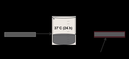
The dopamine solution (10 mM) was prepared using 0.1 g of dopamine in 50 mL of tris- and maintained at pH 8.5, as described in a recently published paper [33]. During preparation, the liquor ratio of PP mesh materials to dopamine solution was 1:100. PP mesh devices were soaked in a prepared solution of dopamine. The solution was stirred at 80 rpm in a controlled environment at 37 °C for 12–24 h. After the required duration time (12–24 h), PP mesh devices were taken out and hot rinsed (50 °C) with distilled water several times and dried in an oven at 40 °C. All PP mesh devices were functionalized with the same concentration of solution, but different time durations (12 and 24 h).

3. Characterization

3.1. SEM & EDX

PP control and surface functionalized PP meshes were coated using platinum (pt). Platinum coated PP meshes were scanned for surface morphology, using a scanning electron microscope (SEM, Quanta SEM 250, and FEITM). Moreover, for element analysis, Energy Dispersive (ISIS 300, Oxfordshire, UK) X-ray spectroscopy was used, while an EDX component was attached with SEM.

3.2. FTIR

PP control and dopamine surface functionalized PP meshes were analyzed using Fourier Transform Infrared Spectroscopy (ATR) by Nicolet 6700, Waltham, MA, USA. The samples were scanned in the range of 500–4000 wavenumber cm−1.

3.3. XRD and DSC Analysis

XRD was used to scan PDA functionalized and untreated samples in the range of 2θ (5°–60°). Thus, an X-ray diffractometer made in Tokyo Japan (Rigaku D/MAX 2550/PC) was used at a scanning rate of 0.02°/min. Moreover, a differential scanning calorimeter (Pyris, Perkin Elemer 4000, Grove, IL, USA) was used in the heating range of 25–250 °C to obtain the melting temperature of untreated and PDA functionalized PP meshes.

3.4. Water Contact Angle

PP meshes were of a 0.1 mm fiber diameter and it was difficult to obtain the contact angle of treated and untreated fibers. Therefore, nonwoven fabrics (melt blown, 23 g/m2) were used to measure the contact angle of PDA treated and untreated fabrics. A dynamic contact angle method using a sessile drop was used to measure the contact angle. Moreover, WCA 20 (software) was employed to calculate the hydrophilic or hydrophobic water contact angle of PDA functionalized and untreated PP fabrics. Each time, three drops (5 µL) were dispensed onto fabric and an average value was calculated.

3.5. Statistical Analysis

The standard deviation and error bars are presented in the figures with symbols (* and —). One way analysis of variance (ANOVA) was employed to analyze the data. The data with *** is less than 0.001 and mentioned as p < 0.001, while the data with (**) represents p < 0.01 and the data displaying (*) means p < 0.05. Thus, the value of p (*) < 0.05 was selected as the confidence interval value.

4. Results and Discussion

4.1. Surface Functionalization of PP Mesh Fibers

PP meshes were surface functionalized with bio-inspired polydopamine (PDA). Thus, PP meshes were soaked in a weak alkaline solution of dopamine and continuously stirred for 12–24 h at 37 °C. However, PDA was expected to coat surfaces of PP mesh by self-polymerization. The process of soaking and PDA structure are shown in Figure 1. According to the literature, PDA can form a thin layer on the surface of any fiber [35]. Thus, we received similar results and PP mesh fibers were successfully surface functionalized with PDA.
Moreover, PP meshes were soaked in the same concentration of dopamine solution, but for different durations of time. We observed a marginal difference in the surface morphology and coating efficiency between the 12 h soaking and 24 h soaking time. The PP meshes soaked for a 24 h dipping time exhibited a rougher surface with more PDA compared to that of PP meshes dipped for 12 h. However, there was not much difference in the yield. During the 12 h dipping duration, 0.2% of an average weight was increased, while during the 24 h dipping time, 0.35% corresponding weight was increased. It was really hard to record an accurate average weight increase; therefore, we considered standard testing atmosphere conditions (37 °C and 65% humidity) and weighed the samples before and after the surface functionalization of PP mesh fibers.

4.2. Surface Morphology of PP Meshes Fibers Before and After Surface Functionalization

Figure 2 displays SEM images of PDA surface functionalized and untreated PP meshes. It can be seen that the PP control (Figure 2a,b) shows smooth surfaces before functionalization, but after PDA treatment for 12 h, a thin layer of PDA coated the surfaces of PP fibers (Figure 2b,c). PDA treated PP mesh surfaces displayed a dark grey color with a shiny surface, rather than a dull grey color. Moreover, small spheres on the surfaces of treated fibers can be observed. PP mesh fibers treated for 24 h (Figure 2e,f) show gamut and thin layer of PDA coating, but have rougher surfaces and white patches along the whole sphere of fibers. In the past, carbon fibers have been successfully coated with PDA [33]. Herein, we also successfully obtained surface changes of PP meshes with a thin layer of PDA coating. Thus, it is proved that bio-inspired PDA can coat PP fibers at room temperature.

4.3. Surface Characterization of Polydopamine Functionalized PP Meshes

Figure 3 shows PP control and PDA functionalized PP meshes.
It can be observed that untreated PP meshes (Figure 3a) displayed a complete peak height and 100% weight of carbon (C) atom within 0.3 keV. Nevertheless, in Figure 3b, PDA functionalized PP meshes demonstrate an additional peak of the oxygen (O) atom within 0.4 keV, and show a 9.95% oxygen atomic weight increase. However, the carbon (C) atom peak is at a similar position, but the weight % of carbon atom is reduced to 90.05%. Thus, it is proved that PP meshes were successfully functionalized with PDA.
The FTIR (ATR) spectra of polydopamine (PDA) functionalized and untreated PP meshes fibers are shown in Figure 4. The untreated PP meshes demonstrate peaks at 2951 cm−1, 2917 cm−1, 1451 cm−1, and 1377 cm−1 [36,37]. Nevertheless, PDA functionalized PP meshes for 12 h exhibit an additional peak of hydroxyl (OH) at 3220 cm−1. Moreover, identical vibration peak bands can be observed at 1624 cm−1 (amide I) and 1535 cm−1 (amide II). Thus, these peaks may be due to the C=O stretching vibrations and C-N stretching and N-H bending. Furthermore, PDA functionalized PP meshes for 24 h also show similar structural results and vibration peaks (1624 cm−1, 1535 cm−1) were observed at a similar wavenumber cm−1.

4.4. Structural and Thermal Properties

Figure 5a displays the crystal structure of untreated and dopamine functionalized PP meshes. The PP control shows five peak (14.20, 17.13, 18.90, 21.41, and 25) lattices within 2θ [38]. Thus, polydopamine (PDA) functionalized PP fibers maintained same peaks. The crystallinity of PP untreated, PDA treated for 12 h (PDA-12), and PDA treated for 24 h (PDA-24) was 61.2%, 61.31%, and 61.42%, respectively. Therefore, it can be summarized that PDA treatment had no significant effect on the crystallinity of PP fibers.
Figure 5b shows the thermal properties of PP meshes before and after polydopamine treatment. The melting temperature of untreated, and polydopamine functionalized (PDA-12) and (PDA-24) samples were 147.8 °C, 147.6 °C, and 147.9 °C, respectively. It can be noticed that there was no significant change in the melting temperature of treated and untreated mesh samples. Thus, thermal properties before and after treatment were almost similar. The reason for this may be that dopamine functionalized PP meshes had a very thin layer which may not have had a significant impact on the thermal properties.

4.5. Water Contact Angle

The water contact angles of polydopamine (PDA) functionalized and untreated PP meshes were revealed (Figure 6) by the sessile drop method. As presented in Figure 6A, the water contact angle of PP before surface functionalization was 138.9°, but after 12 h treatment (PDA-12) in dopamine solution, the contact angle decreased to 90.7°.
Moreover, the contact angle of PP fabric treated with PDA for 24 h (PDA-24) dramatically decreased up to 74.1°. Thus, an average contact angle difference of untreated (Figure 6B) to PDA-12 and PDA-24 was 34.65% and 47.22%, respectively. However, these results of the contact angle of PDA functionalization are in accord with a recently published paper on polydopamine coating [31]. The reason for contact angle reduction is mainly based on the surface amount of PDA on the PP surfaces. As the functionalization efficiency increased, the contact angle decreased. This is due to the fact that PP was functionalized with PDA and many hydrophilic groups (OH) are present on the surfaces of PP fibers.

5. Conclusions

Polypropylene mesh devices were successfully surface functionalized with polydopamine (PDA). FTIR results evidenced that bio-inspired PDA functionalized the PP mesh surfaces. It was proved by XRD patterns and thermal properties (DSC) that there was no impact of PDA surface functionalization on PP meshes. Thus, the melting temperature and their structure were very similar before and after surface functionalization.
Moreover, surface functionalized PP devices were found to be highly hydrophilic, which is an advantage of PP mesh hernia devices as they absorb soluble antimicrobial drugs during mesh implantation. Thus, PP meshes modified with bio-inspired polydopamine could be a valuable addition to hernia mesh implantation.

Author Contributions

N.S. and F.W. conceived and designed the experiments; N.S. and X.S. performed the experiments; M.P., A.H., and N.S. analyzed the data; N.S. and L.W. wrote the paper.

Funding

This research was funded by 111 project “Biomedical Textile Material Science and Technology” (grant No. B07024), the National Key Research and Development Program of China (Grant No. 2016YFB0303300-03), and the Fundamental Research Funds for the Central Universities (Grant No. 17D110111 & 2232018G-01).

Conflicts of Interest

The authors declare no conflict of interest.

References

  1. Baylon, K.; Rodriguez-Camarillo, P.; Elias-Zuniga, A.; Diaz-Elizondo, J.A.; Gilkerson, R.; Lozano, K. Past, Present and Future of Surgical Meshes: A Review. Membranes 2017, 7, 47. [Google Scholar] [CrossRef] [PubMed]
  2. Biondo-Simoes, M.L.; Carvalho, L.B.; Conceicao, L.T.; Santos, K.B.; Schiel, W.A.; Arantes, M.; Silveira, T.D.; Magri, J.C.; Gomes, F.F. Comparative study of Polypropylene versus Parietex composite(R), Vicryl(R) and Ultrapro(R) meshes, regarding the formation of intraperitoneal adhesions. Acta Cir. Bras. 2017, 32, 98–107. [Google Scholar] [CrossRef] [PubMed]
  3. Miao, L.; Wang, F.; Wang, L.; Zou, T.; Brochu, G.; Guidoin, R. Physical Characteristics of Medical Textile Prostheses Designed for Hernia Repair: A Comprehensive Analysis of Select Commercial Devices. Materials 2015, 8, 8148–8168. [Google Scholar] [CrossRef] [PubMed] [Green Version]
  4. Kalaba, S.; Gerhard, E.; Winder, J.S.; Pauli, E.M.; Haluck, R.S.; Yang, J. Design Strategies and Applications of Biomaterials and Devices for Hernia Repair. Bioact. Mater. 2016, 1, 2–17. [Google Scholar] [CrossRef] [PubMed]
  5. Sanbhal, N.; Miao, L.; Xu, R.; Khatri, A.; Wang, L. Physical structure and mechanical properties of knitted hernia mesh materials: A review. J. Ind. Text. 2017. [Google Scholar] [CrossRef]
  6. Guillaume, O.; Perez-Tanoira, R.; Fortelny, R.; Redl, H.; Moriarty, T.F.; Richards, R.G.; Eglin, D.; Petter Puchner, A. Infections associated with mesh repairs of abdominal wall hernias: Are antimicrobial biomaterials the longed-for solution? Biomaterials 2018, 167, 15–31. [Google Scholar] [CrossRef]
  7. Kokotovic, D.; Bisgaard, T.; Helgstrand, F. Long-term Recurrence and Complications Associated With Elective Incisional Hernia Repair. JAMA 2016, 316, 1575–1582. [Google Scholar] [CrossRef]
  8. Kulaga, E.; Ploux, L.; Balan, L.; Schrodj, G.; Roucoules, V. Mechanically Responsive Antibacterial Plasma Polymer Coatings for Textile Biomaterials. Plasma Process. Polym. 2014, 11, 63–79. [Google Scholar] [CrossRef]
  9. Knetsch, M.L.W.; Koole, L.H. New Strategies in the Development of Antimicrobial Coatings: The Example of Increasing Usage of Silver and Silver Nanoparticles. Polymers 2011, 3, 340–366. [Google Scholar] [CrossRef] [Green Version]
  10. Deeken, C.R.; Lake, S.P. Mechanical properties of the abdominal wall and biomaterials utilized for hernia repair. J. Mech. Behav. Biomed. Mater. 2017, 74, 411–427. [Google Scholar] [CrossRef]
  11. Poussier, M.; Deneve, E.; Blanc, P.; Boulay, E.; Bertrand, M.; Nedelcu, M.; Herrero, A.; Fabre, J.M.; Nocca, D. A review of available prosthetic material for abdominal wall repair. J. Visc. Surg. 2013, 150, 52–59. [Google Scholar] [CrossRef]
  12. Coda, A.; Lamberti, R.; Martorana, S. Classification of prosthetics used in hernia repair based on weight and biomaterial. Hernia 2011, 16, 9–20. [Google Scholar] [CrossRef]
  13. Shankaran, V.; Weber, D.J.; Reed, R.L., 2nd; Luchette, F.A. A review of available prosthetics for ventral hernia repair. Ann. Surg. 2011, 253, 16–26. [Google Scholar] [CrossRef] [PubMed]
  14. Greca, F.H.; de Paula, J.B.; Biondo-Simões, M.L.; da Costa, F.D.; da Silva, A.P.; Time, S.; Mansur, A. The influence of differing pore sizes on the biocompatibility of two polypropylene meshes in the repair of abdominal defects. Hernia 2001, 5, 59–64. [Google Scholar] [PubMed]
  15. Hazebroek, E.J.; Ng, A.; Yong, D.H.; Berry, H.; Leibman, S.; Smith, G.S. Evaluation of lightweight titanium-coated polypropylene mesh (TiMesh) for laparoscopic repair of large hiatal hernias. Surg. Endosc. 2008, 22, 2428–2432. [Google Scholar] [CrossRef] [PubMed]
  16. Jerabek, J.; Novotny, T.; Vesely, K.; Cagas, J.; Jedlicka, V.; Vlcek, P.; Capov, I. Evaluation of three purely polypropylene meshes of different pore sizes in an onlay position in a New Zealand white rabbit model. Hernia 2014, 18, 855–864. [Google Scholar] [CrossRef] [PubMed]
  17. Labay, C.; Canal, J.M.; Modic, M.; Cvelbar, U.; Quiles, M.; Armengol, M.; Arbos, M.A. Antibiotic-loaded polypropylene surgical meshes with suitable biological behaviour by plasma functionalization and polymerization. Biomaterials 2015, 71, 132–144. [Google Scholar] [CrossRef]
  18. Mazaki, T.; Mado, K.; Masuda, H.; Shiono, M. Antibiotic prophylaxis for the prevention of surgical site infection after tension-free hernia repair: A Bayesian and frequentist meta-analysis. J. Am. Coll. Surg. 2013, 217, 788–801. [Google Scholar] [CrossRef]
  19. Mazaki, T.; Mado, K.; Masuda, H.; Shiono, M.; Tochikura, N.; Kaburagi, M. A randomized trial of antibiotic prophylaxis for the prevention of surgical site infection after open mesh-plug hernia repair. Am. J. Surg. 2014, 207, 476–484. [Google Scholar] [CrossRef]
  20. Rosen, M.J. Polyester-based mesh for ventral hernia repair: Is it safe? Am. J. Surg. 2009, 197, 353–359. [Google Scholar] [CrossRef]
  21. Bringman, S.; Conze, J.; Cuccurullo, D.; Deprest, J.; Junge, K.; Klosterhalfen, B.; Parra-Davila, E.; Ramshaw, B.; Schumpelick, V. Hernia repair: The search for ideal meshes. Hernia 2010, 14, 81–87. [Google Scholar] [CrossRef] [PubMed]
  22. Brown, C.N.; Finch, J.G. Which mesh for hernia repair? Ann. R. Coll. Surg. Engl. 2010, 92, 272–278. [Google Scholar] [CrossRef] [PubMed] [Green Version]
  23. Falagas, M.E.; Kasiakou, S.K. Mesh-related infections after hernia repair surgery. Clin. Microbiol. Infect. 2005, 11, 3–8. [Google Scholar] [CrossRef] [PubMed] [Green Version]
  24. Bilsel, Y.; Abci, I. The search for ideal hernia repair; mesh materials and types. Int. J. Surg. 2012, 10, 317–321. [Google Scholar] [CrossRef] [PubMed] [Green Version]
  25. Harth, K.C.; Rosen, M.J.; Thatiparti, T.R.; Jacobs, M.R.; Halaweish, I.; Bajaksouzian, S.; Furlan, J.; von Recum, H.A. Antibiotic-releasing mesh coating to reduce prosthetic sepsis: An in vivo study. J. Surg. Res. 2010, 163, 337–343. [Google Scholar] [CrossRef] [PubMed]
  26. Faulk, D.M.; Londono, R.; Wolf, M.T.; Ranallo, C.A.; Carruthers, C.A.; Wildemann, J.D.; Dearth, C.L.; Badylak, S.F. ECM hydrogel coating mitigates the chronic inflammatory response to polypropylene mesh. Biomaterials 2014, 35, 8585–8595. [Google Scholar] [CrossRef] [PubMed] [Green Version]
  27. Perez-Kohler, B.; Fernandez-Gutierrez, M.; Pascual, G.; Garcia-Moreno, F.; San Roman, J.; Bellon, J.M. In vitro assessment of an antibacterial quaternary ammonium-based polymer loaded with chlorhexidine for the coating of polypropylene prosthetic meshes. Hernia 2016, 20, 869–878. [Google Scholar] [CrossRef]
  28. Bellon, J.M.; Rodriguez, M.; Garcia-Honduvilla, N.; Pascual, G.; Bujan, J. Partially absorbable meshes for hernia repair offer advantages over nonabsorbable meshes. Am. J. Surg. 2007, 194, 68–74. [Google Scholar] [CrossRef]
  29. Patel, H.; Ostergard, D.R.; Sternschuss, G. Polypropylene mesh and the host response. Int. Urogynecol. J. 2012, 23, 669–679. [Google Scholar] [CrossRef]
  30. Perez-Kohler, B.; Bayon, Y.; Bellon, J.M. Mesh Infection and Hernia Repair: A Review. Surg. Infect. 2016, 17, 124–137. [Google Scholar] [CrossRef]
  31. Liu, Y.; Fang, Y.; Qian, J.; Liu, Z.; Yang, B.; Wang, X. Bio-inspired polydopamine functionalization of carbon fiber for improving the interfacial adhesion of polypropylene composites. RSC Adv. 2015, 5, 107652–107661. [Google Scholar] [CrossRef]
  32. Dung The Nguyen, T.H.A.N. Surface modification of polyamide thin film composite membrane by coating of titanium dioxide nanoparticles. J. Sci. 2016, 1, 468–475. [Google Scholar] [CrossRef] [Green Version]
  33. Lei, Z.; Yu, Z.; Xi-Cang, R.; Cheng-Yun, N.; Guo-Xin, T.; Ying, T.A.N. Bioinspired Polydopamine Functionalization of Titanium Surface for SilverNanoparticles Immobilization with Antibacterial Property. J. Inorg. Mater. 2014, 29, 1320. [Google Scholar] [CrossRef]
  34. Zhang, R.-X.; Leen, B.; Liu, T.-Y.; Luis Alconero, P.; Wang, X.-L.; Van der Bruggen, B. Remarkable Anti-Fouling Performance of TiO2-Modified TFC Membranes with Mussel-Inspired Polydopamine Binding. Appl. Sci. 2017, 2017, 81. [Google Scholar] [CrossRef]
  35. Wei, Q.; Haag, R. Universal polymer coatings and their representative biomedical applications. Mater. Horizons 2015, 2, 567–577. [Google Scholar] [CrossRef]
  36. Nava-Ortiz, C.A.; Alvarez-Lorenzo, C.; Bucio, E.; Concheiro, A.; Burillo, G. Cyclodextrin-functionalized polyethylene and polypropylene as biocompatible materials for diclofenac delivery. Int. J. Pharm. 2009, 382, 183–191. [Google Scholar] [CrossRef]
  37. Sarau, G.; Bochmann, A.; Lewandowska, R.; Christianse, S. From Micro– to Macro–Raman Spectroscopy: Solar Silicon for a Case Study. In Advanced Aspects of Spectroscopy; InTech: Vienna, Austria, 2012. [Google Scholar] [Green Version]
  38. Lin, J.-H.; Pan, Y.-J.; Liu, C.-F.; Huang, C.-L.; Hsieh, C.-T.; Chen, C.-K.; Lin, Z.-I.; Lou, C.-W. Preparation and Compatibility Evaluation of Polypropylene/High Density Polyethylene Polyblends. Materials 2015, 8, 8850–8859. [Google Scholar] [CrossRef] [Green Version]
Figure 1. Schematic of PP meshes soaking process in dopamine and PDA structure.
Fibers 07 00006 g001
Figure 2. SEM imaged (a,b) untreated PP mesh fibers, (c,d) PDA treated PP meshes for 12 h, and (e,f) PDA treated PP meshes for 24 h.
Fibers 07 00006 g002
Figure 3. EDX spectra (a) weight % of untreated PP mesh fibers and (b) after surface functionalization with polydopamine (PDA-24).
Fibers 07 00006 g003
Figure 4. FTIR (ATR) spectra without treatment (PP control), PP meshes functionalized with polydopamine (PDA) for 12 h and 24 h.
Fibers 07 00006 g004
Figure 5. Structural and thermal properties of polydopamine functionalized and untreated PP meshes (a) XRD patterns; (b) DSC of PP control and PDA modified samples.
Fibers 07 00006 g005
Figure 6. (A) Water contact angle drops (a) PP untreated, (b) PDA-12, and (c) PDA-24. (B) Average water contact angle of untreated and polydopamine treated PP meshes.
Fibers 07 00006 g006