Article

Retinol Has a Skin Dehydrating Effect That Can Be Improved by a Mixture of Water-Soluble Polysaccharides

1
BotanicalsPlus®, Fairfield, NJ 07004, USA
2
Jeen® International, Fairfield, NJ 07004, USA
*
Author to whom correspondence should be addressed.
Cosmetics 2020, 7(4), 80; https://doi.org/10.3390/cosmetics7040080
Submission received: 22 September 2020 / Revised: 13 October 2020 / Accepted: 23 October 2020 / Published: 27 October 2020

Abstract

:
It is common that retinoids used in skincare can cause skin dryness, irritation and redness which is a complaint for the use of these molecules in skincare formulations. Objective: to investigate the influence of a mixture of polysaccharides to improve retinol-based formulations in a 12-day inner volar forearm study. Methods: in total, 22 inner volar forearms were treated over a 12-day topical application of a Placebo formulation containing 0.5% retinol verses a formulation containing 0.5% retinol and 3.0% of a complex of polysaccharides. Application occurred 2X/day in the morning and evening. Skin testing included barrier disruption, erythema, and skin hydration. After a 3-day regression of treatment, skin hydration was measured again. Results: the 0.5% retinol Placebo formulation showed a significant impact on skin dehydration compared to untreated control or polysaccharide-treated areas. The formulation containing retinol and 3.0% of the polysaccharides, maintained skin hydration levels comparable to the untreated control. Neither formulation had a statistically significant impact on skin erythema or barrier disruption. After the 3-day regression, the polysaccharide mixture continued to demonstrate significant moisturization benefits superior to the untreated and active-treated sites. Conclusions: a mixture of polysaccharides was able to mitigate the short-term skin drying effects of retinol and continued to moisturize the skin after a 3-Day regression.

Graphical Abstract

1. Introduction

Trans-retinoic acid, known as tretinoin, is a staple ingredient for people suffering from severe acne and other skin-related maladies [1,2,3]. As the acid form, retinoic acid is a very aggressive skin exfoliating agent that also has interesting skin-improving benefits. These include smoothing of the skin’s stratum corneum and thickening of the skin’s epidermis as well as effective restructuring of the underlying dermal and epidermal matrix proteins such as elastin and collagen. The ingredient is also used in many strong skin brightening formulations often in combination with steroids and hydroquinone [4]. Because retinoic acid is a known teratogen, its use is tightly controlled, and it can only be purchased with a doctor’s prescription.
Retinol has become a very popular proxy for retinoic acid. Retinol is a retinoic acid precursor that shares many of the same skin benefits as retinoic acid, but without the aggressive aspects often associated with the use of retinoic acid [5,6]. In addition, even milder forms of retinol are known, such as retinol esters like the very popular retinyl palmitate, that are much milder, do not share the teratogenic aspects of retinoic acid and can be sold in basic skin care products without a doctor’s prescription. As one moves from retinoic acid, to retinol and then to more milder forms such as retinyl palmitate, while the aggressive skin irritating effects are diminished, often the beneficial skin improving effects are tempered by the mildness of the retinoic acid derivatives. Nonetheless, retinol and its corresponding esters are very popular skin care active ingredients with well-established, clinically proven skin benefits.
Many people who use retinoid-based products will report that the active ingredient is irritating to their skin. However, this claim of irritation, as it relates to retinol and the retinol esters, is more anecdotal than factual. There are few vigorous clinical scientific studies that conclusively demonstrate that retinol, applied topically without occlusion, irritates the skin. One clinical study undertaken to show the advantages of a conjugated retinoid derivatives verses retinol at 1.0% and retinoic acid at 0.025% demonstrated some advantages in using retinol for skin hydration vs retinoic acid, noting that in early use, the retinol was more drying than the conjugated retinoid, but significantly less drying than retinoic acid [7]. In this study, however, the participants could continue to use skin moisturizers which may have influenced the skin hydration results.
Recently, a blend of polysaccharides with a demonstrated ability to control skin inflammation via reductions in release of active Caspase-1 from inflammasome-induced inflammation was shown to also have what appeared to be a profound ability to maintain skin hydration for an extend period of time after topical application [8,9]. Because retinol was expected to be somewhat aggressive to the skin, it was felt that examination of this blend of polysaccharides in a clinical study employing non-occlusively applied retinol formulations might be an interesting test of the polysaccharide’s ability to control inflammation and moisturize the skin. In this study, 22 individual forearms were employed to examine two formulations containing 0.5% retinol and to see how the formulations impacted skin barrier disruption (Transepidermal Water Loss (TEWLmeter), erythema (Chromameter) and hydration (Corneometer).

2. Materials and Methods

For this study, 11 volunteers (6 females, 5 males, ages 35–60, Fitzpatrick Skin Types II-III) were recruited to participate and each signed Informed Consent to participate in the study. All 11 volunteers allowed both arms to be employed in the studies which effectively allowed for examination of 22 individually treated forearms. Two skin lotion formulations were created for the study using the ingredients shown in Table 1 that were identical to the polysaccharides reported in the earlier studies [8,9].
The lotions were prepared immediately before the study began and were placed into air-tight pump dispensers to minimize as much as possible the potential air oxidation of the retinol over the course of the study. Individual forearms were marked with indelible ink with a spot as shown below in Figure 1.
The individuals were provided with two lotion bottles marked simply as Lotion 1 (in a black bottle) and Lotion 2 (in a white bottle). It was important for the individuals not to mix up the bottles or treatments which would take place at home during the 12 days of the study. For this reason, the bottles were differentiated by color and the participants were provided with a copy of Figure 1 to use as a guide. The participants were asked to apply approximately a dime sized quantity of product to each treatment site and to rub the product in well using separate fingers in an area of approximately 4 × 4 cm taking care not to place product into the areas of the control measurements or to cross over the indelible ink mark. The ink mark, which was maintained throughout the study, allowed constant control of testing site measurement locations.
Prior to commencing the study, baseline measurements were taken of each of the treatment and the control areas to establish a baseline for statistical analyses. For the entire study, the Cortex DermaLab Combo (Hadsund, Denmark) was employed using the instruments designed to measure Transepidermal Water Loss (TEWLmeter), Erythema (Chromameter) and Hydration (Corneometer) [10,11]. The instrument detectors were calibrated just prior to the study. The individual measurements were done in a temperature and humidity-controlled room set at 22–25 °C and 55–60% RH. Participants sat for 10 min prior to each set of measurements to allow their skin to equilibrate to the surrounding room temperature and humidity. After the initial baseline measurements were completed, each participant was provided with their lotions and shown how to carefully apply the products to each treatment site, how much product to use, and most importantly, how to rub the product in with separate fingers so as not to cross contaminate the formulations.
For the TEWL measurements, each site was tested four (4) times at different locations until a steady state reading was achieved at each site and the four values were averaged for a final TEWL reading. For the Chromameter measurements, the DermaLab instrument allows four individual readings which are automatically averaged by the instrument. The values measuring erythemal response (redness) were examined in this study. The Corneometer allows for eight individual measurements that are subsequently averaged by the instrument for a final hydration value. The measurements followed the pattern: TEWL, Chromameter, and then Corneometer.
During the study, the individuals took the products home and applied them 2X per day, in the morning after their washing routines and in the evening prior to going to bed. To keep as much standardization as possible in the study, the individuals were instructed not to use any surfactant-containing ingredients when rising the test sites, only warm water. Application of any other products was restricted including sunscreens (very important as the study took place over the Fourth of July Holiday) and other kinds of moisturizing products. Because the study was run on the inner volar forearm, the impact of potential UV radiation on the treated sites would also be minimized as compared, for example, to a study run on the face. In addition, all the volunteers noted that they would avoid any type of tanning activities during the length of the study. Each volunteer noted that they had not used a retinoid-containing product on their skin or orally in the last six months.
Final data were analyzed using Microsoft Excel statistical software which included Paired T-Test analyses. Prior to analyzing all datapoints, the individual TEWL, Chromameter, and Corneometer measurements were baseline normalized to account for the variations seen across the various treated and non-treated sites.
At the end of the 12-day treatment period, the individuals did one final application in the morning, and a measurement of all parameters was done in the morning. The individuals were instructed not to use surfactants or other products on their arms over the weekend and when they returned on Monday morning a final analysis of skin hydration was done on each treatment site. This allowed for a regression analysis of the treatments for skin moisturization.
To compare the results of the product treatments, the accumulated testing data was normalized to baseline measurements and the difference in the values of the final measurement, T(4), and the initial measurement, T(0), were determined. Statistical analyses were done on the baseline normalized values. After the completion of the regression study, a final moisture measurement, T(5), was completed and analyzed in a similar fashion.

3. Results and Discussion

3.1. Transepidermal Water Loss (TEWL) Response

Results from the impact of the treatments on Transepidermal Water Loss (TEWL) are shown in Figure 1. From the data neither the active retinol-containing formulation, or the active polysaccharide containing formulation impacted skin barrier function significantly compared to the untreated control site, Figure 2.

3.2. Erythema Response

Results from the measurement of erythema are shown in Figure 3.
While it appears that the placebo-treated site was starting to cause some possible redness compared to the control site (p = 0.068), the value did not reach statistical significance within the 12-days of the study. More importantly, if an erythemal response was occurring none of the volunteers noted or could see any indications of redness occurring on their skin. The Chromameter may have detected an early indication of a potential for erythema, but within the 12-days of this study, it was not significant enough to be measured.

3.3. Skin Hydration Response

Results of the skin hydration measurements are shown in Figure 4.
From the results of the skin hydration measurements the placebo formulation containing the 0.5% retinol had a strong skin dehydrating effect compared to both the control site and to the active formulation containing the polysaccharides. Importantly, the polysaccharide-containing formulation was able to maintain skin hydration comparable to the untreated control site indicating that while the 0.5% retinol is very aggressively drying to the skin, the polysaccharides are able to overcome this drying effect and maintain normal skin hydration.

3.4. Extended Skin Hydration Regression Study Response

In earlier work examining the moisturizing benefits of the polysaccharides, it was noted that in a skin hydration study that included a single application of product containing 3% of the polysaccharides, skin moisturization was improved verses placebo-treated sites over a 48-h time frame [8,9]. It was hypothesized that the reason for this benefit was that the blend of polysaccharides was a high molecular weight mixture of strong water-binding polysaccharides that were not naturally recognized by the skin and so subsequently would not be quickly degraded and removed by normal skin biological responses. It was noted that to attain longer term hydration, ingredients applied to the skin would likely need to be higher molecular polymers or polysaccharides that are not degraded by the skin’s normal biological processes as smaller molecules such as pyrrolidone carboxylic acid (PCA), lactic acid, urea or glycerin would be. However, the original study, while compelling, was not done in the typical fashion that extended skin moisturizing studies are run. Typically, to examine extended skin moisturization, a regression study is required. During this study, an opportunity to examine skin moisturization via a regression study presented itself. Results of this study are shown in Figure 5.
The results from the 3-day regression study demonstrated that the Placebo-treated site which had earlier been statistically lower than the control site was now within statistical significance to the control site (p = 0.434) indicating that the skin was rehydrating itself after the retinol treatment had stopped. However, the site treated with the 3% polysaccharide mixture showed statistically significant improvements to the control site (p = 0.047) and to the Placebo-treated site (p = 0.006) indicating that the mixture of polysaccharides was able to extend moisture benefits to the skin for three days post final application. These data support the earlier published findings that indicated that a single treatment of a formulation containing the polysaccharides was able to effectively hydrate the skin for at least 48-h. The current results continue to support the hypothesis that to gain meaningful extended skin hydration, it is important to employ high molecular weight, water-binding polymers that have excellent compatibility with the skin and can remain bound to the skin for extended periods of time. It was noted that the overall moisture of the skin increases from Day(0) to Day(4) and into T(5) after cessation of the product treatments during the time course of the studies as seen in Figure 4 and Figure 5. This change is curious but not necessarily unusual and is likely due to possible changes in daily environmental humidity during the course of the studies which took place, as noted, over the July 4th holiday in New Jersey. What is apparent from the skin hydration data is that the treatment with 0.5% retinol is diminishing the skin’s hydration and this diminishment can be overcome by the 3% polysaccharide blend. The reasons why this might be happening will be discussed more below.
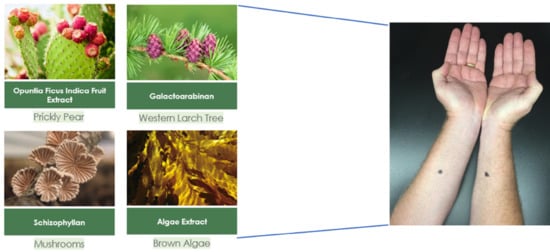
The polysaccharides that comprise the mixture employed in this study are derived from several sources and are known individually to have benefits for controlling inflammation and boosting immunity [12]. The blend has previously been shown to suppress the expression of active Caspase-1 from NLRP-activated inflammasomes in Normal Human Epidermal Keratinocytes [8,12]. The Laminaria extract is a Brown-alga extract that are known to contain unique exopolysaccharides such as fucoidan and laminarin, which are both shown to have benefits in skin health [13,14]. Opuntia ficus-indica is often referred to as Prickly Pear and the plant is known to contain unique anti-inflammatory and water-binding polysaccharides [15]. The galactoarabinan is a purified polysaccharide isolated from Western Larch trees and has been sold commercially for it benefits in human immune health [16]. Mushrooms are a well-known source of a water-soluble form of β-(1,3)-glucan which has demonstrated anti-inflammatory and immune boosting benefits [17].
Both formulations, the placebo and 3% polysaccharide formulations, employed 0.5% retinol in a very simple lotion base without known lipophilic occlusive ingredients such as mineral oil or petroleum jelly. The only fundamental difference in the formulations was the presence of the 3% polysaccharide. The molecular weight (Mw) distribution of the polysaccharides is significantly over the typical 500 Dalton Mw that is anticipated to allow appreciable penetration of the polysaccharides into unperturbed skin. It is anticipated, therefore, that the polysaccharides are principally located at the surface of the stratum corneum. It may be that the polysaccharides could also be inhibiting the penetration of the retinol into the skin as would be required for the retinol to reach the viable epidermis. While this cannot be conclusively ruled out, the overall concentration of the polysaccharides on the treated skin is quite low given the large surface area of the available skin. As skin biopsies or tape-stripping of the volunteer’s skin was not done and all skin measurements were done using non-invasive measuring techniques, we cannot exclusively rule out the possibility that the polysaccharides may inhibit some penetration of the retinol. Retinol is effective as a topical treatment at levels as low as 0.1%. It would be surprising if the polysaccharides were able to effectively minimize all penetration of the retinol given their relatively low abundance on the skin.
The possibility that the polysaccharides might sequester the retinol in a kind of molecular complex within the overall formulation has been suggested. For example, it has been demonstrated that certain oligosaccharide/polyethylenimine copolymers can form sequestered complexes with retinol in aqueous solution [18]. The behavior of the retinol in aqueous solution would be quite different than its behavior in the colloidal emulsified formulations used in the study. In such formulations, the highly lipophilic retinol, which is provided as a solution in soybean oil, would likely remain highly concentrated in the oil phase of the oil-in-water emulsions used in the study. As such, unlike the complexes described in the work by Bekhradnia et al., the retinol would not be available to interact well with the entirely aqueous solubilized polysaccharides. The retinol would remain intimately dissolved in the soybean oil even after application and so would likely have little chance to interact with the hydrophilic polysaccharides even after application. It remains unlikely that the polysaccharides are interfering with the skin penetration of the retinol which would have a strong affinity for the more lipophilic components of the skin’s lipid barrier.
The polysaccharide blend also contains a small amount of glycerin (less than 5%) which helps to stabilize the mixture and keeps the viscosity of the four-polysaccharide hydrogel complex pourable. It is unlikely that the levels of glycerin found in the 3% of the polysaccharide blend employed in the study would contribute to the moisturizing benefits. In previous studies, it was demonstrated that the blend of polysaccharides worked to extend hydration for 48 h even in the presence of much higher levels of glycerin [9].
It is interesting that in the shorter duration of this study, that is 12 days, the topical treatment of the skin with 0.5% retinol appears to influence skin hydration most profoundly. In the lengthier study reported by McDaniel et al., it was noted that retinoic acid is quite dehydrating to the skin and that retinol, while not being as dehydrating, still indicated a drying effect [7]. It has been reported that retinol can upregulate both the Aquaporin-3 and Hyaluronic Acid Synthase genes in skin mimics [19,20]. This might suggest that upregulation of these genes should increase skin hydration. The Aquaproin-3 gene/protein is a moisture regulator, and it could be that upregulation of this gene may influence skin hydration by pulling skin moisture from the surface of the skin into the epidermis. Upregulation of the HAS gene suggests increased formation of hyaluronic acid in the skin’s epidermis which could increase skin plumpness but not necessarily the skin’s surface moisture content. Furthermore, the timeframe for formation of appreciable amounts of HA within the epidermis may be longer than the 12-days of this study and may not manifest itself in any appreciable way with increased skin surface moisture content as would be measured by a Corneometer. It is important to note as well that most studies looking at retinol’s influence typically focus on the longer term (8–12 weeks) anti-aging benefits and less on the shorter-term moisturizing effects on the skin. In addition, the studies are most often run on the face and not on the inner volar forearm where the thickness of the skin’s stratum corneum may play an influencing role in all the parameters examined in this study.

4. Conclusions

This study examined the impact of formulations containing 0.5% retinol to see how the formulations impacted skin hydration, skin barrier function and skin redness over a 12-day application timeframe with product application 2X/day. It was found that formulations containing 0.5% retinol were not overly irritating or damaging to the skin’s barrier function over the 12-day treatment regimen. However, 0.5% retinol did prove to be very drying to the skin, a condition which upon longer application periods might begin to lead to some barrier disruption and skin irritation. It was found that a unique blend of polysaccharides previously shown to influence the skin’s innate immune response to external threats that prime skin cells NLRP inflammasomes (keratinocytes) to release active Caspase-1 was able to not only maintain skin hydration during the retinol treatments, but also to extend skin hydration out for 3-days after a final treatment and regression. The study supports an emerging hypothesis about how to build and maintain extended skin hydration by employing larger, natural, water-loving polysaccharides that can remain bound to the skin over extended timeframes.

Author Contributions

Conceptualization, J.V.G., V.S. and J.R.; Methodology, J.V.G., V.S. and J.R.; Writing—original draft, J.V.G., V.S. and J.R.; Writing—review & editing, J.V.G., V.S. and J.R. All authors have read and agreed to the published version of the manuscript.

Funding

The research was funded by BotanicalsPlus®.

Conflicts of Interest

The authors of the paper are all employees of BotanicalsPlus®. No other Conflicts of Interest are reported. The funders had no role in the design of the study; in the collection, analyses, or interpretation of data; in the writing of the manuscript, or in the decision to publish the results.

References

  1. Fisher, G.J.; Esmann, J.; Griffiths, C.E.; Talwar, H.S.; Duell, E.A.; Hammerberg, C.; Elder, J.T.; Finkel, L.J.; Karabin, G.D.; Nickokoloff, B.J. Cellular, Immunologic and Biochemical Characterization of Topical Retinoic Acid-Treated Human Skin. J. Investig. Dermatol. 1991, 96, 699–707. [Google Scholar] [CrossRef] [PubMed] [Green Version]
  2. Fluhr, J.W.; Vienne, M.P.; Lauze, C.; Dupuy, P.; Gehring, W.; Gloor, M. Tolerance Profile of Retinol, Retinaldehyde and Retinoic Acid Under Maximized and Long-Term Clinical Conditions. Dermatology 1999, 199 (Suppl. 1), 57–60. [Google Scholar] [CrossRef] [PubMed]
  3. Mukherjee, S.; Date, A.; Patravale, V.; Korting, H.C.; Roeder, A.; Weindl, G. Retinoids in the Treatment of Skin Aging: An Overview of Clinical Efficacy. Clin. Interv. Aging 2006, 1, 327–348. [Google Scholar] [CrossRef] [PubMed]
  4. Kligman, A.M.; Willis, I.A. New Formula for Depigmenting Human Skin. Arch. Dermatol. 1975, 111, 40–48. [Google Scholar] [CrossRef] [PubMed]
  5. Kang, S.; Duell, E.A.; Fisher, G.J.; Datta, S.C.; Wang, Z.Q.; Reddy, A.P.; Tavakkol, A.; Yi, J.Y.; Griffiths, C.E.M.; Elder, J.T.; et al. Application of Retinol to Human Skin In Vivo Induces Epidermal Hyperplasia and Cellular Retinoid Binding Proteins Characteristic of Retinoic Acid but Without Measurable Retinoic Acid Levels or Irritation. J. Investig. Dermatol. 1995, 105, 549–556. [Google Scholar] [CrossRef] [PubMed] [Green Version]
  6. Shao, Y.; He, T.; Fisher, G.J.; Voorhees, J.J.; Quan, T. Molecular Basis of Retinol Anti-Aging Properties in Naturally Aged Human Skin In Vivo. Int. J. Cosmet. Sci. 2017, 39, 56–65. [Google Scholar] [CrossRef] [PubMed] [Green Version]
  7. McDaniel, D.H.; Mazur, C.; Wortzman, M.S. Efficacy and tolerability of a double-conjugated retinoid cream vs 1.0% retinol cream or 0.025% tretinoin cream in subjects with mild to severe photoaging. J. Cosmet. Dermatol. 2017, 16, 542–548. [Google Scholar] [CrossRef] [PubMed] [Green Version]
  8. Gruber, J.V.; Stojkoska, V. NLRP Inflammasomes and Induced Skin Inflammation, Barrier Recovery and Extended Skin Hydration. Int. J. Cosmet. Sci. 2020, 42, 68–78. [Google Scholar] [CrossRef] [PubMed]
  9. Gruber, J.V. Inflammasomes: Key to the Engine of Skin Inflammaging. Pers. Care N. Am. 2020, 3, 27–33. [Google Scholar]
  10. Gankande, T.U.; Duke, J.M.; Wood, F.; Wallace, H.J. Interpretation of the DermaLab Combo® Pigmentation and Vascularity Measurements in Burn Scar Assessment: An Exploratory Analysis. Burns 2015, 41, 1176–1185. [Google Scholar] [CrossRef] [PubMed]
  11. Hua, W.; Fan, L.M.; Dai, R.; Luan, M.; Xie, H.; Li, A.Q.; Li, L. Comparison of two series of non-invasive instruments used for the skin physiological properties measurements: The DermaLab® from Cortex Technology vs the series of detectors from Courage & Khazaka. Skin Res. Technol. 2017, 23, 70–78. [Google Scholar] [PubMed]
  12. Gruber, J.V.; Holtz, R. In vitro expression of NLRP inflammasome-induced active Caspase-1 expression in normal human epidermal keratinocytes (NHEK) by various exogenous threats and subsequent inhibition by naturally derived ingredient blends. J. Inflamm. Res. 2019, 12, 219–230. [Google Scholar] [CrossRef] [PubMed] [Green Version]
  13. Hwang, P.A.; Yan, M.D.; Lin, H.T.V.; Li, K.L.; Lin, Y.C. Toxicological evaluation of low molecular weight Fucoidan in vitro and in vivo. Mar. Drugs 2016, 14, 121. [Google Scholar] [CrossRef] [PubMed] [Green Version]
  14. Ozanne, H.; Toumi, H.; Roubinet, B.; Landemarre, L.; Lespessailles, E.; Daniellou, R.; Cesaro, A. Laminarin effects, a β-(1,3)-Glucan, on skin cell inflammation and oxidation. Cosmetics 2020, 7, 66. [Google Scholar] [CrossRef]
  15. Petruk, G.; Di Lorenzo, F.; Imbimbo, P.; Silipo, A.; Bonina, A.; Rizza, L.; Piccoli, R.; Monti, D.M.; Lanzetta, R. Protective effect of Opuntia ficus-indica L. cladodes against UVA-induced oxidative stress in normal human keratinocytes. Bioorg. Med. Chem. Lett. 2017, 27, 5485–5489. [Google Scholar] [CrossRef] [PubMed] [Green Version]
  16. Kelly, G.S. Larch arabinogalactan: Clinical relevance of a novel immune enhancing polysaccharide. Altern. Med. Rev. 1999, 4, 96–103. [Google Scholar] [PubMed]
  17. Novak, M.; Vetvicka, V. Beta-glucans, history, and the present: Immunomodulatory aspects and mechanisms of action. J. Immunotoxicol. 2008, 5, 47–57. [Google Scholar] [CrossRef] [PubMed]
  18. Bekhradnia, S.; Naz, I.; Lund, R.; Effenberg, C.; Appelhaus, D.; Sande, S.A.; Nystrom, B. Characterization of oligosaccharide-functionalized hyperbranched poly(ethylene imine) and their complexes with retinol in aqueous solution. J. Colloid Interface Sci. 2015, 458, 178–186. [Google Scholar] [CrossRef] [PubMed]
  19. Chaudhuri, R.K.; Bojanowski, K. Bakuchiol: A retinol-like functional compound revealed by gene expression profiling and clinically proven to have anti-aging effects. Int. J. Cosmet. Sci. 2014, 36, 221–230. [Google Scholar] [CrossRef] [PubMed]
  20. Li, W.H.; Wong, H.K.; Serrano, J.; Randhawa, M.J.; Kaur, S.; Southhall, M.D.; Parsa, R. Topical stabilized retinol treatment induces the expression of HAS genes and HA production in human skin In vitro and in vivo. Arch. Dermatol. Res. 2017, 309, 275–283. [Google Scholar] [CrossRef] [PubMed]
Figure 1. Arrangement of three treatment sites on each arm. Control measurements were taken near the wrist and near the elbow as to generate control measurements.
Cosmetics 07 00080 g001
Figure 2. Results of Transepidermal Water Loss (TEWL) measurements (g/m2/h). Dark grey is the untreated control, black is the retinol placebo-treated and light grey is the polysaccharide active-treated sites. N = 22, p ≤ 0.05.
Cosmetics 07 00080 g002
Figure 3. Results of erythema measurements. Dark grey is the untreated control, black is the retinol placebo-treated and light grey is the polysaccharide active-treated sites. N = 22, p ≤ 0.05.
Cosmetics 07 00080 g003
Figure 4. Results of skin hydration (µSiemans) measurements. Dark grey is the untreated control, black is the retinol placebo-treated and light grey is the polysaccharide active-treated sites. N = 22, p ≤ 0.05.
Cosmetics 07 00080 g004
Figure 5. Results of 3-day skin hydration regression study. Dark grey is the untreated control site, black is the retinol placebo-treated site and light grey is the polysaccharide active-treated site. N = 22, p ≤ 0.05).
Cosmetics 07 00080 g005
Table 1. Ingredients employed in formulations used in the study.
IngredientINCI NameQuantity (%)
Placebo
WaterAqua95
Jeesperse®ICE-T®-LB-T-NSSodium Acrylate/Sodium Acrylol Dimethyl Taurate Copolymer (and) Cetyl Alcohol (and) Glycol Stearate (and) Glyceryl Stearate (and) Caprylic/Capric Triglyceride3.5
Jeecide® PhenoxyPhenoxyethanol1
Retinol 10 SURetinol (and) Glycine Soja (Soybean) Oil0.5
Active
WaterAqua92
Jeesperse®ICE-T®-LB-T-NSSodium Acrylate/Sodium Acrylol Dimethyl Taurate Copolymer (and) Cetyl Alcohol (and) Glycol Stearate (and) Glyceryl Stearate (and) Caprylic/Capric Triglyceride3.5
Jeecide® PhenoxyPhenoxyethanol1
Retinol 10 SURetinol (and) Glycine Soja (Soybean) Oil0.5
StrataPhix™ POLYWater (and) Glycerin (and) Laminaria Digitata Extract (and) Opuntia Ficus-Indica Stem Extract (and) Galactoarabininan (and) Schizophyllan3
Publisher’s Note: MDPI stays neutral with regard to jurisdictional claims in published maps and institutional affiliations.